7 Simple Rules for Parasite Control in Racing Yards

Article by James Gibbons

FWEC testing for worms in racehorses

When Benjamin Franklin wrote, in 1789, ‘in this world nothing can be said to be certain, except death and taxes’, he could, perhaps, have added another certainty to his list – worms in horses. Unlike most other infectious diseases of horses, such as strangles or influenza—which infect a small number of horses relative to the entire horse population at any one time—worms are present in almost all horses all of the time.  This fact leads to two obvious conclusions: first, it is not possible to eradicate parasites and the threat of parasitic disease from our horses; second, worm control is vital in any environment where horses are kept.  

The threat posed by intestinal worms to racehorse performance has long been recognised by horsemen the world over. For many years, regular treatment with anthelmintic drugs (‘dewormers’) was the mainstay of worm control in racing yards, as it was in most in areas of horse breeding and production. The emergence of resistance to these deworming drugs (referred to as ‘anthelmintic resistance’), in the last 10 years in particular, has meant that such regular treatments may no longer be effective and may in fact make the resistance situation in the yard or farm worse. With this in mind, it is important that we consider how best to control worms while preserving the efficacy of the few deworming drugs available to us. This can be achieved using control programs comprised of drug- and non-drug control measures. This article sets out seven points/rules to consider and implement when developing such a worm control programme for racing yards. 

1. Know your enemy

It is not possible to draw up a parasite control program without considering which worms it is you are trying to control. The three main types of worms we are concerned with are strongyles (large and small redworms), ascarids (roundworms) and tapeworms. The role of tapeworms in equine intestinal disease is debated, but they appear to be linked to certain forms of colic when present in high numbers. Notably, there is no evidence of age-related immunity to tapeworms. Ascarids are a cause of disease in foals primarily and yearlings and so are likely to be of less concern in most racing yards. However, unlike redworms, which are transmitted almost exclusively at grass, ascarids can be transmitted in the stable; and ascarid eggs survive for years in the environment so that a single infected animal can infect other young horses for years to come.  

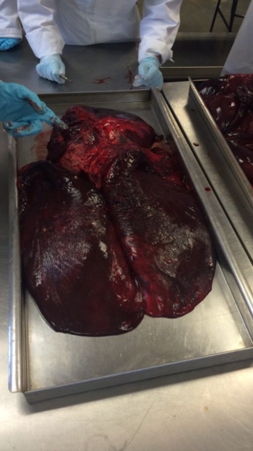
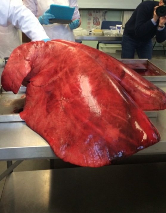
Redworms are the most important parasite of horses and are found in horses of all ages where they can cause anaemia, weight loss, ill-thrift and diarrhoea. Large redworms can burrow into the walls of blood vessels that supply blood to the gut causing a very severe form of colic.  Small red worms can lie dormant in the gut wall for extended periods then emerge en masse to cause an acute shock-like syndrome with severe diarrhoea, which is often fatal.  

Identifying worms through FWEC testing

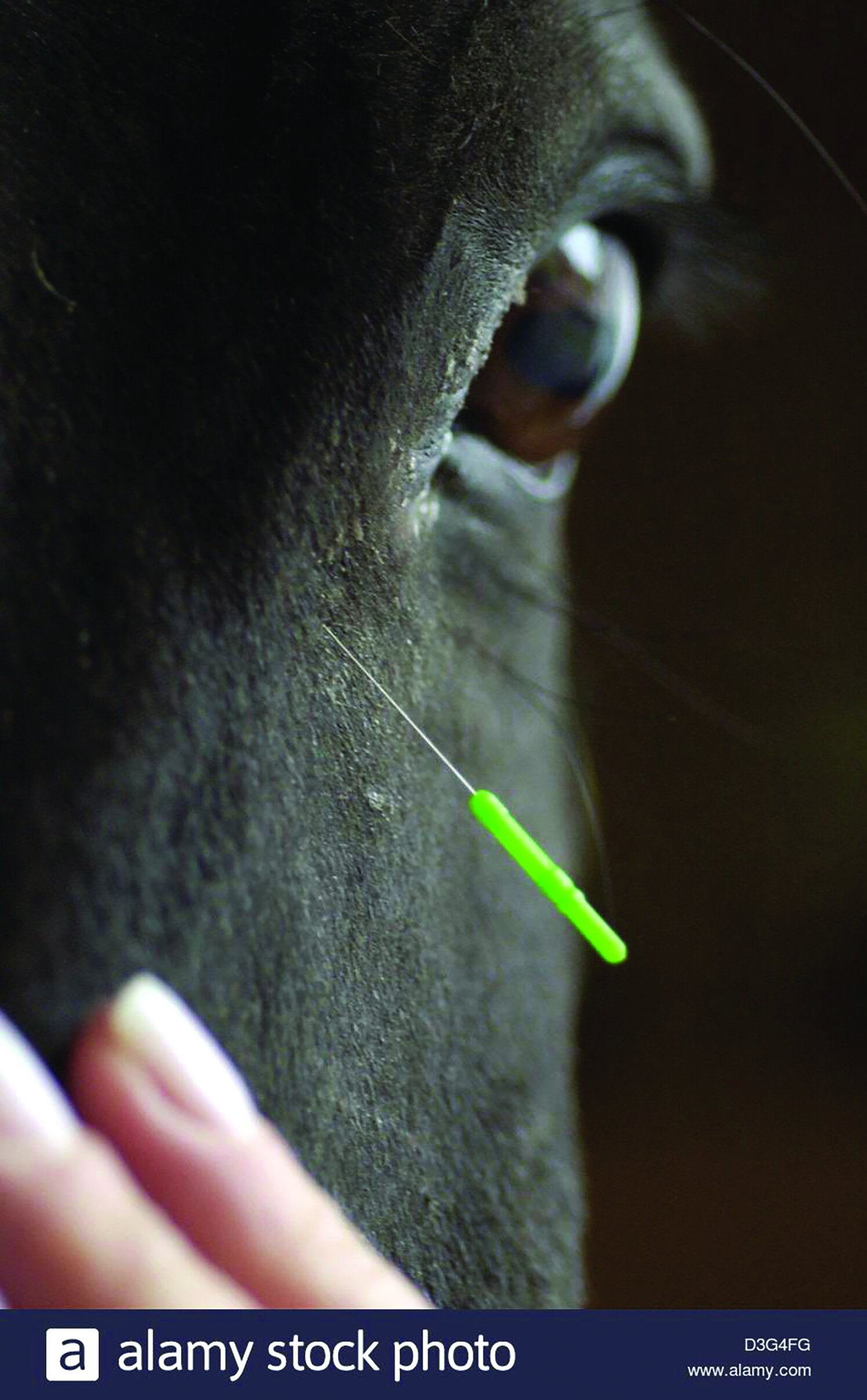
Faecal egg count (FEC) testing will identify if redworms or ascarid eggs are present in your horse’s droppings and this, in turn, tells you that the adult worm of that species is present in your horse’s gut. Tapeworm eggs can be detected by FEC testing, but it is not the most reliable method for their detection as the shedding of these eggs is not consistent. As redworms are the most relevant worm present in most yards, most of the information in this article relates specifically to the control of redworms rather than ascarids or tapeworms.

2. Know your horses’ risk

All trainers pride themselves on their knowledge of almost every aspect of their horses’ anatomy and physiology, but not all could tell you which of their horses are at greater or lesser risk of worms. As mentioned above, all horses are likely to carry some worms, but the adult worm burden and the number of worm eggs shed in faeces is far greater in younger horses than in mature stock.  Most horses between the ages of 5 and 15 years will have a lower worm burden, and a lower risk of parasitic disease, than horses below this age due to a degree of age-related immunity. Foals and yearlings can carry particularly high worm burdens and shed large numbers of eggs into the environment to infect other horses. 

It follows that any yard with yearlings—two- or three-year-olds—will need a comprehensive worm control programme; while yards with older stock may get away with less strenuous controls. Horses over the age of 15 may also have higher worm burdens. And while these are unlikely to be in training, they may be used as a riding horse or companion animal and act as a potential source of infection for the string.

3. Know your yard’s weak points

Paddock maintenance to decrease worm burdens

It is not unusual for our lab to get a call from a racehorse trainer wishing to express their disbelief that the faecal egg count test from their horses has tested positive for redworm eggs despite their horses having no access to grass. In these situations, careful questioning as to how the yard operates will usually reveal the use of turn-out paddocks for a short period at some point during the week. Invariably, these are shared, often quite small, paddocks which host many horses over time; and so they are more likely to be contaminated with worm eggs.  

While the intention is for the horse to get ‘a pick of grass’, it may be that it is more ‘a pick of worms’ they are getting in such paddocks! As part of a worm control plan, it is important to first identify high-traffic areas, which may be a pinch-point for worm transmission.  Once identified, the key to reducing the worm burden on such paddocks is ideally through the removal of droppings. This can be a labour-intensive exercise, but it only needs to be done twice weekly rather than daily. And there are now more automated methods for cleaning paddocks than the more traditional wheelbarrow and spade!  

4. Identify high shedders 

FEC testing not only tells you what type of worm is present in your horse’s droppings but also how many eggs there are per gramme of faeces.  Repeated FEC testing allows you to build up a picture of the shedding patterns of the horses within your stable. Horses in racing yards should have FEC testing carried out every three months. It is generally accepted that the shedding of worm eggs in horses follows the 80/20 rule; that is, 20% of the horses shed 80% of the eggs.  

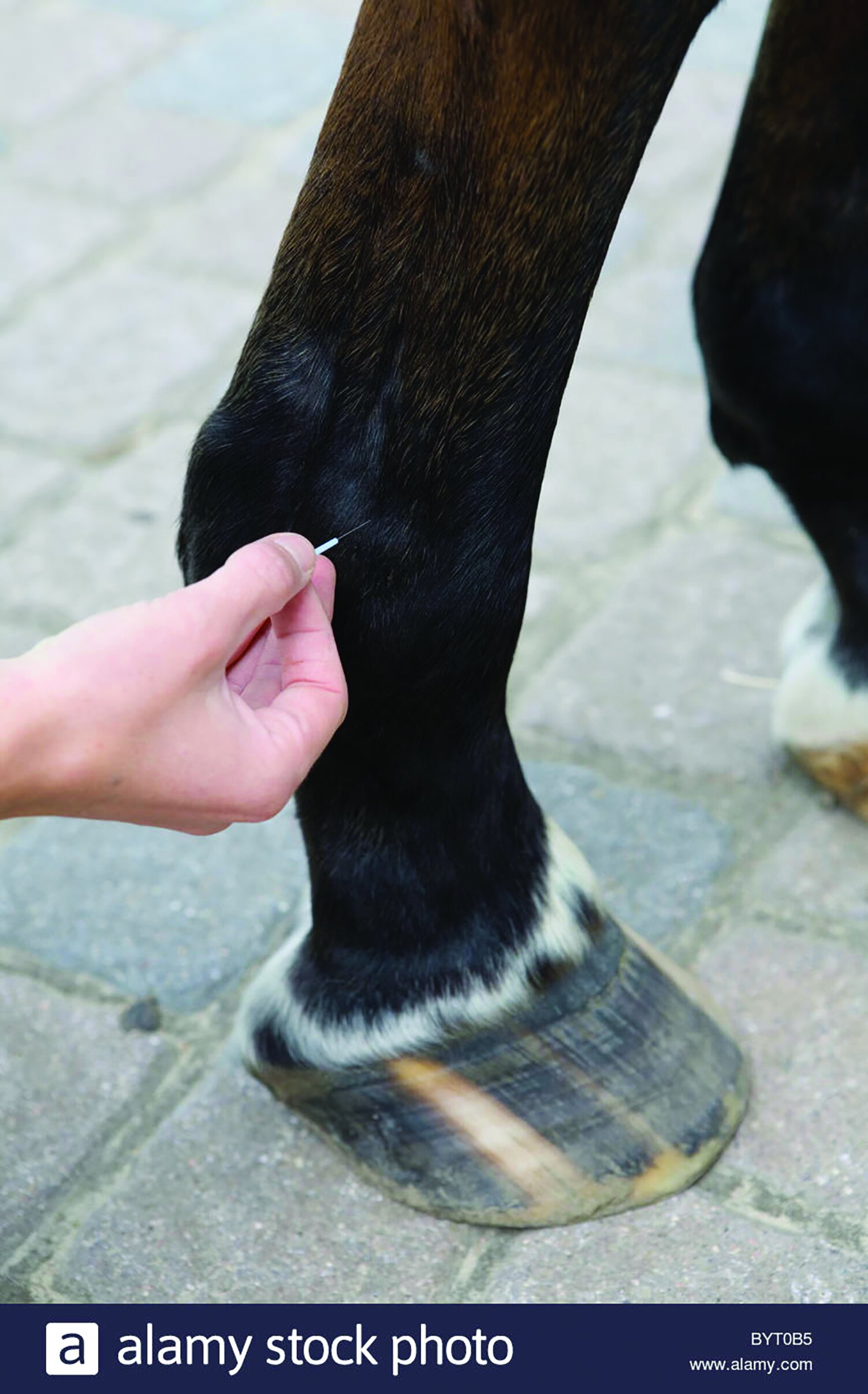
If these high shedders in the group can be identified, then targeted treatment of them may be more beneficial (and cost-effective) than blanket treatment of the entire group.    Horses with a strongyle egg count in excess of 250 eggs per gramme (EPG) on repeated testing may be considered high shedders and require more frequent egg counts and treatment.  It is important to state that this figure of 250EPG is not absolute, and the threshold above which animals are considered high shedders or requiring treatment should be set in conjunction with your vet. Worm eggs are not distributed evenly within the droppings, so when collecting samples for FEC testing, make sure to take at least three faecal balls—each from a different area of the pile. 

FEC testing can only detect egg-laying adult worms, but the egg count is not a reliable indicator of the adult worm burden of the horse, i.e. a horse with a high FEC does not necessarily have a greater worm burden than a horse with a lower FEC; but the horse with the high FEC is more significant in terms of worm transmission to other horses. Immature worms that may be present in the horse are not detected by FEC testing. In recent years, new tests have been developed that can detect antibodies to tapeworms and small redworm in blood and/or saliva. These tests are a useful addition to any worm control programme. Regardless of the type of test used, dewormers are still necessary as part of any worm control programme, in foals and yearlings in particular, but also in high-risk environments, and in order to control the disease risks posed to horses of all ages by large strongyles.  

5. Know which drugs work

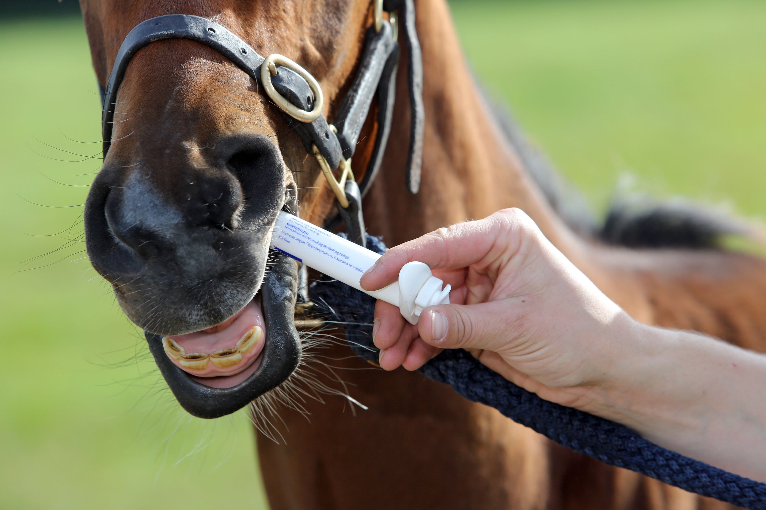
While it might seem like there is an endless range of deworming products for horses on the market, the number of active ingredients in these products is very limited with only four drugs (fenbendazole, ivermectin, moxidectin and pyrantel) available for the treatment of redworms and ascarids. With such limited availability, and no prospect of new worm treatments entering the market anytime soon, it is vital that we use the existing drugs judiciously so as to preserve their effectiveness into the future.  

The threat posed by anthelmintic resistance is very real. Already fenbendazole resistance in redworms is widespread, and ivermectin/moxidectin resistance in ascarids is becoming more common. This means that there are only one or two effective treatments remaining for these resistant worms. While thoughts of anthelmintic resistance may not keep any racehorse trainer awake at night, we do expect that the deworming treatments we use to be effective, not least because they cost quite a lot of money!  The only way to determine if our treatments are indeed effective is to carry out a faecal egg count reduction test (FECRT). 

This test follows on from the FEC test; any horse with a FEC of 500EPG or more should be treated with a dewormer and have a follow-up FEC carried out 10–14 days after that treatment. The percentage reduction in egg numbers is calculated and should be greater than 90–95% (depending on the drug). Reductions less than this are suggestive of a resistance problem. Because of the existing resistance situation, FECRTs should be carried out before using fenbendazole to treat redworms or ivermectin/moxidectin to treat ascarids. Equally, if you have any suspicion that a deworming product is not working effectively in your yard, then a FECRT for that drug should be carried out to investigate this.   

6. Don’t overtreat

Not overtreating with worming products to stop resistance

Aside from the risk of anthelmintic resistance development and the significant costs incurred by frequent or blanket deworming of all horses in the yard, such treatments can have other effects on the horse, which may ultimately impact performance. Research has shown that anthelmintic administration is associated with a decrease in the diversity and abundance of certain bacteria in the horse’s gut, which may impact digestion and other metabolic processes. 

In humans, changes in the composition of the gut bacterial population have been shown to impact physiology, immunity and behaviour; and, while the structure of the gut may differ greatly between human and horses, there is no reason to believe the equine gut bacteria do not play a similarly important role in horse health. In recent years, the role of parasites in human health has been revaluated. And while they are still recognised as an important cause of disease, it is also accepted that they can play a beneficial role in the development of immunity and the prevention of some diseases. While we don’t have similar evidence of a benefit to horse health from parasites, it is not impossible that such benefits do really exist.  

A study of standardbred trotters in Denmark in 2011 found that horses with higher egg counts had better race finishing position than those with lower egg counts. So,  the presence of parasites does not always lead to poor performance! It was standard practice, in some racing yards, to deworm horses every eight weeks, but this seems excessive when one considers the biology of the worms involved. The small redworms take approximately eight weeks to develop to maturity in the horse, while the large redworms take approximately six months to do so. Treatment every eight weeks could only be justified if there were evidence of overwhelming worm exposure on an ongoing basis—something which is unlikely in a modern racing yard. Even if that were the case, other non-pharmaceutical controls would be necessary to get the situation under control.

7. Manage new arrivals

One of the challenges of worm control in a racing yard is the constant turnover of horses with some leaving the yard for rest or recovery from injury, others returning from such breaks and new horses joining the yard. When a horse leaves the yard, they leave the worm control programme as well. Evem if the yard’s deworming programme is followed while they are away, the risk factors in their new environment may be totally different and require an entirely different approach to treatment. More worrisome, the arrival of new horses or the return of others also risks the introduction of new or resistant parasites to the yard. In order to minimise this risk, all new arrivals should be isolated, tested and treated. Ideally, all new arrivals would be isolated for two weeks while a FECRT is carried out to ensure treatment has been effective.  

This may not be practical in all yards, but an isolation of period of at least three days after deworming should be observed to ensure all worms are shed before the horse joins the group. Treatment without isolation is not recommended as treated horses can shed viable eggs for a few days after deworming. There may be a temptation to let horses out to turnout paddocks for exercise or grass during the isolation period, but we have seen cases where this has led to the introduction of new parasites to the yard despite the horse being treated before it was turned out.   

There was a time when parasite control in racing yards was relatively simple and relied primarily on the regular use of deworming drugs. As with many aspects of horse racing, what was once considered acceptable is no longer so. Effective parasite control is not as simple anymore, and an overall plan designed to meet the needs of the individual yard is required. That is not to say that any trainer should be daunted by the prospect of drawing up such a plan. The seven points discussed here provide a basis to work from and your vet and laboratory are well placed to assist in building on this.  

De-worming treatment

Equine Neck CT: Advancing diagnostic precision in racehorses

Article by Rachel Tucker MRCVS

Diagnosing neck conditions in racehorses with CT imaging

Introduction

When considering neck disorders in the racehorse, we most commonly think of severe conditions such as acute neck trauma and cervical vertebral myelopathy (Wobbler Syndrome). These represent the most severe end of the scale of orthopaedic and neurologic injury to the neck; and a diagnosis, or at least prognosis, is usually clear. However, neck conditions encompass a far wider range of clinical presentations. 

horse positioned in CT scanner

Horse positioned in ct scanner

At the milder end of the scale, signs may be subtle and easily missed, whilst still being responsible for discomfort and reduced performance. The recent ability to perform a computed tomography (CT) scan of a horse’s neck represents a major advancement in our ability to diagnose neck conditions. Timely and accurate diagnosis allows efficient and targeted treatment, the ability to plan schedules and improvement in welfare through the provision of appropriate treatment and earlier return to function. 

In neurologic cases, an accurate diagnosis facilitates risk management for both the horse, their handlers and riders, while improving safety for all.  As conditions and injuries of the neck are being better characterised using CT, new medical and surgical treatment options are being developed, giving the potential for improved outcomes and fewer losses from the racing industry. 

This article summarises how CT is being increasingly used by vets to diagnose conditions of the neck and how it is revealing previously unknown information and providing exciting new treatment opportunities. 

Presentation

Conditions of the neck can cause a range of signs in the horse, which are wide-ranging in their presentation and variable in their severity. The manner in which these conditions present depends on which anatomical structures are affected. Issues affecting the bones, joints and/or soft tissues of the neck can all cause neck pain, which can manifest in a number of ways. Cases of neck pain can be severe, resulting in a horse with a rigid, fixed neck carriage, an unwillingness to walk and struggling to eat, perhaps due to a traumatic event. Neck fractures are thankfully uncommon but can be catastrophic. 

Palpating horses neck to diagnose injury

More moderate signs might be displayed as a stiff neck, with reduced range of movement and resentment of ridden work. There may be pain on palpation of the neck and changes in the neck musculature. Increasingly, we are seeing horses with far more subtle signs, which are ultimately revealed to be due to neck pain and neck pathology. Typically, these horses might have an acceptable range of motion of their neck under most circumstances, but they suffer pain or restriction in certain scenarios, resulting in poor performance. This may be seen as tension through the neck, resisting rein contact, a reluctance to extend the neck over fences, or they may struggle on landing. 
Riders might report a feeling of restriction or asymmetry in the mobility of the neck. In addition, these horses may be prone to forelimb tripping or show subtle forelimb lameness. 

Any condition, which causes injury or disease to the spinal cord or nerves within the neck, also causes a specific range of neurologic signs. Compression of the spinal cord is most commonly caused by malformations or fractures of the cervical vertebrae, or enlargement of the adjacent articular process (facet) joints. This results in classic ‘Wobbler’ symptoms, which can range from subtle weakness and gait abnormalities, through to horses that are profoundly weak, ataxic and uncoordinated. This makes them prone to tripping, falling, or they may even become recumbent. 

Peripheral nerve deficits are uncommon but become most relevant if they affect the nerves supplying the forelimbs, which can result in tripping, forelimb lameness, or local sensory deficits. This lameness might be evident only in certain circumstances, such as when ridden in a rein contact. This lameness is difficult to pinpoint as there will be no abnormality to find in the lame limb, indeed a negative response to nerve and joint blocks (diagnostic analgesia) will usually be part of the diagnostic process.

Horses can present with varying combinations and severities of neck pain, neurologic signs and peripheral nerve deficits, creating a wide range of manifestations of neck related disease. 

Diagnosis

A diagnosis of neck pain is based on careful static and dynamic clinical examination and may be supported by seeing a positive response to treatment. Neurologic deficits are noted during a specific neurologic assessment, which includes a series of provocation tests such as asking a horse to walk over obstacles, back up, turn circles and walk up and down a hill. Confirming neck pathology as the cause of signs can be difficult. Until recently, radiography has been the mainstay imaging modality. Radiographs are useful for assessment of the cervical vertebrae and continue to play an important role in diagnosis; however the complex 3-dimensional shape of these bones, the large size of the neck and an inability to take orthogonal (right-angled) x-ray views means that this 2-dimensional imaging modality has significant limitations. High quality, well-positioned images are essential to maximise the diagnostic potential of radiographs. 

A turning point in our diagnostic ability and understanding of neck dysfunction has been the recent adaptation of human CT scanners to allow imaging of the horse’s neck. A number of equine hospitals across the United Kingdom and Northern Europe now offer this imaging modality. We have been providing this service at Liphook Equine Hospital since 2017, with over 150 neck scans performed to date.

The CT procedure

A computed tomography (CT) scan combines a series of x-ray images taken from different angles around the area of interest to create a 3-dimensional volume of imaging data. This data is presented as a grayscale image which can be viewed in any plane and orientation. It provides excellent bone detail, and post processing techniques can provide information on soft tissue structures too. Additional techniques can be employed such as positive contrast myelography to provide greater detail about soft tissue structures. Myelography delineates the spinal cord using contrast medium injected into the subarachnoid space and is indicated in any case showing neurologic signs suggestive of spinal cord compression. 

Neck CT is performed under a short general anaesthetic. Scans without myelography typically take less than 20 minutes to complete. The entire neck is imaged, from the poll to the first thoracic vertebra. The procedure is non-invasive and low risk, with anaesthetic-related complication presenting the main risk factor to the procedure. We have not encountered any significant complications in our plain CT scan caseload to date. Horses showing ataxia, weakness and incoordination (Wobbler’s), undergo CT myelography which adds around 20 minutes to the procedure. These horses are exposed to a greater level of risk due to their neurologic condition, the injection of a contrast agent and the increased chance of destabilising a more severe lesion during the procedure. 

Sagittal and transverse CT myelogram images of a young racing thoroughbred showing neurologic (Wobbler) signs.  shows narrowing of the spinal canal at the base of the neck (arrow)

Sagittal and transverse CT myelogram images of a young racing thoroughbred showing neurologic (Wobbler) signs. shows narrowing of the spinal canal at the base of the neck (arrow)

CT is revealing more detailed information about ways that spinal cord compression can occur in Wobbler cases, about compression of spinal nerves resulting in forelimb gait deficits and precise detail about fracture configurations. It gives us detailed images of articular process joint disease, intervertebral disc disease, developmental conditions and anatomic variations. It is also revealing information about rare diseases such as vertebral abscesses or spinal neoplasia. As our caseload and confidence in the imaging modality grows, we are learning more about the value of CT in examining more subtle neck conditions. We are also bringing the benefit of a more accurate diagnosis, allowing precise targeted treatment and a better ability to provide a prognosis about outcomes—likely progression or safety factors. CT myelography allows circumferential imaging of the spinal canal and yields significantly more information than traditional x-ray myelography. As a result, we hope to enable better case selection of horses that may benefit from Wobbler surgery, with the goal of resulting in improved success rates of the surgery.

Innovations in treatment

image shows a fracture of the articular process of the 3rd cervical vertebrae in the mid neck, which was not visible on x-ray. 

image shows a fracture of the articular process of the 3rd cervical vertebrae in the mid neck, which was not visible on x-ray. 

New treatment options are emerging as a result of our more accurate diagnoses of neck pathology. Of the first 55 horses which underwent neck CT at our hospital, we were surprised to discover that 13 (24%) had some form of osteochondral fragmentation within the articular process joints of their neck. Some of these horses were young Thoroughbreds, bred to race but showing Wobbler signs. These tended to have convincing CT evidence of type 1 CVM (Wobbler Syndrome) and osteochondrosis affecting their neck. Others had fragments which were larger and more discrete, with evidence of articular process joint enlargement/arthritis but no other bony lesions. These horses were typically older and of a range of breeds and uses. 

In those horses presenting with signs of neck pain but no neurologic deficits, surgical removal of these fragments was proposed. Following further consideration and cadaver training, we have begun to offer this surgery for horses that fit the appropriate criteria and have surgically accessible fragments.  We have performed arthroscopic or arthroscopic-assisted fragment removal from eight articular process joints in six horses to date. No intraoperative or postoperative complications have been encountered; and five of six horses showed complete resolution of neck pain. In the sixth horse, full recovery was not anticipated due to the presence of additional neck pathology, but partial improvement occurred for two years.  Fragment removal has relieved signs of neck pain and stiffness and caused improved performance in these horses. 


Two procedures that are emerging to treat spinal nerve root impingement are a targeted peripheral nerve root injection and a keyhole surgical procedure to widen the intervertebral foramen. Nerve root injection is performed in the standing sedated horse under ultrasound guidance. Surgery is performed under anaesthesia, using specialised minimally invasive equipment to widen the bony foramen using a burr. This surgery is in its infancy but offers an exciting treatment option.  

Additionally, CT gives us the ability to better plan for fracture repair, undoubtedly improving our case selection for Wobbler surgery; it more accurately guides intra-articular injection of the articular process joints. 

Summary

Computed tomography is transforming our ability to diagnose conditions of the horse’s neck. The procedure is low risk and now widely available in the UK and other parts of Europe. It is driving the innovation of novel treatment options with the goal of improving outcomes and reducing losses to conditions of the neck. Our CT findings are posing new questions about neck function, pain and neurologic disease and is an active area of ongoing research.

Anatomy of the cervical vertebrae

The neck vertebrae of the horse anatomy

The neck vertebrae of the horse

The neck consists of seven cervical vertebrae which form a gentle S-shaped curve to link the skull at the poll to the thoracic vertebrae of the chest. Its primary functions are to protect the spinal cord, support the heavy weight of the head and to allow a large range of movement so that a horse can monitor his environment and run at speed, both being vital to this prey species. 

The first (atlas) and second (axis) cervical vertebrae are highly adapted to allow head mobility. The third to sixth vertebrae are very similar in shape, whereas the seventh is shortened as it starts to show similar features to the vertebrae of the thorax. The typical cervical vertebra consists of a cylindrical column of bone (vertebral body) articulating with adjacent vertebrae via an intervertebral disc. 

Cross section through a cervical vertebra

Running along the upper surface of this bony column is a bony canal created by the vertebral arch. This canal protects the spinal cord and associated structures that run through the centre. Above and to the side of the spinal canal sit the paired articular processes and below; and to the side sit the transverse processes. These are bony prominences to which muscles attach. The soft tissues of the neck are complex and intrinsically linked to the forelimbs and entire axial skeleton.  

The articular processes also form the articular process joints (facet joints) which create an additional two joints between each vertebra. Although highly adapted, these joints are similar to others in the body consisting of cartilage-lined surfaces, a joint capsule and joint fluid. The joint surfaces are oval in shape, approximately 3-4cm in diameter and sit at an oblique angle to the neck. They provide important additional support and mobility to the neck.

References:

Schulze N., Ehrle, A., Beckmann, I and Lischer, C. (2021) Arthroscopic removal of osteochondral fragments of the cervical articular process joints in three horses. Vet Surg. ;1-9. 

Swagemakers J-H, Van Daele P, Mageed M. Percutaneous full endoscopic foraminotomy for treatment of cervical spinal nerve compression in horses using a uniportal approach: Feasibility study. Equine Vet J. 2023. 

Tucker R, Parker RA, Meredith LE, Hughes TK, Foote AK. Surgical removal of intra-articular loose bodies from the cervical articular process joints in 5 horses. Veterinary Surgery. 2021;1-9.

Wood AD, Sinovich M, Prutton JSW, Parker RA. Ultrasonographic guidance for perineural injections of the cervical spinal nerves in horses. Veterinary Surgery. 2021; 50:816–822. 


Treating 'bucked shins' in the thoroughbred racehorse

Article by Adam Jackson MRCVS 

Bucked shins veterinary perspective

One of the most common causes of lost days to training and racing in racehorses is dorsal metacarpal disease (DMD), which is often referred to as “bucked shins” or “sore shins”.  

Often a frustration to trainers and owners, this problem rears its ugly head at the time of highest expectations, such as arising the last day of work before a horse’s first race; right after a horse’s first victory; or after a horse was purchased at a two-year old sale.

This disease presents with heat, pain with or without inflammation (swelling) on the dorsal (front) surface or the dorsomedial (front inside) surface of the third metacarpal bone (cannon) referred to as acute periostitis. With rest and reduced exercise, the condition can improve, but catastrophic fractures of the cannon may occur at the site of previous DMD episodes.  A good understanding of this disease and strategies of prevention are vital in order to improve the welfare of the horse and reduce the potential expenses to all shareholders.  

Introduction

Cannon bone structure in racehorses

The cannon bone is an important structure in the weight-bearing and absorbing shock. As the horse moves, the bone bends a little and then returns to its original shape like an elastic band, often referred to as elastic deformation.  In addition, it has been observed that horses that work slowly have tension on the front of the cannon bone; in other words, the bone is stressed by a stretching force rather than a compressing force. However, at higher speeds, these forces change from stretching to compressing forces.

Repeated bending forces (stress cycle) on the cannon bone causes dorsal metatarsal disease. When the horse is young, it has a thin bone cortex. As the horse grows and is repeatedly subjected to these forces, the bones remodel and the cortex thickens, making it stronger. However, if the bending forces exceed the bone’s ability to remodel, then this leads to stress fatigue and bone damage.

The occurrence of bucked shins is most common when horses are developing, typically two–three years old as training becomes more intensive. But it must be noted that if the horse is not bone fit, any aged racehorse is susceptible to these diseases when they begin training. Roughly at the age of five years old when a horse is fit, they are at a low risk of this disease. Within the first six months of training, DMD may present in one or both front limbs. If the condition does occur in both front limbs and the horse is being trained on a circular track, then it is likely the inside leg is where it will occur first.  In other words, if the training tends to be in a counterclockwise training circuit, then there are greater forces on the left limb than the right; thus the left is more likely to develop the disease before the right limb.

Risk Factors of DMD

Risk factors of DMD in racehorses

Age: DMD occurs most commonly in 2–3 year olds, often within their first 6 months of training. It is rarely seen in horses with a mature skeleton (age 4 and over). However, this disease has been seen in 5 year olds especially if they have been stalled for a long amount of time after weaning and not racing until that age.

Gender: It is believed that the gender of the horse does not alter its risk to DMD.

Breed: Most common in thoroughbreds but may be seen in both standardbreds and quarter horses. 

Surface impact on bucked shins in Thoroughbreds

Genetics: The risk of DMD is influenced by genetics as variation in limb bone geometry (inherited) behaves differently to force/strains on the bone. In addition, the longer the cannon bone, the greater the load is at flexion of the dorsal cortex of the bone, making it more susceptible to DMD.

Training and racing surfaces: The different types of training and racing surface alter the risk to DMD because there are variations in the force applied to limbs as well as the acceleration rates of hoof impact.  Furthermore, the impact of these forces is increased with greater speed.  Dirt tracks tend to be the hardest surface, whereas synthetic tracks reduce hoof and limb impact and loading force.  However, it is important to remember that the hardness of all of these surfaces can be altered by a number of other factors such as:

Forces applied to the cannon bone impacting bucked shins from different surfaces
  1. Different surface materials

  2. Changes in weather, temperature and humidity

  3. Surface maintenance (i.e., soaking, harrowing)

  4. Changes in horse body weight

  5. Age of surface – wear and tear of surface

  6. Human opinion of track’s condition

Training: The length of time for bones to respond to different training practices is unknown. Although further research is required, it is suggested that fast work should be avoided in the early stages of training as it is thought that high-speed exercise introduced too quickly (within 1 month) was detrimental to bone health.


Direction of training: Track direction varies globally. Thoroughbreds tend to lead with the inside forelimb around turns then switch to the outer forelimb on the straight. It has been suggested that due to greater forces on the leading limb on the turn, that limb is more at risk of bucked shins. However, more research is required to make accurate conclusions.


Speed: Current research is contradictory. Some research indicates a reduction in the risk of DMD if the horse is trained at high speeds with every extra mile worked and canter work increases the risk. However, other research suggests that short periods of work (< 1 month) at high speed increases the risk of DMD.


Camber:  European tracks, with turf being the prevalent surface, tend to vary in their design, often including slopes, twists, turns, uphill sections, and cambers. In addition, races may be run straight, clockwise or counterclockwise. This is in contrast to the USA where the tracks are usually flat. Although it is known that this variation in track characteristics alters the horse’s gait, thus altering forces on the forelimbs, further research is needed to understand if these variations increase the risk of DMD.

How does DMD develop?

Buck shin is the formation of tiny stress fractures on the front or inside of the cannon bone of the horse’s front legs. DMD occurs when the stress on the legs with high-speed training exceeds the bone’s ability to adapt to those stresses. 

Bone is a dynamic tissue that is constantly adapting its structure. Once the bone is formed in immature animals, the bone grows and changes shape by a process called modelling. Bone remodelling is different from modelling in that its function is to renew the skeleton and involves both bone resorption and formation to occur at the same location in a sequential manner.   

With high-speed training, there is high-strain fatigue, which causes excessive compression of the bone. During this compression, there is insufficient amount of bone remodelling at the point of stress. At this site, this new bone is much weaker; thus, it is susceptible to inflammation and pain and may lead to fractures.

Treatment of dorsal metacarpal disease

Treatment of DMD is designed to alleviate pain and inflammation while allowing the remodelling process of the bone to catch up with the damage that has been caused from stress cycling.

The core of the treatment is rest and providing pain relief, followed by a slow and gradual increase in exercise levels. 

Lower limb x-rays in horses to diagnose bucked shins

Fractures of the bone cortex can be treated with surgery using lag screw fixation and osteostixis. Osteostixis is the drilling of many holes around the site of fracture in order to promote bone healing. Lag screw fixation is the drilling of a screw across the fracture line to compress and stabilise the bone. However, fracture recurrence is common with both techniques and requires 5–6 months out of training.

There are additional treatments that may be used to complement core treatments. Extracorporeal shock wave therapy (ESWT) is commonly used for treatment and involves a highly concentrated, powerful acoustic (sound) energy source being applied to the site of injury. The rationale is that ESWT increases blood flow, increases growth of new blood vessels and increases the production of natural healing factors in the treated area. The research findings are limited on its effectiveness but anecdotally amongst the veterinary profession, it seems to work on bucked shins and stress fractures. 

Shock wave therapy for horses to treat bucked shins

In Europe, horses must not have had shock wave therapy on the day of racing, or on any of the five days before the race day in which the horse is declared to run. In North America, horses are not permitted to race or breeze for 30 days following treatment as per the Horseracing Integrity and Safety Authority’s (HISA) rulings. 

With all treatment options, there must be a careful and considered discussion with the veterinarian and all stakeholders on the desired outcome while bearing in mind the important factor of the horse’s welfare and wellbeing.

What about bisphosphonates?

Some clinicians are using a combination of shockwave and bisphosphonates (Tildren TM, OsPhos, TM) to treat DMD. Bisphosphonates were first seen in human medicine and used for osteoporosis. Bones are constantly remodelling in a process that removes old bone cells and deposits new ones. Bisphosphonates help prevent bones from losing calcium and other minerals by slowing or stopping that natural process that dissolves bone tissue, thus, helping bones remain strong and intact. Veterinary surgeons report mixed results with these therapies, and long-term use of bisphosphonates is expensive and has serious consequences. Bisphosphonates are toxic to the gastrointestinal and renal systems, thus, potentially causing colic and kidney disease. Their safety has not been evaluated for the use in horses younger than four years old nor in pregnant and lactating mares.

RULES ARE CHANGING - Bisphosphonates

Bisphosphonates are not to be administered to a racehorse under the age of three years and six months as determined by its recorded date of birth, on the day of the race or on any of the 30 days before the day of the race in which the horse is declared to run as per The International Federation of Horseracing Authorities rulings for Europe. 

In America, HISA’s Anti-Doping and Medication Control (ADMC) Program came into effect on March 27 and with it, new regulations regarding the presence and use of bisphosphonates.

The Horseracing Integrity & Welfare Unit (HIWU) states “The ADMC Program regulations categorise bisphosphonates as a Banned Substance, meaning that they are prohibited from being administered to, or present in, covered horses at any time. Covered horses that test positive for bisphosphonates under the ADMC Program are subject to lifetime ineligibility, and associated covered persons may incur an Anti-Doping Rule Violation.”

“HIWU will not pursue disciplinary action against Covered Horses or their associated covered person(s) for the presence of bisphosphonates if the covered person(s) can provide documentation (e.g. medical records or a positive test result) to HIWU of the administration or presence of bisphosphonates prior to the implementation date of the ADMC Program.” 

Training regimens

Training regimens for horses recovering from bucked shins

With DMD, it must be remembered that it is an appropriate response for new bone formation when the cannon endures cyclic stress and injury. This injury cannot be ignored but addressed to reduce the risk of serious consequences.  Exercise is the root of the problem; therefore, the solution is to alter the patterns of exercise.   

Dr David Nunamaker DVM of the University of Pennsylvania has developed a training programme, which is believed to reduce the risk of DMD. The rationale when developing this modified training programme is that horses are not born with the right bone structure for racing. The bones are to develop and adapt to racing. By providing training programmes that mimic racing, the bones can adapt to the forces that are applied during racing, thus reducing the risk of developing bucked shins.

When initiating this training regimen, it is assumed that young horses are broken to ride in autumn and able to gallop a mile by January so that training can start. 

Stage 1 (5 week duration) – Horses finish the gallops two times a week with the last 1/8th of the mile (last 200 metres of 1600 metres) completed in an open gallop in 15 seconds.

Stage 2 (5 week duration) – Twice a week open gallops for ¼ of a mile (400 metres of 1600 metres) in 30 seconds, including a 1 mile (1600 metres) gallop.

Stage 3 (7 week duration) – The addition of speed work once per week.  Breezing (moderate speed) for ¼ mile (400 metres) and daily gallops lengthened to 1 ¼ miles twice per week for 4 weeks. The following 3 weeks, the ¼ mile breeze is continued with a strong gallop out for another furlong (roughly 40 seconds total for a breeze).


Conclusion

The findings of exercise research are often varied and contradictory due to many research variables making comparisons and conclusions difficult. In addition, most of the research of musculoskeletal issues in racehorses uses racing data, but most injuries occur during training

Because more research is needed, there remain conflicting views of the effects of racing on horses before skeletal maturity and the most effective and safe way to introduce speed exercise. At present, the data suggests that distance and speed be implemented gradually and should include high-speed work at full racing speed.

The racing industry must continue to work cooperatively to address the welfare concerns associated with horses experiencing DMD.

Racing with DMD / bucked shins

The importance of the Sacroiliac joint

Article by Annie Lambert

Horses that present as sore in the hindquarters can be perplexing to diagnose. Sometimes the problem is found in the last place you look – the sacroiliac joint.

Location of the sacroiliac joint on the horse

Even though the sacroiliac joint (SI) was on veterinary radars long ago, due to its location buried under layers of muscle in the equine pelvic region, the joint and surrounding ligaments were tough to diagnose and treat.

The sacroiliac joint is often a source of lower back discomfort in race and performance horses. Trainers may notice several clinical signs of a problem. These hints include sensitivity to grooming, objections to riders getting legged up, stiffness of motion, pain to manual palpation of the rump or back, resistance to being shod behind and poor performance.

Of course, those symptoms could describe other hind limb soundness issues, making the origin of the problem arduous to ascertain. A thorough physical examination with complete therapeutic options can relieve sacroiliac pain. The treatments are complicated, however, by the anatomy of the SI area. 

Location of the sacroiliac joint on the horse

The equine pelvis is composed of three fused bones: ilium, ischium and pubis. The sacrum, the lower part of the equine back, is composed of five fused vertebrae. The sacroiliac joint is located where the sacrum passes under the top of the pelvis (tubera sacrale). The dorsal, ventral and interosseous sacroiliac ligaments help strengthen the SI joint. 

The SI and surrounding ligaments provide support during weight bearing, helping to transfer propulsive forces of the hind limbs to the vertebral column—creating motion much like the thrust needed to break from the starting gate.

Sound complicated? It certainly can be.

Diagnosing Dilemmas

It wasn’t until modern medical technology advanced that the SI could be explored seriously as a cause of hind lameness.

Dr Michael Manno

“The sacroiliac is one of the areas that’s very hard to diagnose or image,” explained Dr. Michael Manno, a senior partner of San Dieguito Equine Group in San Marcos, California. “[Diagnostics] of the area probably correlated with bone scans or nuclear scintigraphy. You can’t really use radiographs because the horse is so massive and there is so much muscle, you can’t get a good image.

“About the only time you can focus on the pelvis and get a decent radiograph is if the horse is anesthetized—you have a big [x-ray] machine and could lay the horse down. But, it’s hard because with anything close to a pelvic injury, the last thing you want to do is lay them down and have them have to get back up.”  

The nuclear scintigraphs give a good image of hip, pelvis and other anatomical structures buried deep in the equine body, according to Manno, a racetrack practitioner. “Those images can show areas of inflammation that could pretty much be linked right to the SI joint.”

Ultrasound scan of the Sacroiliac joint

The other modern technological workhorse in the veterinary toolbox is the digital ultrasound machine. Manno pointed out that veterinarians improved diagnostics as they improved their ultrasounding skills and used those skills to ultrasound areas of the body they never thought about before. Using different techniques, frequencies and various heads on the machine’s probe, the results can be fairly remarkable.

“The ultrasound showed you could really image deeper areas of the body, including an image of the sacroiliac joint,” Manno said. “It can also show some ligament issues.”

Where the SI is buried under the highest point of a horse’s rump, and under heavy gluteal muscles, there are two sets of ligaments that may sustain damage and cause pain. The dorsal sacroiliac ligaments do not affect the sacroiliac joint directly, but help secure the ilium to the sacral spine. The ventral sacroiliac ligaments lie deeper, in the sacroiliac joint area, which they help stabilize. These hold the pelvis tight against its spine. The joint itself, being well secured by these ligaments, has little independent movement and therefore contains only minimal joint fluid.

Trotting up for diagnostic purposes

Diagnosing the SI can be complex because horses often travel their normal gait with no change from normal motion—no signs of soreness. Other horses, however, are sore on one leg or another to varying degrees, sometimes with a perceptible limp. 

“I don’t know that there is a specific motion,” Manno explained. “You just know that you have a hind end lameness, and I think a lot of performance horses have mildly affected SI joints. 

“The horses that are really severe become acutely lame behind, very distinct. You go through the basic diagnostics, and I think most of these horses will show you similar signs as other issues behind. We palpate along the muscles on either side of their spine and they are sore, or you palpate over their croup and you can get them to drop down—that kind of thing. Other times you do an upper limb flexion on them and they might travel weird on the opposite leg. So, it can be a little confusing.” 

In the years prior to the early 2000s, the anatomical location of the SI hindered a definite diagnosis; decisions on hind soreness were more of a shrug, “time and rest” treatment evaluation. As one old-time practitioner called it, a SWAG – “Scientific Wild Ass Guess.”

Even with modern tools, making a conclusive diagnosis can be opaque.

“The less affected horses, through exercise and with medications like Robaxin [muscle relaxer] or mild anti-inflammatories, seem to be able to continue to perform,” Manno said. “I don’t know how you can be perfectly sure of an inside joint unless you try to treat it and get results.”

“That’s why bone scans came into play and are really helpful,” Manno added. “You can image that [SI] area from different angles with the machine right over the path of the pelvis, looking down on it or an angle view into it, and then you see it from the side and the back very often. We can get an idea from the different views and angles of where the inflammation is and pinpoint the problem from that.” 

Once Manno has a generalized idea of where the problem is, he fine-tunes his hypothesis using more diagnostics with a digital ultrasound machine. 

“You can ultrasound from up above and see the joint that way,” he said. “As ultrasound has progressed, we’ve found that the rectal probes the breeding vets have used can also be tuned in to start looking for other things. If you turn them upwards, you can look at the bottom of the pelvis and the SI joint. You can see things through the rectum by just looking straight up. That is a whole new thing that we probably never thought about doing. I don’t profess to be very great at it; it’s not something I do a lot, but there are people that are just wonderful at it.” 

Treating a Theorem

But, if the diagnosis is incorrect, the prescribed treatment may be anything but helpful.

Palpation of the sacroiliac joint

“In many cases, if a horse is really sore, you need to be very careful,” cautioned Manno. “What you don’t want to do is go from a strain or some sort of soft tissue injury into a pelvic fracture by trying to keep them going. In many cases you are back in the old rest and time type of treatment.”

Manno pointed out one treatment that has advanced over many years is injecting the SI joint directly. There are a couple of techniques used when injecting the SI. With a blind injection the practitioner directs a long, straight needle into the joint by relying solely on equine anatomy. The other technique employs an ultrasound machine to guide the placement of the needle into the joint.

“Normally we are just injecting cortisone in those cases,” Manno noted. “We are trying to get the inflammatory response to settle down. Hopefully that gives the horse some relief so that they’re a bit more relaxed in their musculature. You know how it is when you get a sore back; it’s hard to keep yourself from cramping, which makes everything worse.”

A slight tweak of that technique is to use a curved needle. When you are positioning the curved needle, it follows the curve of the horse’s anatomy and helps the practitioner direct the injection into the joint.

“It curves right into position for you; it gives you a little help,” Manno confirmed of the curved needle. “Some people are really good with that technique; others still like to go to the straight needle. [The curved needle] helps you approach the site without interference from the bones in that area.”

SI joint injuries affect most performance horses, including Standardbred trotters and pacers, Western performance athletes as well as hunters, jumpers and dressage horses.

The older show horses are often diagnosed with chronic SI pain, sometimes complicated by arthritis. These chronic cases—and admittedly some racehorses—are treated with different therapies. These conservative, nonsurgical treatments have been proven effective.

In addition to stall rest and anti-inflammatories, physical training programs can be useful in tightening the equine patient’s core and developing the topline muscles toward warding off SI pain. Manno, a polo player who also treats polo ponies, believes the hard-working ponies avoid having many SI injuries due to their fitness levels. 

“I think these polo horses are similar to a cross between a racehorse and a cutting horse,” Manno opined. “They are running distances and slide stopping and turning.”

Other treatments utilized include shockwave, chiropractic, acupuncture, therapeutic laser and pulsed electromagnetic therapy.

Superior Science

With the new diagnostic tools and advanced protocols in their use, veterinarians can pinpoint the SI joint and surrounding areas much closer. This gives them an improved indication that there definitely is an issue with the sacroiliac. 

When there is a question about what is causing hind end lameness, most practitioners begin with blocking from the ground up.

Flexion test for diagnostic purposes

“In many cases with hind end lameness that we can’t figure out, we block the lower leg; if it doesn’t block out down low, we conclude the problem is up high,” Manno said. “Once you get up to the hock you’re out of options of what you can figure out. You start shooting some x-rays, but by the time you get to the stifle, you’re limited. Bone scans and ultrasounds have certainly helped us with diagnosing.”

Manno doesn’t see a lot of SI joint injuries in his practice, but he noted there were cases every now and again. He also opined that there were probably other cases that come up in racehorses on a short-term basis. He also noted that, although it may not be a real prominent injury, that’s not to say it has not gone undiagnosed.

“I think we realize, in many of the horses we treat, that the SI joint is something that may have been overlooked in the past,” Manno concluded. “We just didn’t have the ability to get any firm diagnosis in that area.”

International Codes of Practice on equine disease for 2023

Article by Victoria Colgate and Richard Newton

Horses are one of the most internationally travelled species, second only after humans, and this mobility, both between and within countries, means that the spread of equine infectious diseases is a very real and ever-present threat.

Indeed, infectious disease outbreaks are often related to new arrivals at, or movement of animals on and off premises, such as to competitions and race meetings. As well as having a negative impact on horse health and welfare, disease outbreaks can have further reaching consequences in terms of treatment costs, economic losses due to movement restrictions and an inability to compete, as well as disruption to the breeding schedule, which may have effects in racing in future years. Additionally, restrictions imposed in the face of the diagnosis of certain diseases can prevent the free export of horses between countries, impacting trade and equestrian sport. Infectious diseases are truly trans-boundary, and such a problem necessitates global cooperation and communication, echoing the mantra that ‘prevention is better than cure’. 

The origins and evolution of the Codes of Practice

International codes of practice 2023

figure 1

In 1977 in Newmarket, UK, there was widespread development of vulval discharge in thoroughbred mares post-covering that adversely affected their fertility and to an extent that caused temporary closure of stallion barns and stud farms. Initially unknown, the cause of this outbreak was later identified as the bacterium Tayorella equigenitalis, the causative agent of contagious equine metritis (CEM) and often referred to as the CEM organism, or CEMO. 

The impact on the 1977 breeding season was significant enough for the Horserace Betting Levy Board (HBLB) to consider a serious need to control CEM in future years and in reaction to this, the Codes of Practice (CoP) were created. First developed by HBLB committee discussion in the summer of 1977 and then formally published in 1978, the HBLB CoP outlined swabbing protocols in the weeks prior to covering in an attempt to prevent the venereal transmission of CEM. 

Despite the perceived draconian nature of these first codes, compliance was high and overall were highly successful—with CEM cases drastically falling following their introduction. In subsequent years, the CEM CoP was extended to include control measures on reproductive disease caused by the other venereal bacterial pathogens: Klebsiella pneumoniae (capsule types 1, 2 and 5) and Pseudomonas aeruginosa. Codes on the prevention and management of equine herpesvirus-1 (EHV-1) and equine viral arteritis (EVA) were also added following disease outbreaks of significant concern to the thoroughbred breeding industry in subsequent years. 

Today the CoP are referred to as the ‘HBLB International Codes of Practice’, with the 2020 CoP being the inaugural internationally branded edition and representing a comprehensive manual outlining a series of voluntary standards (codes) and advisories (guidelines), with accompanying appendices. They are intended to assist breeders, trainers and horse owners (in collaboration with their veterinary surgeons) to control and prevent a range of important infectious diseases in equids. The CoP have a broad application among thoroughbred breeders; and the UK, Ireland, France, Germany and Italy are all signatories. Additionally, in a translated form, they have formed the basis of written equine infectious disease advice in many other countries. The initial reduction and then sustained maintenance of low levels of infectious disease outbreaks experienced after the CoP’s introduction is testament to the document’s effectiveness and importance.  

The HBLB International Codes of Practice for the 2023 breeding season

The CoP are reviewed annually by a group of international veterinary breeding and infectious disease experts and stakeholder representatives. This review ensures that all advice is as current as possible regarding the latest scientific evidence and global disease situation. The CoP convey practical recommendations gained considering recent experiences with the occurrence and control of relevant outbreaks. 

The 2023 edition of ‘The CoP Manual’ (title cover, above Fig 1 and table of contents, below Fig 2) comprises:

The 2023 edition of ‘The CoP Manual’

figure 2

  • Six Codes on the following diseases: CEM (covering CEMO, Klebsiella pneumoniae and Pseudomonas aeruginosa), EVA, EHV-1, equine coital exanthema (ECE, caused by EHV-3), equine infectious anaemia (EIA) and dourine 

  • Five Guidelines on equine influenza (EI), piroplasmosis, strangles, West Nile Fever (WNF) and artificial insemination (AI) 

  • Eleven Appendices with a range of  supporting information and guidance 

The CoP are published on the HBLB website (https://codes.hblb.org.uk/); a PDF download  (https://codes.hblb.org.uk/downloads/2023/Codes%20of%20Practice%202023.pdf) is also available. In Great Britain, the Thoroughbred Breeders Association (TBA) produces a spiral-bound printed hard copy for its members. 

The CoP outline each disease sequentially, using a standardised format of sections which include information on notification procedures, clinical signs, transmission, prevention, diagnosis, control, treatment, freedom from disease and export requirements. It is a document that continues to evolve over time in terms of both the diseases included and the expert advice imparted. 

Why ‘codes’ and ‘guidelines’?

Although the logic behind the distinction as to why specific diseases in the CoP are covered by a guideline rather than a code is not necessarily immediately initiative. It is worth remembering that a Code of Practice may be defined as ‘A documented set of recommended or preferred processes, actions or organisational structures to be applied in a given setting’, whereas a guideline is: ‘A general rule, principle or piece of advice’.  Therefore, the diseases applied as codes are those that directly relate to, and have an impact on, breeding and that necessitate particular actions either to prevent or control disease, should they occur. The guidelines, in contrast, are merely advisory measures to those involved in thoroughbred breeding businesses, but cover diseases and practices that are also highly applicable to other populations of horses. The remainder of the article outlines several of the important codes and guidelines.

Strangles

Strangles, caused by the bacterium Streptococcus equi, is a disease affecting the lymph nodes of the upper respiratory tract. Although endemic within the UK horse population, it only occasionally affects thoroughbred stud farms. Accordingly, it was first included in the CoP as an advisory guideline in 2004 and has since been periodically updated in line with developments in diagnostic testing and vaccination. Due to the ability of Streptococcus equi to develop persistent infection, remaining within material (pus/chondroids) in the guttural pouches long after resolution of clinical signs, one of the key elements in prevention and control is identification and treatment of these carrier animals. This involves performing guttural pouch endoscopy and lavage of strangles cases around 30 days after clinical recovery from infection, and also of any new arrivals at a premises during the quarantine period, before they are allowed to mix with the resident herd. 

Equine infectious anaemia (EIA)

Equine infectious anaemia (EIA) code of practice 2006

figure 3

EIA, also known as swamp fever, is caused by the equine infectious anaemia virus (EIAV) and is transmitted between horses by the transfer of infected blood, either by insect vectors or contaminated veterinary equipment or through administration of infected blood products (e.g., plasma or whole blood transfusion). It is found in thoroughbred and non-thoroughbred populations worldwide, including parts of mainland Europe; but it is currently not present in Great Britain, where any suspicion of disease is notifiable by law to the Animal and Plant Health Agency (APHA) and would result in compulsory slaughter of the affected animal. Following an outbreak in Ireland in 2006, a CoP for EIA was developed by Defra in the UK in August that year and was included as an addendum for the 2007 breeding season (Figure 3) and subsequently as a full code from 2008 onwards. EIA has been included as a full CoP since 2008, and it recommends pre-breeding testing of all mares and stallions prior to commencement of the covering season as the best way to establish and maintain freedom from infection. 

Dourine 

A new code on dourine was added in 2012, following its re-identification in Sicily and the Italian mainland during 2011, which necessitated pre-import screening of horses arriving from this area. A notifiable venereal disease caused by the protozoan parasite Trypansoma equiperdum, once widespread, had largely undergone eradication and of late had only been reported in Asia, Africa, South America, Eastern Europe, Mexico and Russia. There is no cure for dourine, and euthanasia is usually the advised treatment option on the grounds of animal welfare and population health. As investigations into the 2011 Italian outbreak indicated subclinical seropositivity to dourine in many regions of Italy, it was evident that the disease was closer to our shores than anticipated. That led to its addition to the CoP in order to keep all owners/breeders informed and appropriately advised. 

Equine influenza (EI)

EI was added to the CoP in 2020 as an advisory guideline following the 2019 European epidemic, which saw a major outbreak in the UK (Figure 4), including cases of clinical disease in vaccinated thoroughbreds. This led to the cancellation of British horseracing for six days in February 2019 as a pre-emptive control measure, but still at significant economic cost to the industry. 

Equine influenza (EI) major outbreak in the UK graph

Figure 4

Although clinical EI is usually fairly mild and self-limiting, the resulting damage to the respiratory epithelium can impact performance for up to six months and leaves the horse vulnerable to secondary bacterial infections and potential pneumonia. Appropriate vaccination, biosecurity protocols and effective quarantining of new arrivals are outlined in the Code as the cornerstone to EI control. With such a highly contagious virus capable of spreading over large distances and with great speed, especially in the immunologically naïve, awareness and prevention are key.

West Nile fever (WNF)

WNF, caused by West Nile virus (WNV), is an infectious but non-contagious disease transmitted through the bite of an infected mosquito. Although the natural host-vector cycle is between birds and mosquitoes, horses and indeed humans can become infected but act as so-called incidental or ‘dead-end’ hosts; importantly, not presenting is a risk for onward transmission as there is insufficient WNV in their bloodstream. Many horses infected with WNV remain without signs, but approximately 10% will develop neurological disease, which can be fatal. Historically confined to Africa and the East, it entered North America in 1999 leading to widespread infection with many equine and human fatalities. Since then it has become endemic in the USA and continues to spread further into northern Europe as climate change alters vector habitats and life cycles. After the development of equine cases in Germany in 2018 and evidence of human and bird cases in the Netherlands in 2020, WNV was considered to pose an increasing threat to the UK horse population, especially animals that travelled overseas for competition and breeding purposes. WNF was therefore added as a guideline to the CoP in 2021. 

Piroplasmosis

The latest disease addition to the CoP was an advisory guideline on piroplasmosis in 2022, following concern that the disease was becoming increasingly important among the international equine population. Piroplasmosis is a tick-borne disease caused by the intracellular parasites Babesia caballi and Theileria equi. Although the UK is currently considered to be free from locally acquired endemic disease (referred to as ‘autochthonous’), cases have occasionally been confirmed in the UK and are endemic in other European countries. With no formal requirements for pre-import screening, infection could re-enter the UK through importation of infected horses. The code gives informative background information to raise awareness among thoroughbred breeders and owners/keepers of other horse populations.  

Improving accessibility and applicability of the CoP

Another way in which the CoP have more recently evolved is through the mechanisms of delivery to stakeholders. In July 2016, the accessibility and reach of the CoP took a further leap with the generation of the smartphone EquiBioSafe app (https://play.google.com/store/apps/details?id=com.veterinaryadvances.android.equibiosafe&hl=en_GB&gl=US&pli=1). With the HBLB Codes of Practice and National Trainers Federation Codes of Practice précised into key elements for the control and prevention of infectious diseases, the app allows interactive and stable-side access to advice, as well as assisting trainers to comply with sporting authority vaccine regulations and disease notification procedures. With the ability to send emergency notifications in the event of heightened disease threats in a particular area, the app provides real-time relevant information to assist with implementation of proactive biosecurity measures. This helps safeguard horse health and the socioeconomic livelihoods of all those involved in equestrian sport. Like the CoP, the EquiBiosafe app has mainly been targeted to a European audience, but downloads recorded from North America, Asia and Latin America demonstrate its international application. 

The HBLB International Codes of Practice act as broad, minimum requirement recommendations for the identification, treatment, prevention and control of a range of important equine infectious diseases—equally relevant across international borders and from pleasure to elite competition horses. They are also dynamic, evolving over time in line with the ever-changing disease situation, and therefore acting as a vital education and reference resource to all those involved in the equestrian industry. They form a user-friendly instruction manual of exactly ‘how (not) to’ allow infectious diseases to fulfil their devastating potential.  

EIPH - could there be links to sudden death and pulmonary haemorrhage?

Dr Peter W. Physick-Sheard, BVSc, FRCVS, explores preliminary research and hypotheses, being conducted by the University of Guelph, to see if there is a possibility that these conditions are linked and what this could mean for future management and training of thoroughbreds. 

EIPH / bleeders in Thoroughbreds.jpg

"World's Your Oyster,” a three-year-old thoroughbred mare, presented at the veterinary hospital for clinical examination. She won her maiden start as a two-year-old and placed once in two subsequent starts. After training well as a three-year-old, she failed to finish her first start, easing at the top of the stretch, and was observed to fade abruptly during training. Some irregularity was suspected in heart rhythm after exercise. Thorough clinical examination, blood work, ultrasound of the heart and an ECG during rest and workout revealed nothing unusual. 

Returning to training, Oyster placed in six of her subsequent eight starts, winning the last two. She subsequently died suddenly during early training as a four-year-old. At post-mortem, diagnoses of pulmonary haemorrhage and exercise-induced pulmonary haemorrhage were established—a very frustrating and unfortunate outcome.

Across the racing world, a case like this probably occurs daily. Anything that can limit a horse's ability to express its genetic potential is a major source of anxiety when training. The possibility of injury and lameness is the greatest concern, but a close second is respiratory disease, with bleeding  from the lungs (most often referred to as exercise induced pulmonary [lung] haemorrhage or EIPH) being high on the list. 

EIPH is thought to occur in as many as 85 percent of racehorses, and may initially be very mild without obvious clinical consequences. In some cases it can be associated with haemorrhage of sufficient severity for blood to appear at the nostrils, even at first occurrence. In many racing jurisdictions this is a potentially career-ending problem. In these horses, an impact on performance is unquestionable. Bleeding from the lungs is the reason for the existence of ‘Lasix programs,’ involving pre-race administration of a medication considered to reduce haemorrhage. Such programs are controversial—the justifications for their existence ranging from addressing welfare concerns for the horse to dealing with the performance impacts. 

Much less frequently encountered is heavy exercise-associated bleeding from the nostrils (referred to as epistaxis), which can sometimes be accompanied by sudden death, during or shortly after exercise. Some horses bleed heavily internally and die without blood appearing at the nostrils. Haemorrhage may only become obvious when the horse is lying on its side, or not until post-mortem. Affected animals do not necessarily have any history of EIPH, either clinically or sub-clinically. There is an additional group of rare cases in which a horse simply dies suddenly, most often very soon after work and even after a winning performance, and in which little to nothing clearly explains the cause on post-mortem. This is despite the fact most racing jurisdictions study sudden death cases very closely.

EIPH is diagnosed most often by bronchoscopy—passing an endoscope into the lung after work and taking a look. In suspected but mild cases, there may not be sufficient haemorrhage to be visible, and a procedure called a bronchoalveolar lavage is performed. The airways are rinsed and fluid is collected and examined microscopically to identify signs of bleeding. Scoping to confirm diagnosis is usually a minimum requirement before a horse can be placed on a Lasix program. 


Are EIPH, severe pulmonary haemorrhage and sudden death related? Are they the same or different conditions? 

Thoroughbredlungs being scoped.jpg

At the University of Guelph, we are working on the hypothesis that most often they are not different—that it’s degrees of the same condition, or closely related conditions perhaps with a common underlying cause. We see varying clinical signs as being essentially a reflection of severity and speed of onset of underlying problems. 

Causes in individual cases may reflect multiple factors, so coming at the issues from several different directions, as is the case with the range of ongoing studies, is a good way to go so long as study subjects and cases are comparable and thoroughly documented. However, starting from the hypothesis that these may all represent basically the same clinical condition, we are approaching the problem from a clinical perspective, which is that cardiac dysfunction is the common cause. 

Numerous cardiac disorders and cellular mechanisms have the potential to contribute to transient or complete pump (heart) failure. However, identifying them as potential disease candidates does not specifically identify the role they may have played, if any, in a case of heart failure and in lung haemorrhage; it only means that they are potential primary underlying triggers. It isn't possible for us to be right there when a haemorrhage event occurs, so almost invariably we are left looking at the outcome—the event of interest has passed. These concerns influence the approach we are taking.


Background

The superlative performance ability of a horse depends on many physical factors:

  • Huge ventilatory (ability to move air) and gas exchange capacity

  • Body structure including limb length and design - allows it to cover ground rapidly with a long stride

  • Metabolic adaptations - supports a high rate of energy production by burning oxygen, tolerance of severe metabolic disruptions toward the end of race-intensity effort

  • High cardiovascular capacity - allows the average horse to pump roughly a brimming bathtub of blood every minute

At race intensity effort, these mechanisms, and more, have to work in coordination to support performance. There is likely not much reserve left—two furlongs (400m) from the winning post—even in the best of horses. There are many wild cards, from how the horse is feeling on race day to how the race plays out; and in all horses there will be a ceiling to performance. That ceiling—the factor limiting performance—may differ from horse to horse and even from day to day. There’s no guarantee that in any particular competition circumstances will allow the horse to perform within its own limitations. One of these factors involves the left side of the heart, from which blood is driven around the body to the muscles.


A weak link - filling the left ventricle

The cardiovascular system of the horse exhibits features that help sustain a high cardiac output at peak effort. The feature of concern here is the high exercise pressure in the circulation from the right  ventricle, through the lungs to the left ventricle. At intense effort and high heart rates, there is very little time available to fill the left ventricle—sometimes as little as 1/10 of a second; and if the chamber cannot fill properly, it cannot empty properly and cardiac output will fall. The circumstances required to achieve adequate filling include the readiness of the chamber to relax to accept blood—its ‘stiffness.’ Chamber stiffness increases greatly at exercise, and this stiffened chamber must relax rapidly in order to fill. That relaxation seems not to be sufficient on its own in the horse at high heart rates. Increased filling pressure from the circulation draining the lungs is also required. But there is a weak point: the pulmonary capillaries.

These are tiny vessels conducting blood across the lungs from the pulmonary artery to the pulmonary veins. During this transit, all the gas exchange needed to support exercise takes place. The physiology of other species tells us that the trained lung circulation achieves maximum flow (equivalent to cardiac output) by reducing resistance in those small vessels. This process effectively increases lung blood flow reserve by, among other things, dilating small vessels. Effectively, resistance to the flow of blood through the lungs is minimised. We know this occurs in horses as it does in other species; yet in the horse, blood pressure in the lungs still increases dramatically at exercise. 

If this increase is not the result of resistance in the small vessels, it must reflect something else, and that appears to be resistance to flow into the left chamber. This means the entire lung circulation is exposed to the same pressures, including the thin-walled capillaries. Capillaries normally work at quite low pressure, but in the exercising horse, they must tolerate very high pressures. They have thin walls and little between them, and the air exchange sacs in the lung. This makes them vulnerable. It's not surprising they sometimes rupture, resulting in lung haemorrhage.

Recent studies identified changes in the structure of small veins through which the blood flows from the capillaries and on toward the left chamber. This was suspected to be a pathology and part of the long-term consequences of EIPH, or perhaps even part of the cause as the changes were first identified in EIPH cases. It could be, however, that remodelling is a normal response to the very high blood flow through the lungs—a way of increasing lung flow reserve, which is an important determinant of maximum rate of aerobic working. 

The more lung flow reserve, the more cardiac output and the more aerobic work an animal can perform. The same vein changes have been observed in non-racing horses and horses without any history or signs of bleeding. They may even be an indication that everything is proceeding as required and a predictable consequence of intense aerobic training. On the other hand, they may be an indication in some horses that the rate of exercise blood flow through their lungs is a little more than they can tolerate, necessitating some restructuring. We have lots to learn on this point.

If the capacity to accommodate blood flow through the lungs is critical, and limiting, then anything that further compromises this process is likely to be of major importance. It starts to sound very much as though the horse has a design problem, but we shouldn't rush to judgement. Horses were probably not designed for the very intense and sustained effort we ask of them in a race. Real-world situations that would have driven their evolution would have required a sprint performance (to avoid ambush predators such as lions) or a prolonged slower-paced performance to evade predators such as wolves, with only the unlucky victim being pushed to the limit and not the entire herd. 

Thoroughbred lungs.jpg

Lung blood flow and pulmonary oedema

There is another important element to this story. High pressures in the capillaries in the lung will be associated with significant movement of fluid from the capillaries into lung tissue spaces. This movement in fact happens continuously at all levels of effort and throughout the body—it's a normal process. It's the reason the skin on your ankles ‘sticks’ to the underlying structures when you are standing for a long time. So long as you keep moving a little, the lymphatic system will draw away the fluid. 

In a diseased lung, tissue fluid accumulation is referred to as pulmonary oedema, and its presence or absence has often been used to help characterise lung pathologies. The lung lymphatic system can be overwhelmed when tissue fluid is produced very rapidly. When a horse experiences sudden heart failure, such as when the supporting structures of a critical valve fail, one result is massive overproduction of lung tissue fluid and appearance of copious amounts of bloody fluid from the nostrils. 

The increase in capillary pressure under these conditions is as great as at exercise, but the horse is at rest. So why is there no bloody fluid in the average, normal horse after a race? It’s because this system operates very efficiently at the high respiratory rates found during work: tissue fluid is pumped back into the circulation, and fluid does not accumulate. The fluid is pumped out as quickly as it is formed. An animal’s level of physical activity at the time problems develop can therefore make a profound difference to the clinical signs seen and to the pathology.

Usual events with unusual consequences 

If filling the left ventricle and the ability of the lungs to accommodate high flow at exercise are limiting factors, surely this affects all horses. So why do we see such a wide range of clinical pictures, from normal to subclinical haemorrhage to sudden death? 

Variation in contributing factors such as type of horse, type and intensity of work, sudden and unanticipated changes in work intensity, level of training in relation to work and the presence of disease states are all variables that could influence when and how clinical signs are seen, but there are other considerations.

Although we talk about heart rate as a fairly stable event, there is in fact quite a lot of variation from beat to beat. This is often referred to as heart rate variability. There has been a lot of work performed on the magnitude of this variability at rest and in response to various short-term disturbances and at light exercise in the horse, but not a lot at maximal exercise. Sustained heart rate can be very high in a strenuously working horse, with beats seeming to follow each other in a very consistent manner, but there is in fact still variation. 

Some of this variation is normal and reflects the influence of factors such as respiration. However, other variations in rate can reflect changes in heart rhythm. Still other variations may not seem to change rhythm at all but may instead reflect the way electrical signals are being conducted through the heart. 

Thoroughbred lungs inflated.jpg

These may be evident from the ECG but would not appear abnormal on a heart rate monitor or when listening. These variations, whether physiologic (normal) or a reflection of abnormal function, will have a presently, poorly understood influence on blood flow through the lungs and heart—and on cardiac filling. Influences may be minimal at low rates, but what happens at a heart rate over 200 and in an animal working at the limits of its capacity.

Normal electrical activation of the heart follows a pattern that results in an orderly sequence of heart muscle contraction, and that provides optimal emptying of the ventricles. Chamber relaxation complements this process. 

An abnormal beat or abnormal interval can compromise filling and/or emptying of the left ventricle, leaving more blood to be discharged in the next cycle and back up through the lungs, raising pulmonary venous pressure. A sequence of abnormal beats can lead to a progressive backup of blood, and there may not be the capacity to hold it—even for one quarter of a second, a whole cardiac cycle at 240 beats per minute. 

For a horse that has a history of bleeding and happens to be already functioning at a very marginal level, even minor disturbances in heart rhythm might therefore have an impact. Horses with airway disease or upper airway obstructions, such as roarers, might find themselves in a similar position. An animal that has not bled previously might bleed a little, one that has a history of bleeding may start again, or a chronic bleeder may worsen. 

Relatively minor disturbances in cardiac function, therefore, might contribute to or even cause EIPH. If a horse is in relatively tough company or runs a hard race, this may also contribute to the onset or worsening of problems. Simply put, it's never a level playing field if you are running on the edge.

Severe bleeding

It has been suspected for many years that cases of horses dying suddenly at exercise represent sudden-onset cardiac dysfunction—most likely a rhythm disturbance. If the rhythm is disturbed, the closely linked and carefully orchestrated sequence of events that leads to filling of the left ventricle is also disturbed. A disturbance in cardiac electrical conduction would have a similar effect, such as one causing the two sides of the heart to fall out of step, even though the rhythm of the heart may seem normal. 

The cases of horses that bleed profusely at exercise and even those that die suddenly without any post-mortem findings can be seen to follow naturally from this chain of events. If the changes in heart rhythm or conduction are sufficient, in some cases to cause massive pulmonary haemorrhage, they may be sufficient in other cases to cause collapse and death even before the horse has time to exhibit epistaxis or even clear evidence of bleeding into the lungs. 

EIPH and dying suddenly

If these events are (sometimes) related, why is it that some horses that die of pulmonary haemorrhage with epistaxis do not show evidence of chronic EIPH? This is one of those $40,000 questions. It could be that young horses have had limited opportunity to develop chronic EIPH; it may be that we are wrong and the conditions are entirely unrelated. But it seems more likely that in these cases, the rhythm or conduction disturbance was sufficiently severe and/or rapid in onset to cause a precipitous fall in blood pressure with the animal passing out and dying rapidly. 

In this interpretation of events, the missing link is the heart. There is no finite cutoff at which a case ceases to be EIPH and becomes pulmonary haemorrhage. Similarly, there is no distinct point at which any case ceases to be severe EIPH and becomes EAFPH (exercise-associated fatal pulmonary haemorrhage). In truth, there may simply be gradation obscured somewhat by variable definitions and examination protocols and interpretations.

The timing of death

It seems from the above that death should most likely take place during work, and it often does, but not always. It may occur at rest, after exercise. Death ought to occur more often in racing, but it doesn't. 

The intensity of effort is only one factor in this hypothesis of acute cardiac or pump failure. We also have to consider factors such as when rhythm disturbances are most likely to occur (during recovery is a favourite time) and death during training is more often a problem than during a race. 

A somewhat hidden ingredient in this equation is possibly the animal's level of emotional arousal, which is known to be a risk factor in humans for similar disturbances. There is evidence that emotions/psychological factors might be much more important in horses than previously considered. Going out for a workout might be more stimulating for a racehorse than a race because before a race, there is much more buildup and the horse has more time to adequately warm up psychologically. And then, of course, temperament also needs to be considered. These are yet further reasons that we have a great deal to learn.

Our strategy at the University of Guelph

Heart rate being taken.jpg

These problems are something we cannot afford to tolerate, for numerous reasons—from perspectives of welfare and public perception to rider safety and economics. Our aim is to increase our understanding of cardiac contributions by identifying sensitive markers that will enable us to say with confidence whether cardiac dysfunction—basically transient or complete heart failure—has played a role in acute events. 

We are also looking for evidence of compromised cardiac function in all horses, from those that appear normal and perform well, through those that experience haemorrhage, to those that die suddenly without apparent cause. Our hope is that we can not only identify horses at risk, but also focus further work on the role of the heart as well as the significance of specific mechanisms. And we hope to better understand possible cardiac contributions to EIPH in the process. This will involve digging deeply into some aspects of cellular function in the heart muscle, the myocardium of the horse, as well as studying ECG features that may provide insight and direction. 

Fundraising is underway to generate seed money for matching fund proposals, and grant applications are in preparation for specific, targeted investigations. Our studies complement those being carried out in numerous, different centres around the world and hopefully will fill in further pieces of the puzzle. This is, indeed, a huge jigsaw, but we are proceeding on the basis that you can eat an elephant if you're prepared to process one bite at a time.

How can you help? Funding is an eternal issue. For all the money that is invested in horses there is a surprisingly limited contribution made to research and development—something that is a mainstay of virtually every other industry; and this is an industry. 

Look carefully at the opportunities for you to make a contribution to research in your area. Consider supporting studies by making your experience, expertise and horses available for data collection and minimally invasive procedures such as blood sampling. 

Connect with the researchers in your area and find out how you can help. Watch your horses closely and contemplate what they might be telling you—it's easy to start believing in ourselves and to stop asking questions. Keep meticulous records of events involving horses in your care— you never know when you may come across something highly significant. And work with researchers (which often includes track practitioners) to make your data available for study. 

Remember that veterinarians and university faculty are bound by rules of confidentiality, which means what you tell them should never be ascribed to you or your horses and will only be used without any attribution, anonymously. And when researchers reach out to you to tell you what they have found and to get your reactions, consider actually attending the sessions and participating in the discussion; we can all benefit—especially the ultimate beneficiary which should be the horse. We all have lots to learn from each other, and finding answers to our many challenges is going to have to be a joint venture.  

Finally, this article has been written for anybody involved in racing to understand, but covering material such as this for a broad audience is challenging. So, if there are still pieces that you find obscure, reach out for help in interpretation. The answers may be closer than you think!

Oyster

And what about Oyster? Her career was short. Perhaps, had we known precisely what was going on, we might have been able to treat her, or at least withdraw her from racing and avoid a death during work with all the associated dangers—especially to the rider and the associated welfare concerns. 

Had we had the tools, we might have been able to confirm that whatever the underlying cause, she had cardiac problems and was perhaps predisposed to an early death during work. With all the other studies going on, and knowing the issue was cardiac, we might have been able to target her assessment to identify specific issues known to predispose. 

In the future, greater insight and understanding might allow us to breed away from these issues and to better understand how we might accommodate individual variation among horses in our approaches to selection, preparation and competition. There might be a lot of Oysters out there!


For further information about the work being undertaken by the University of Guelph

Contact - Peter W. Physick-Sheard, BVSc, FRCVS.

Professor Emeritus, Ontario Veterinary College, University of Guelph - pphysick@uoguelph.ca

Research collaborators - Dr Glen Pyle, Professor, Department of Biomedical Sciences, University of Guelph - gpyle@uoguelph.ca

Dr Amanda Avison, PhD Candidate, Department of Biomedical Sciences, University of Guelph. ajowett@uoguelph.ca


References

Caswell, J.I. and Williams K.J. (2015), Respiratory System, In ed. Maxie, M. Grant, 3 vols., 6th edn., Jubb, Kennedy and Palmer’s Pathology of Domestic Animals, 2; London: Elsevier Health Sciences, 490-91.

Hinchcliff, KW, et al. (2015), Exercise induced pulmonary hemorrhage in horses: American College of Veterinary Internal Medicine consensus statement, J Vet Intern Med, 29 (3), 743-58.

Rocchigiani, G, et al. (2022), Pulmonary bleeding in racehorses: A gross, histologic, and ultrastructural comparison of exercise-induced pulmonary hemorrhage and exercise-associated fatal pulmonary hemorrhage, Vet Pathol, 16:3009858221117859. doi: 10.1177/03009858221117859. Online ahead of print.

Manohar, M. and T. E. Goetz (1999), Pulmonary vascular resistance of horses decreases with moderate exercise and remains unchanged as workload is increased to maximal exercise, Equine Vet. J., (Suppl.30), 117-21.

Vitalie, Faoro (2019), Pulmonary Vascular Reserve and Aerobic Exercise Capacity, in Interventional Pulmonology and Pulmonary Hypertension, Kevin, Forton (ed.), (Rijeka: IntechOpen), Ch. 5, 59-69.

Manohar, M. and T. E. Goetz (1999), Pulmonary vascular resistance of horses decreases with moderate exercise and remains unchanged as workload is increased to maximal exercise, Equine Vet. J., (Suppl.30), 117-21.

Small wounds leading to synovial infections

Operating on a synovial infection wound.jpg

Article by Peter Milner

Most experienced trainers will know from bitter experience that a seemingly tiny wound can have a big impact if a horse is unlucky enough to sustain a penetrating injury right over a critical structure like a joint capsule or tendon sheath. Collectively, joints and tendon sheaths are called synovial structures, and synovial infection is a serious, potentially career-ending and sometimes life-threatening problem. 

A team of veterinary researchers from Liverpool University Veterinary School, published a study in Equine Veterinary Journal that examined factors influencing outcome and survival. This article was first published in European Trainer (issue 50 - summer 2015) but is being republished due to popular demand.

What is synovial infection?

Infection involving a synovial cavity, such as a joint or tendon sheath, is a common and potentially serious injury for the horse. The most prevalent cause is a wound, although a smaller proportion of cases result following an injection into a joint or tendon sheath, or after elective orthopaedic surgery to the area. Additionally, infection can occur via the bloodstream, particularly in foals that have not received enough colostrum.  Left untreated, the horse will remain in pain, and ongoing infection and inflammation can result in permanent damage. This can ultimately result in euthanasia on welfare grounds. 

What factors are important for horse survival?

synovial infections in Thoroughbred's.jpg

When a synovial infection occurs there is a huge inflammatory response, leading to swelling and pain. The horse usually shows severe lameness but following a good clinical examination, the cause is often quickly identified.  Prompt veterinary recognition of involvement of a joint or tendon sheath and aggressive treatment (involving flushing the affected synovial cavity and the correct use of systemic and local antibiotics) will often result in a good outcome for the horse.  Flushing removes inflammatory debris including destructive enzymes and free radicals, and it eliminates contaminating bacteria in most cases. This is performed most effectively by arthroscopic guidance (“keyhole” surgery) under general anaesthesia. Using a “scope” to do this is considered superior to flushing through needles because arthroscopy allows the inside of the problem area to be inspected, foreign material (for example, dirt or splinters of wood) to be removed, and any concurrent damage (such as damage to the cartilage or a cut into a tendon) to be evaluated. In addition, targeted high volume lavage is best achieved via arthroscopy. 

Survival following arthroscopic treatment of synovial sepsis is good – approximately 80-90% of adult horses undergoing a flush are discharged from hospital.  In foals, however, the figure is much lower, at around 55%, and this likely due to complicating factors such as concurrent sepsis involving multiple organs.  Our study, recently published in Equine Veterinary Journal, investigated what factors might be involved in determining survival to hospital discharge in 214 horses undergoing arthroscopic treatment for synovial sepsis. We used statistical modelling to evaluate the interactions with different factors at three key time points during the management of the condition at Liverpool Veterinary School, one of the leading UK referral veterinary hospitals. Information collected on admission to the hospital included when the horse was last seen to be normal, the cause of the infection, the degree of lameness present, and the level of white blood cells and protein in synovial fluid collected from the infected joint or tendon sheath. These lab tests are an important method which veterinarians use to determine how severe the infection is. Additional data collected included whether the surgery was performed out-of-normal working hours, if foreign material was present, the amount of inflammation present in the area, and whether any additional cartilage or tendon damage was found at surgery. Post-operative information gathered included what the levels of white blood cells and protein were in the synovial fluid after surgery and whether the horse needed further surgical treatment.

All horses in this study were greater than six months old and the majority had sustained a wound that communicated with a joint or tendon sheath.  Eighty-six per cent of the 214 horses admitted to the hospital survived to hospital discharge.  Of the 31 horses that did not survive, 27 were euthanised due to persistent infection or lameness.

An angry, protein-soup

A high level of protein in the synovial fluid of the affected joint or tendon sheath on admission and levels that remained high after surgery were strongly associated with a poor outcome and loss of the horse.  Protein concentrations are normally fairly low in a normal joint or tendon sheath, but protein leaks into the synovial cavity from surrounding blood vessels when inflamed. Protein is also produced by cells in the synovial cavity when they are activated in response to a severe insult such as infection. Protein clots trap bacteria in the joint, making it harder to remove infection. The protein soup also includes lots of inflammatory mediators such as enzymes and signalling molecules, and these cause further inflammation, tissue damage, and sensitise pain receptors in the synovial cavity magnifying the inflammatory response and increasing the pain experienced by the horse. Unchecked, this angry, inflamed environment can result in cartilage degeneration, bone damage, and adhesion (scar) formation. This fits well with another observation from this study linking the presence of moderate or severe synovial inflammation at surgery as a negative factor for survival. 

Small wounds can lead to big trouble

Interestingly, horses presenting with an obvious wound (as opposed to a small penetrating injury or no visible wound) were more likely to survive to hospital discharge. This may be due to the injury being noticed earlier and hence prompting earlier veterinary intervention. Alternatively, open wounds may allow drainage of inflammatory synovial fluid and lessen the detrimental effects of increased pressure within the joint as well as reducing ongoing exposure to inflammatory mediators. This finding highlights the fact that trainers should act promptly when faced with a wound – it is easy to underestimate just how much damage may be going on under the surface.

arthroscopic treatment of synovial sepsis.jpg

Horses undergoing surgical treatment of a joint or tendon sheath infection out-of-hours (for example in the middle of the night) were three times less likely to survive to hospital. Often, horses with a synovial infection arrive stressed and painful and not in an ideal state for having an anaesthetic. Early identification of an infection and appropriate management is important but stabilisation of the horse and preparation for surgery appear to outweigh any perceived benefits of undertaking immediate surgery.  This is borne out by the finding that time from initial injury to treatment was not associated with outcome and is in agreement with previous findings from other researchers. It is important to reiterate that prompt recognition and treatment of a horse with an infection in a synovial cavity is essential but that surgical management within 12-24 hours of diagnosis, so that the horse is in the best condition for undergoing anaesthesia, does not affect outcome. 

Do horses return to work after a synovial infection?

arthroscopic treatment of synovial sepsis.jpg

The big question that owners and trainers want to know is whether the horse will regain full function of the joint or tendon sheath after having an infection. Figures for return to function following surgical (arthroscopic) treatment for a synovial infection vary between 54-81%.  Various factors appear to relate to outcome but when looking at a predominately thoroughbred racing population, the statistic for return to training appears to be at the higher end of this range. Factors associated with failure to return to athletic performance include the presence of thickened inflammatory tissue (known as pannus) at the time of surgery and that may relate to the development of fibrous adhesions and scar tissue within joint or tendon sheath longer-term. Some structures are particularly likely to compromise future function, and horses with an infection of the navicular bursa in the foot following a nail penetration generally do worse. 

Take home message

Horses sustaining an infection to a joint or tendon sheath have a good chance of the infection clearing up and surviving the injury, with the likelihood of racing as high as around 80%.  Our key message for trainers from this study is that it is essential that they recognise early when an infection involves one of these structures and have a veterinarian fully evaluate the injury. Aggressive treatment is important and involves flushing the synovial cavity using a “scope” under anaesthesia to remove as much inflammatory and infective debris as possible. 

Equine Veterinary Journal - Wiley online library.jpg

Equine infectious disease surveillance in Northern Europe

By Fleur Whitlock


Equine infectious disease occurrences remain an ever-present threat, irrespective of the country where a horse resides. With climate change and increased international horse movements, monitoring and surveillance of infectious disease is more important than ever. But how is this conducted in our equine population?

In Northern Europe and the majority of countries worldwide, there are three infectious respiratory diseases commonly found to be circulating in horse populations (referred to as ‘endemic’). These include the viral diseases: equine influenza and equine herpes; and the bacterial disease: strangles. It is essential that horse keepers and their veterinary surgeons remain vigilant and knowledgeable around how these diseases present to ensure rapid implementation of control measures if they occur and more importantly what actions to take to prevent them in the first place. 

Why is surveillance vital?

Identifying and controlling infectious diseases when they occur is important to limit both the number of infected horses on a premises and the disruptive and costly effects that disease can have on commercial enterprises,  as well as avoiding the spread of infection to the wider horse population. To optimise control and prevention measures, diseases are monitored at a national and international level, through surveillance activities. 

How is surveillance conducted?

There are two main ways that equine surveillance is conducted:

1. Statutory reporting of notifiable diseases 

Diseases that are notifiable under veterinary or human health legislation in horses may include (but are not limited to and will have country-specific designation): African Horse Sickness (AHS), Contagious Equine Metritis (CEM), Dourine, Equine Viral Arteritis (EVA), Equine Infectious Anaemia (EIA), Glanders and West Nile fever. These diseases have been designated as notifiable, either due to their potential implications for human health, equine health or trade. Statutory reporting is required if a disease is suspected due to suspicious clinical signs or confirmed through diagnostic testing, such as those recommended by the HBLB International Codes of Practice before horses are bred each year. The specific approaches to their occurrences will be disease- and country-dependent but may include movement restrictions and testing of the in-contact population as a minimum. Information gathered from these outbreak investigations is evaluated and shared with the wider industry, through platforms such as the World Organisation for Animal Health - World Animal Health Information System (OIE-WAHIS) (https://wahis.oie.int). 

2. Voluntary disease investigation and reporting of positive laboratory test results for non-notifiable diseases

If a horse is examined and undergoes confirmatory diagnostic testing through a laboratory and either the veterinary surgeon or laboratory contributes to surveillance initiatives, the diagnosis may be reported (Figure 1). Alongside this, epidemiological data relating to horse-specific factors such as the horse’s age, breed, vaccination status and specific factors such as approximate geographical location, number of resident horses on the premises and history of recent horse movements may also be available. In addition to this, to increase our understanding about pathogens and how they are changing over time, further analysis of the pathogen isolated from the infected horse(s) may be conducted to determine factors such as the particular strain of the pathogen. Information such as this can then be utilised to inform factors such as vaccination requirements. However, given the necessary voluntary steps that are required for a confirmed disease diagnosis to reach the reporting stage through this surveillance method, reported cases may not reflect the true extent of disease in a particular region or country. Also, some bias in the type of outbreaks that get reported may exist as detection and reporting may favour more severe cases, particular groups that undergo required testing or be more likely to be sampled due to subsidised testing costs existing in a particular country. 

Figure 1: The pathway of surveillance.

Examples of surveillance initiatives 

Country-specific initiatives may be available to encourage diagnostic testing of suspect infectious cases through incentives such as subsidised laboratory fees. In the UK, equine vets can submit nasopharyngeal swab samples from horses with signs that could be indicative of equine influenza, for free PCR testing at a designated laboratory—with this scheme funded by the Horserace Betting Levy Board (HBLB) and overseen by Equine Infectious Disease Surveillance (EIDS), University of Cambridge, United Kingdom. In addition to this scheme, EIDS maintains a surveillance network of all commercial laboratories conducting equine influenza testing in the UK, encouraging the voluntary reporting of positive samples and the sharing of associated epidemiological and virological information. Both schemes enable monitoring of equine influenza in the UK, and this is essential given that equine influenza viruses naturally change and adapt, giving the potential for new strains to be more infectious or to emerge beyond the protection imparted by current vaccines. In addition to equine influenza, the UK closely monitors laboratory-confirmed occurrences of equine herpes virus-1 (EHV-1), given its ability to cause neurological signs and abortion in pregnant mares and death of newborn foals. Strangles is also under surveillance with epidemiological and bacteriological data collected and analysed to improve our understanding of this frustrating contagious disease and contribute to improving its control and prevention.

What disease reporting platforms are available?

Figure 3: Examples of the international and country-specific reporting platforms monitored by the International Collating Centre (ICC): an interim email report issued by ICC and a recent embedded disease alert.

Country-specific reporting platforms exist worldwide, and these predominantly notify stakeholders—usually through email alerts—about laboratory-confirmed disease occurrences in the reporting country. Examples in Europe include France’s Réseau D'épidémio-Surveillance En Pathologie Équine (RESPE, www.respe.net), the Netherlands Surveillance Equine Infectious Disease Netherlands (SEIN, www.seinalerts.nl), Belgium’s Equi Focus Point Belgium (EFPB, www.efpb.be) and Switzerland’s Equinella (www.equinella.ch). 

Figure 3: Examples of the international and country-specific reporting platforms monitored by the International Collating Centre (ICC): an interim email report issued by ICC and a recent embedded disease alert.

Complementary to this, the International Collating Centre (ICC) is overseen by EIDS and supported by the International Thoroughbred Breeders’ Federation (ITBF) and International Equestrian Federation (FEI) and has for over 30 years collated outbreak reports from available country-specific reporting contacts and platforms worldwide. In addition, EIDS receives reports directly from veterinary surgeons and diagnostic laboratories (Figure 2). Collated reports are sent to registered subscribers on an almost daily basis through an email that contains embedded links to specific ICC outbreak alerts. A quarterly summary report is also produced and emailed to subscribers four times a year and is available in the resources and archive section of the ICC website (https://equinesurveillance.org/iccview/). Reported outbreaks are predominately made up of at least one case that has had the diagnosis confirmed through laboratory testing. It is therefore expected that those outbreaks that reach the reporting stage by the ICC will not reflect true infectious disease frequency within the international equine population; and a country with no reported outbreaks of a disease does not necessarily mean that the disease is not present in that country. 

Figure 3: Examples of the international and country-specific reporting platforms monitored by the International Collating Centre (ICC): an interim email report issued by ICC and a recent embedded disease alert.

There is an interactive ICC website enabling analysis of all international infectious disease outbreaks reported through the ICC, which was launched in August 2019; and outbreak data for all of 2019 onwards is available through this platform. Through the ICC, infectious disease outbreak information is shared with stakeholders throughout the world, ensuring people remain up to date through this active communication network. 

In addition to the ICC, EIDS has an equine influenza-specific platform, EquiFluNet (www.equinesurveillance.org/equiflunet), which presents influenza outbreak reports for the UK and worldwide.

A summary of the recent findings of surveillance initiatives

Equine influenza 

Figure 2: European countries reporting equine influenza outbreaks in Europe through the ICC for 2019, 2020 and 2021.

During 2019, Europe experienced an epidemic of equine influenza with widespread welfare and economic effects, including the temporary ceasing of horseracing in the UK. During 2021, influenza occurrences in Europe reported by the ICC returned to a more expected level (Figure 3). However, the potential of viral strain changes alongside international horse movements makes monitoring and surveillance of this virus essential.


Equine herpesvirus-1 (EHV-1)

EHV-1 is endemic in Europe, and the ICC regularly reports on occurrences of EHV-1 disease. An example of an ICC report released during 2020 detailed an outbreak of EHV-1 neurological disease on a premises in Hampshire, United Kingdom, with multiple equine fatalities (Figure 4 – left panel). Another example of an ICC report included a widespread neurological EHV-1 outbreak that occurred in Spain at several international show jumping events during 2021 (Figure 4 – right panel).

Figure 4: The International Collating Centre(www.equinesurveillance.org/iccview) reports detailing outbreaks of equine herpesvirus-1 neurological disease in Hampshire, United Kingdom in January 2020 (left) and Valencia, Spain in February 2021 (right). 

West Nile fever (WNF)

Figure 5: European countries reporting equine WNV outbreaks in Europe reported through the ICC from 2019, 2020 and 2021.

WNF is caused by West Nile virus (WNV) by biting mosquitoes, with birds acting as sources of the virus. It is a zoonotic disease, meaning humans can become infected if bitten by an infected mosquito. The ICC has reported equine cases across Europe over recent years (Figure 5). Given that many countries in Europe remain ‘free’ from WNV, it is still possible  for horses to have neurological signs such as weakness and incoordination or even death following infection. Humans can also be affected if bitten by an infected mosquito, so monitoring and surveillance of equine WNF occurrences, alongside mosquito, bird and human surveillance are essential. By way of example, WNV was confirmed in birds and humans in the Netherlands for the first time in 2020, but as of March 2022, it has not yet been confirmed in horses.

Summary

Having an appreciation of how and why surveillance of equine infectious diseases are conducted helps to improve engagement with and encourage an increased contribution to surveillance initiatives. A well-informed view on equine infectious disease outbreaks worldwide ensures continued advancements in enhancing control and prevention measures. This in turn will help reduce the risk from disease outbreaks and ensure the industry can continue to operate to its full potential.


Sources for further information about equine infectious diseases and their control and prevention are available

More information about equine infectious diseases and prevention/control:

  • Horserace Betting Levy Board’s International Codes of Practice 2021: https://codes.hblb.org.uk/

  • Equine Infectious Disease Surveillance (EIDS) website, hosting the International Collating Centre (ICC) and Equiflunet: www.equinesurveillance.org

  • Sign up to receive International Collating Centre reports by contacting EIDS: equinesurveillance@gmail.com

IF YOU LIKE THIS ARTICLE

WHY NOT SUBSCRIBE - OR ORDER THE CONTENT FROM THIS ISSUE IN PRINT?

Racecourse Fracture Support System

By Ian Wright


Figure 1: The fracture support system is provided in two mobile impact-resistant carrying boxes that protect the equipment and allow it to be checked before racing. All boots and splints are permanently labelled with individual racecourse identification to ensure return of equipment that may have left the racecourse. 

The year 2022 heralds a major step forward in racehorse welfare and a world first for British racecourses. With a generous grant from the Racing Foundation and additional support from the RCA, ARVS and NTF, all British racecourses are to be provided with fracture support systems (Figure 1). These consist of two compression boots and two flexion splints—both for use in the forelimbs—and a set of aluminium modular adjustable splints. One size of each compression boot and flexion splint fits the majority of flat racehorses and the other larger jump racehorses. Together, these provide appropriate rigid external support for the vast majority of limb fractures that occur during racing. The general principles are that management of all fractures is optimised by applying rapid and appropriate support to provide stability, reduce pain and relieve anxiety. 

In the last 25 years, there have been major improvements in fracture treatment due to significant advances in surgical techniques (particularly with internal fixation), minimally invasive approaches (arthroscopy) and the use of computed tomography (CT). Arthroscopy and CT allow accurate mapping and alignment of fractures, which is important for all horses and critical for athletic soundness. All have contributed to improving survival rates; and it is now safe to say that with correct care, the vast majority of horses that sustain fractures in racing can be saved. Equally importantly, many can also return to full athletic function including racing. 

Fracture incidences and locations vary geographically and are influenced by race types, track surfaces and conditions. There is good evidence that the majority of non-fall–related fractures (i.e., those occurring in flat racing and between obstacles in jump racing) are caused by bone fatigue. This is precipitated by the absolute loads applied to a bone, their speed/frequency and the direction of force application. As seen with stress or fatigue, failure in other high-performance working materials such as aeroplanes or formula one cars—in which applied forces are relatively consistent—fractures in racehorse bones occur at common sites, in particular configurations and follow similar courses. Once the fracture location has been identified, means of counteracting forces that distract (separate) the bone parts can therefore be reliably predicted and countered. 

Worldwide, the single most common racing fracture is that of the metacarpal/metatarsal condyles (condylar fracture). In Europe, the second most common fracture is a sagittal/parasagittal fracture of the proximal phalanx (split pastern). Both are most frequent in the forelimbs. In the United States, particularly when racing on dirt, fractures of the proximal sesamoid bones (almost always in the forelimbs) are the most common reason for on-course euthanasia. They occur less frequently when racing on turf but are seen at increased frequency on all-weather surfaces in the UK. 

There is no specific data documenting outcomes of horses with sustained fractures on racecourses. However, there is solid data for the two commonest racing injuries. The figures below are a meta-analysis of published data worldwide.

CONDYLAR FRACTURES

  • Repaired incomplete fractures; 80% returned to racing

  • Complete non-displaced fractures; 66% of repaired fractures returned to racing

  • Displaced fractures; 51% raced following repair

  • Propagating fractures; 40% raced following repair

SPLIT PASTERN

  • Short incomplete fractures; 65% returned to racing

  • Long incomplete fractures; 61% returned to racing

  • Complete fractures; 51% returned to racing

  • Comminuted fractures in most circumstances end racing careers but with appropriate support and surgical repair, many horses can be saved. There is only one comprehensive series of 64 cases in the literature of which 45 (70%) of treated cases survived. 

Figure 2: Newmarket Compression Boot.

The science behind the development of fracture support systems comes from two directions. The first is data collected from racecourse injuries and the second, improved understanding of fracture courses and behaviour. Data collected from UK flat racecourses between 2000 and 2013 demonstrated that 66% of fractures occurred in the lower limb (from knee and hock down); and of that, over 50% of fractures involved the fetlock joints. Condylar fractures are most common, representing 27% of all reported fractures; and of these, approximately two-thirds occurred in the forelimbs. Split pasterns were the second most common, accounting for 19% of all fractures with three quarters of these occurring in the forelimbs. These fractures have predictable planes and courses which means that once recognised, they can effectively be immobilised in a standard manner that is optimal for each fracture type. For condylar fractures and split pasterns, this principally involves extension of the fetlock joint. By contrast, in order to preserve soft tissues and blood supply to the lower limb, fractures of the sesamoid bones (which represent 7% of recorded fractures in UK flat racing) require fetlock flexion. 

Figure 3: Compression boot fitted to a horse with a condylar fracture, allowing safe comfortable movement.

The compression boot (Figure 2) is readily applied “trackside” and can be used to stabilise most distal forelimb fractures sufficiently for horses to be moved off of the course. It is the temporary immobilisation of choice for forelimb condylar fractures and split pasterns (Figure 3). Radiographs can be taken with the boot in place (Figure 4), and this can be maintained for transport. The boot is a rigid construct of fibreglass made from a single mould. The divided front portion is contiguous with a foot plate on which the back of the boot is hinged. Removable foot inserts are provided to make minor adjustments for hoof size. The boot is lined with foam rubber and has a rubber sole plate, which protects the shell and provides a cushion grip for the foot. When the boot is opened, the injured limb is placed into the front of the boot while the back is closed and secured by sequential adjustment of ski boot clips. When the boot edges are opposed (it cannot be over-tightened), immobilisation is secure. It is made with a fixed fetlock angle of 150o which counteracts distracting forces and allows horses to weight-bear and load the limb to walk. 

Figure 4: X-ray of horse with a condylar fracture (arrows) taken with a compression boot fitted.


Figure 5: a & b) Fitted flexion splint. c & d)  X-rays of horse with bilateral sesamoid fractures taken before (c) and after (d) fitting a flexion splint, correcting hyperextension (dropping) of the fetlock and closing the fracture gap.

Flexion splints (Figure 5) are critical for the survival of horses with breakdown injuries such as sesamoid fractures. They are also suitable for other lower limb injuries, which are supported by fetlock and pastern flexion. The splints are made of aluminium with a secure footplate and conjoined foam-lined front splint, which is angled at 30o at the level of the coffin joint and extends to the top of the cannon. There is a shallow foam-covered concavity in which the upper cannon sits, allowing the horse to lean into the splint and load the leg while flexed. The splint is secured to the leg with nylon and Velcro straps. 

The aluminium splints (Figure 6) are lightweight, adjustable and modular to fit individual horse and regional needs. They are spring-locked and light but rigid, secure and are tolerated well. In the hindlimb, the reciprocal apparatus that combines stifle, hock and fetlock joint positions precludes use of a compression boot. However, modular splints provide rigid support for condylar fractures and split pasterns in hindlimbs and are secured over a bandage to create a parallel sided tube, on the inside and outside of the limb. The splints can also be adjusted and assembled to splint fractures that occur less commonly above the fetlock (Figure 7). 

Figure 6: Adjustable aluminium splints and application to a hindlimb to splint a condylar fracture.

Figure 7: Modular use of aluminium splint suitable for splinting (a & b) knee and (c) forearm fractures.

Appropriate immobilisation effectively stops fracture progression (i.e,. getting worse), which not only improves the horse's prospects for recovery but also provides effective relief from pain and anxiety. As flight animals, loss of limb control or function is a major contributor to stress. The relief provided by effective immobilisation is substantially greater than provided by any pain killer or sedative. It is also recognised that when fractures occur in the high adrenalin environment of racing, horses exhibit latent pain syndrome. Application of appropriate rigid support at this time (i.e., on the course) limits pain generation and allows humane movement for considered evaluation, X-ray, etc. away from the racetrack. 

Techniques for application of the boots and splints are taught to racecourse veterinary surgeons at annual seminars facilitated by the Association of Racecourse Veterinary Surgeons (ARVS). The RCA has provided forms to record use and to collect data centrally which, in the fullness of time, will determine impact and help guide future welfare strategies. The equipment is currently being rolled out and will be available at all British racecourses at the start of the 2022 flat race season. 

The initiative has been widely welcomed by the industry. “This new equipment will provide the best possible chance for an injury to be properly assessed while discomfort to the horse is significantly reduced [to] give the best chance of future rehabilitation.” Caroline Davies, RCA Racecourse Services Director.

“The fracture support kit is a major advance in the treatment of horses on the racetrack. It allows immediate effective support to be applied to an injured horse, resulting in pain control and stability, facilitating safe transport from the racecourse to a centre of excellence without risk of exacerbating the injury. This will optimise the chance of horses to return to athletic function. This innovation must be seen as a major step forward in horse welfare for the participants in racing and all other equine disciplines.” Simon Knapp, Horse Welfare Board

“As a clerk of the course, my number-one priority is the safety of the horses and riders who participate in racing, and we constantly seek ways to improve in that area. The equine fracture support kits are an excellent addition to the equipment available to racecourse veterinary teams and a vital step forward in horse welfare. It is so important for both the immediate comfort and long-term prognosis of a horse who suffers a fracture that the injury is immobilised, and the fracture support kits provide that stability quickly and effectively.” Andrew Cooper, Clerk of the Course, Sandown Park and Epsom Downs

“The introduction of these boots and splints to all racecourses in Great Britain represents a major advancement for the welfare of racehorses. This demonstrates the collective desire of all the sports participants to show to a wider society the ambition to continually improve racehorse welfare.” James Given, BHA Director of Equine Health and Welfare

“The importance of the fracture support kits cannot be overstated. In providing stability and support, it gives horses the best possible chance of recovery.” Emma Lavelle, NTF President

“I have no doubt that in time no racecourse in the world which purports to take equine welfare seriously will be without a set of fracture support kits.” Marcus Armytage, Daily Telegraph

IF YOU LIKE THIS ARTICLE

WHY NOT SUBSCRIBE - OR ORDER THE CONTENT FROM THIS ISSUE IN PRINT?

Orthopaedic problems in young Thoroughbreds

Helping these future athletes achieve a protective conformation is vital with respect to their welfare, athletic career and sales potential: Orthopaedic conditions have the potential to blight a promising athletic career and prevent young horses reach their full potential. Early diagnosis and management are critical if horses are to be given the best chances of a successful and long career. And this, of course, depends on horsemen being able to pick up on problems as early as possible so they can be dealt with effectively. The Beaufort Cottage Educational Trust is a charity that aims to help disseminate knowledge in the Thoroughbred breeding and racing communities with the ultimate goal of improving horse welfare.

Each year, the charity organises the Gerald Leigh Memorial lectures which are fantastic resources for horsemen. The lecture series is supported by the Gerald Leigh Trust in honour of Mr. Leigh's passion for the Thoroughbred horse and its health and welfare. Most years, the lectures are presented in person in an event at the UK’s National Horseracing Museum in Newmarket; but for 2021, an in-person gathering was not possible and instead, the lectures are available online. For 2021, the charity chose the theme of orthopaedic problems, which are such a common challenge in young Thoroughbreds.

Angular Limb Deformities: Evaluation and treatment in foals and yearlings

Recognising, diagnosing and understanding angular limb deviations in young Thoroughbreds are critical skills for horsemen and an important part of both stud management and veterinary care. Angular limb deformities (ALD) refer to deviation of the limb in its frontal plane, or side to side when evaluating the individual from the front or back. A varus deformity is a medial deviation of the limb below the location of the problem (e.g., toeing in), whereas a valgus deformity is a lateral deviation of the limb below the location of the deformity (e.g., toeing out). Angular limb deformities must be distinguished from a flexural limb deformity, which is in the sagittal plane, i.e., from front to back when evaluating the individual from the side.

Examples of Valgus (left) and Varus (right) ALDs: A Valgus deformity is a lateral deviation of the limb below the location of the deformity (e.g. Toeing out) whereas a Varus deformity is a medial deviation of the limb below the location of the problem (e.g. Toeing in).

Examples of Valgus (left) and Varus (right) ALDs: A Valgus deformity is a lateral deviation of the limb below the location of the deformity (e.g. Toeing out) whereas a Varus deformity is a medial deviation of the limb below the location of the problem (e.g. Toeing in).

Fig 1 right (varus) (1) (1).jpg

How do ALD occur?

ALD can be both congenital and acquired. Congenital means the condition has been present from birth and causes include incomplete ossification or immaturity of the small cuboidal bones, which make up the hocks and knees as well as weakness of the ligaments supporting the joints and periarticular laxity. These issues tend to result in valgus knees and hocks. We also know that ALD can be inherited and that as a breed, Thoroughbreds tend to be varus (toe in). 

Acquired ALD develop after birth and come about through overloading of the physis (growth plate), which is usually caused either from hard ground, an over-conditioned foal or a combination of the two. The biomechanics of equine limb lead horses to bear more weight through the inside of the leg; therefore, the inside of the growth plate, which is inhibited more than the outside and when there is overloading the net effect is that the foal will toe in.

How do ALD impact a foal’s future career?

Carpal and fetlock injuries in racing Thoroughbreds account for a large majority of the reasons racehorses spend time out of training. Intervening while foals are growing and developing to help them achieve a protective conformation gives them the best chance of maximising their potential and enjoying their racing career. 

Diagnosis of ALD

Evaluating young stock is certainly best achieved using a team approach involving owners/managers, farriers and veterinarians. Regular evaluation from a young age is key, as is examination of the foal while static and while walking. Severe deviations should also be evaluated radiographically.

Treatment of ALD

Conservative treatment options can include exercise restriction, corrective farriery and nutritional management. Hoof correction and toe extensions can be extremely helpful in managing foals and yearlings with minor deviations; and farriery can often correct such issues without needing to resort to surgical treatment options.

The surgical treatment of choice for correcting ALD is the transphyseal screw. In general, it achieves the most effective and cosmetic outcome of the surgical options. The procedure involves placing a screw across the growth plate on the side of the leg that is growing too fast. For example, for a foal that is toeing in, the screw is placed on the outside of the leg. This allows the inside of the growth plate to grow faster and so correct the deviation. The screws are placed under a short general anesthetic. The screw does need to be removed to avoid over-correction, but often they can be removed with the horse standing using a mild sedative once the desired correction is achieved.

Radiograph of a foal’s fetlock post surgery; a transphyseal screw was placed on the outside of a front fetlock to correct a varus (teoing in) deviation.

Radiograph of a foal’s fetlock post surgery; a transphyseal screw was placed on the outside of a front fetlock to correct a varus (teoing in) deviation.

Osteochondrosis – recent advances and diagnosis

Osteochondrosis is one of the most important developmental diseases in young athletic horses. It occurs in young, large-breed horses, including Thoroughbreds, and can cause a variety of clinical signs. The age at which the disease starts to cause clinical signs varies from a young foal to horses over 10 years old. This is because lesions can remain silent and only cause clinical signs later on in life. But even in the absence of any clinical signs, the pathological lesions will have been present since the horses reached skeletal maturity.

How does osteochondrosis affect athletes?

Osteochondrosis often starts to cause problems when the horse is put into training—when they are athletically challenged. This age will differ for different populations, starting earlier in Thoroughbred racehorses than in Warmbloods destined for sports horse disciplines. Often the horse will be sound, or can experience different degrees of lameness and may present with joint effusion. This disease affects more than one joint in an individual in over 50% of cases, and it usually occurs in the same joint on the contralateral limb; but it can also affect multiple different joints. 

How does osteochondrosis develop?

In foals, areas of growth cartilage within the joints will continue to ossify (become bone) after birth. When this process is complete and the animal is skeletally mature, a thin layer of normal articular cartilage will remain supported by subchondral bone. Osteochondrosis is caused by a “failure of endochondral ossification,” which simply means the growth cartilage fails to become healthy bone. A defect, with or without a fragment, is then created in the articular surface of the bone. This dynamically changing area is susceptible to trauma or high biomechanical loads. Recent advances in research, carried out in Norway by Dr. Olstad, suggest that failure of endochondral ossification is likely caused by loss of blood supply to these areas of growth cartilage, which prevents it from ossifying. This has been linked to a heritable predisposition, among other factors such as rapid growth, dietary imbalance, exercise, environment and prior joint sepsis.

Diagnosis of osteochondrosis

Thorough clinical examination and radiography remain at the forefront of osteochondrosis diagnosis. This disease occurs at joint-specific predilection sites as a result of site-specific biomechanical forces and differences in the age at which that site becomes skeletally mature. For example, in the femoropatellar joint (pictured), the most common site of osteochondrosis is the lateral trochlear ridge of the femur. This is predilected by the thick cartilage surface, later age of maturation/ossification, and by the shear forces the patella exerts on the ridge as the stifle flexes and extends. Ultrasonography can also be very sensitive in detecting osteochondrosis in the stifle. Research performed by Dr. Martel in Canada suggests early detection of subclinical lesions in the stifle have been found in foals aged 27-166 days old.  

The photograph on the left shows femoropatellar joint effusion of the left stifle. The radiograph on the right shows a large osteochondrosis lesion of the lateral trochlear ridge of the femur within the femoropatellar joint.

The photograph on the left shows femoropatellar joint effusion of the left stifle. The radiograph on the right shows a large osteochondrosis lesion of the lateral trochlear ridge of the femur within the femoropatellar joint.

Management of osteochondrosis

Lesions can spontaneously resolve, and the majority will have done so by 12 months old. Otherwise, management recommendations to limit lesion development include keeping horses exclusively at pasture up to 1 year old, not using rough terrain, in large group sizes (>3 brood mares) or in a large pasture size (large pasture size > 1 hectare before 2 weeks old and > 6 hectare before 2 months old). Strict box rest is discouraged, and a convalescence paddock of 33ft x 56ft (10m x 17m) for 60-90 days may help stabilise lesions. 

CLICK HERE to return to issue contents.

BUY THIS ISSUE IN PRINT OR DOWNLOAD

Print / Online subscription
£26.95 every 12 months

4 x print issue and online subscription to European Trainer & online North American Trainer. Access to all digital back issues of both editions.

Your subscription will start with the July-September 2025 issue - published at the end of June.

If you wish to receive a copy of the most recent issue, please select this as an additional order.

October - December 2021 (PRINT)
£8.95

Experiences with a new surgical technique for ‘Wobblers’ horses

Experiences with a new surgical technique for ‘Wobblers’ horses&nbsp; Lynn Pezzanite &lt;&lt; EVJ logo near here&gt;&gt; Wobbler syndrome, also known as cervical vertebral compressive myelopathy (CVCM),&nbsp; is the most common cause of neurologica…

Wobbler syndrome, also known as cervical vertebral compressive myelopathy (CVCM), is the most common cause of neurological disease in horses and affects many breeds. Although numerous spinal surgeries are performed on humans, this is the only condition of the spinal cord for which surgery in horses is often performed. Wobbler syndrome involves compression of the spinal cord due to narrowing or abnormal development of the spine in the neck, which results in neurologic deficits—specifically ataxia. Ataxia is a term used by veterinarians to describe incoordination and inability of an animal to properly place their legs and maintain balance when they are standing and walking. It is easy, therefore, to see why horsemen describe CVCM horses as “wobblers.” CVCM has been described in many breeds, and it was estimated to affect up to 3% of thoroughbreds in one UK study.

There is a high prevalence in young male horses, and these horses comprise 75 to 80% of cases. The condition negatively affects athletic performance, and up to 2/3 of horses diagnosed with CVCM are euthanised due to severity of the ataxia or perceived poor response to therapy and subsequent loss of use of the horse. Treatment recommendations are controversial due to the fear that horses cannot recover function when diagnosed with this condition, as well as concerns regarding the cost of treatment, its invasiveness and complications associated with current surgical procedures.

Also, at the current time, it is still very unlikely a veterinarian can accurately predict the degree of improvement and prognosis for a specific horse undergoing treatment. Furthermore, veterinarians do not always agree amongst themselves how severe the ataxia is, which makes it even more difficult to measure improvement following treatment and compare treatments. Despite these concerns, there are many horses that do improve and return to athletic use after neck spinal surgery.

What are the current options for spinal surgery?

The goal of spinal surgery for CVCM is to remove the ability of two vertebral bodies to move by fusing the two adjacent bones together. The result is that over time, the two bones and joints will change in configuration, the fused bones shrink and more space becomes available for the spinal cord. By removing the compression of the spinal cord, neurological function improves.

Current surgical treatments for CVCM include methods for ventral interbody fusion: kerf cut cylinders and ventrally placed locking compression plate and dorsal laminectomy (the top portion of the vertebral body is removed entirely to reduce any compression on the spinal cord). Fusion with using the kerf cut cylinder remains the most commonly performed surgical procedure for cervical stabilisation, but this does not provide stability when the spine is in extension.

Locking compression plate technologies are difficult to apply due to the shape of the vertebral body and limited flexibility in placement of the fusion construct and the associated screws. Despite great advancements in equine surgery over the past years, these surgical methods for equine cervical stabilisation require specialised equipment and extensive surgeon experience and still have a high risk of complications, including implant migration or failure and vertebral fracture with a high chance of associated horse fatality. 

Recent developments in spinal surgery

Because CVCM is relatively common and there is huge interest in returning affected horses to athletic function, there is a demand to develop surgical techniques that are less technically challenging while reducing complications associated with surgery to safely return horses affected by CVCM to their intended use. Overall, there remains room for improvement in surgical treatment of CVCM to both increase biomechanical stability and reduce complications associated with implant placement.

A new technique for spinal surgery

In a recent pilot study by our group at the PreClinical Surgical Research Laboratory at Colorado State University (Fort Collins, CO, USA), a new technique using advanced surgical implants known as pedicle screws and connecting rods with an interbody fusion device (IFD) were evaluated as an alternative to current techniques for cervical fusion in horses.

The idea to use these novel implants came from human surgery, where interbody fusion devices are considered the standard technique for lumbar spine fusion in people, resulting in improved success rates in neurologic function and return to activity.

The IFD device was evaluated initially in four horses, showing that the construct integrated with surrounding bone within eight months and did not result in any severe complications, such as implant failure, migration or fracture (as has been reported with other techniques). In addition, we noted that the polyaxial pedicle screw head allowed for increased screw placement options compared to previously described techniques.

In particular, this is an improvement compared to the locking compression plate technology, which is limited by the conformation of the ventral keel of the cervical vertebrae. The results obtained in this pilot study prompted further investigation of polyaxial pedicle screw and rod technology in equine patients clinically affected by CVCM.

shutterstock_1482585995 (1).jpg

CLICK HERE to return to issue contents.

BUY THIS ISSUE IN PRINT OR DOWNLOAD

Print / Online subscription
£26.95 every 12 months

4 x print issue and online subscription to European Trainer & online North American Trainer. Access to all digital back issues of both editions.

Your subscription will start with the July-September 2025 issue - published at the end of June.

If you wish to receive a copy of the most recent issue, please select this as an additional order.

October - December 2021 (PRINT)
£8.95

IF YOU LIKE THIS ARTICLE

WHY NOT SUBSCRIBE - OR ORDER THE CONTENT FROM THIS ISSUE IN PRINT?

Prick test: Could the ancient Chinese therapy of acupuncture be a trainer’s secret weapon?

Prick test: Could an ancient Chinese therapy be a trainer’s secret weapon? At first glance, the Curragh (Ire) based trainer Michael Grassick Jr. may appear to have little in common with NBA legend Shaquille O’Neal. Yet both have embraced a practice …

By Alysen Miller

At first glance, the Curragh (Ire) based trainer Michael Grassick Jr. may appear to have little in common with NBA legend Shaquille O’Neal. Yet both have embraced a practice derived from traditional Chinese medicine in their quests to leave no margin left ungained when it comes to minimising pain and maximising performance.

Acupuncture may be a controversial subject for some within the equestrian community, but its potential to treat illness and injury and alleviate pain in horses is increasingly being recognised.

“My father used to use it a lot when he was training, so when I took over [in 2013], I continued it,” says Grassick Jr. “I found it very successful. If the lads feel something isn’t quite right, like they’re leaning a little bit or hanging a little bit, then you call the physio. He will pinpoint the area, and we’ll work on that area and usually you wouldn’t need him to look at it again.”

“It’s something I was interested to witness—seeing them, how they respond,” he continues. “You’d see they’d be a lot freer in themselves.”

Although acupuncture has been part of the programme for the equine inhabitants of his family Fenpark Stables for a number of years, it was a brush with Bell’s Palsy that finally convinced Grassick of the benefits of the technique. “One side of my face went numb on me about five or six years ago. They put me on drugs, but the only thing that really got it back 100% was acupuncture.”

So what exactly is acupuncture, and how does it work? Here comes the science bit—concentrate. Acupuncture works by stimulating the sensory nerves under the skin and muscles. Tiny intradermal needles penetrate the skin just enough to stimulate collagen and elastin production—two of the main structural proteins in the extracellular matrix. During this process, the acupuncturist may feel the needle being gripped by the surrounding tissue —a phenomenon known as ‘needle grasp’. A 2001 study by the University of Vermont College of Medicine further revealed that gently manipulating the needles back and forth causes connective tissues to wind around the needle—think spaghetti twirling around a fork—and sends a signal to the fibroblasts (a type of cell that produces the structural framework for such tissues) to spread and flatten, promoting wound healing.

But wait, there’s more. Under MRI, it has been shown that acupuncture causes the body to produce pain-relieving endorphins. Furthermore, it is believed that acupuncture stimulates the central nervous system. This, in turn, releases chemicals into the muscles, spinal cord and brain. These biochemical changes may further help the body’s healing process.

So what’s not to like? According to the British-based acupuncturist, Dietrich Graf von Schweinitz, the scientific benefits of acupuncture have been lost in translation. ‘The trouble with acupuncture is that it has a messy historical baggage’, explains Graf von Schweinitz, ‘that led the Western world to believe that this was metaphysical, spiritual, “barefoot doctor’ territory”’. ‘Qi’ (pronounced “chee”) may be best known as the last refuge of a scoundrel in Scrabble, but in traditional Chinese medicine, the concept of qi refers to the vital life force of any living being. Traditional Chinese medicine practitioners believe the human body has more than 2,000 acupuncture points connected by pathways, or meridians. These pathways create an energy flow—qi—through the body which is responsible for overall health. Disruption of this energy flow can, they believe, cause disease. Applying acupuncture to certain points is thought to improve the flow of qi, thereby improving health. Although in this sense, qi is a pseudoscientific, unverified concept; this linguistic quirk has meant that medical science has been slow to embrace the very real physiological benefits of acupuncture. ‘The ability of neuroscience to unravel more and more of acupuncture physiology is becoming quite staggering’, says Graf von Schweinitz.

A softly spoken American, full of German genes whose accent betrays only the slightest hint of a southern drawl, Graf von Schweinitz was an equine vet for 30 years until he sold his practice to focus on animal acupuncture. ‘I grew up on a farm in Georgia. My parents both came from rural farming backgrounds. So I was around horses all my life. I actually had my first taste of acupuncture at vet school. In my final year there was an acupuncture study going on in the clinics on horses with chronic laminitis or chronic navicular’. Like Grassick, he has personally experienced the benefits of the technique. ‘In my first job as a vet, I got kicked and was treated by a client who was an acupressurist [a close cousin of acupuncture that involves pressing the fingers into key points around the body to stimulate pain relief and muscle relaxation]. The result in terms of pain control was so bizarre and staggering I just thought, “I’ve got to know more about this”, and started my mission’.

CLICK HERE to return to issue contents.

BUY THIS ISSUE IN PRINT OR DOWNLOAD

July - September 2021 - issue 74 (PRINT)
£8.95
Print / Online subscription
£26.95 every 12 months

4 x print issue and online subscription to European Trainer & online North American Trainer. Access to all digital back issues of both editions.

Your subscription will start with the July-September 2025 issue - published at the end of June.

If you wish to receive a copy of the most recent issue, please select this as an additional order.

Can we use biomarkers to predict catastrophic racing injuries in thoroughbreds?

Promising developments in quest to prevent catastrophic racehorse injuries

Promising developments in quest to prevent catastrophic racehorse injuriesUniversity of Kentucky study shows association between mRNA biomarkers and catastrophic injuries in Thoroughbred racehorses—a positive step forward in the development of a pre…

University of Kentucky study shows association between mRNA biomarkers and catastrophic injuries in Thoroughbred racehorses—a positive step forward in the development of a pre-race screening tool.

By Holly Weimers

Catastrophic injuries in Thoroughbred racehorses is a top-of-mind concern for the global racing industry and its fans. That sentiment is shared by researchers at the University of Kentucky and their collaborators, who are working to learn more about changes happening at a cellular level that might indicate an injury is lurking before it becomes career or life ending. 

Could it be possible to identify an early marker or signal in horses at risk of catastrophic injury, allowing for intervention before those injuries happen? And, if so, might this type of detection system be one that could be implemented cost effectively on a large scale?

According to Allen Page, DVM, PhD, staff scientist and veterinarian at UK’s Gluck Equine Research Center, the short answer to both questions is that it looks promising. 

To date, attempts to identify useful biomarkers for early injury detection have been largely unsuccessful. However, the use of a different biomarker technology, which quantifies messenger RNA (mRNA), was able to identify 76% of those horses at risk for a catastrophic injury.  

An abstract of this research was recently presented at the American Association of Equine Practitioners’ annual meeting in December 2020 and the full study published January 12 in the Equine Veterinary Journal (https://beva.onlinelibrary.wiley.com/journal/20423306). In this initial research—which looked at 21 different mRNA markers selected for their roles in encoding proteins associated with inflammation, bone repair and remodeling, tissue repair and general response to injury—three markers showed a large difference in mRNA levels between injured and non-injured horses.

For almost four years, Page and his University of Kentucky colleagues have been analyzing blood samples from almost 700 Thoroughbred racehorses. The samples, collected by participating racing jurisdictions from across the United States, have come from both catastrophically injured and non-injured horses in a quest to better understand changes that might be happening at the mRNA level and if there are any red flags which consistently differentiate horses that suffer a catastrophic injury.

According to Page, the ultimate hope is to develop a screening tool that can be used pre-race to identify horses at increased risk for injury. The results of this study, which was entirely funded by the Kentucky Horse Racing Commission’s Equine Drug Research Council, suggest that analysis of messenger RNA expression could be an economical, effective and non-invasive way to identify individual racehorses at risk for catastrophic injury.

Joining Page in the research from UK’s Gluck Center are Emma Adam, BVetMed, PhD, DACVIM, DACVS, assistant professor, research and industry liaison, and David Horohov, PhD, chair of the Department of Veterinary Science, director of the Gluck Center and Jes E. and Clementine M. Schlaikjer Endowed Chair.

Previous research has shown that many catastrophic injuries occur in limbs with underlying and pre-existing damage, leading to the theory that these injuries occur when damage accumulation exceeds the healing capacity of the affected bones over time. Since many of these injuries have underlying damage, it is likely that there are molecular markers of this that can be detected prior to an injury.

The identification of protein biomarkers for these types of injuries has been explored in previous research, albeit with limited success. The focus of this project, measuring messenger RNA, had not yet been explored, however. The overall objective was to determine if horses that had experienced a catastrophic injury during racing would show increased inflammatory mRNA expression at the time of their injury when compared to similar horses who were not injured.

The genetic acronyms: A primer on DNA, RNA, mRNA and PCR

This research leverages advances made in genetics during the last several decades, both in a greater understanding of the field as well as in applying that knowledge to specific issues facing the equine industry, including catastrophic breakdown in racehorses.

The genetic code of life is made up of genes and regulatory elements encoded by DNA, or deoxyribonucleic acid, which is found in the nucleus of cells in all living organisms. It is arranged in a double helix structure, similar to a twisted ladder. The rungs of that ladder are nucleotide base pairs, and the ordering of those base pairs results in the specific genetic code called a gene. The genetic code in the genes and the DNA tell the body how to make proteins. 

_DSC9410.jpg

RNA (ribonucleic acid) is created by RNA polymerases, which read a section of DNA and convert it into a single strand of RNA in a process called transcription. While all types of RNA are involved in building proteins, mRNA is the one that actually acts as the messenger because it is the one with the instructions for the protein, which is created via a process called translation. In translation, mRNA bonds with a ribosome, which will read the mRNA’s sequence. The ribosome then uses the mRNA sequence as a blueprint in determining which amino acids are needed and in what order. Amino acids function as the building blocks of protein (initially referred to as a polypeptide). Messenger RNA sequences are read as a triplet code where three nucleotides dictate a specific amino acid.  After the entire polypeptide chain has been created and released by the ribosome, it will undergo folding based on interactions between the amino acids and become a fully functioning protein. 

While looking at inflammation often involves measuring proteins, Page and his collaborators opted to focus on mRNA due to the limited availability of reagents available to measure horse proteins and concerns about how limited the scope of that research focus would be. Focusing on mRNA expression, however, is not without issues. 

According to Page, mRNA can be extremely difficult to work with. “A normal blood sample from a horse requires a collection tube that every veterinarian has with them. Unfortunately, we cannot use those tubes because mRNA is rapidly broken down once cells in tubes begin to die. Luckily, there are commercially-available blood tubes that are designed solely for the collection of mRNA,” he said.

“One of the early concerns people had about this project when we talked with them was whether we were going to try to link catastrophic injuries to the presence or absence of certain genes and familial lines. Not only was that not a goal of the study, [but] the samples we obtain make that impossible,” Page said. “Likewise for testing study samples for performance enhancing drugs. The tubes do an excellent job of stabilizing mRNA at the expense of everything else in the blood sample.”

In order to examine mRNA levels, the project relied heavily on the ability to amplify protein-encoding genes using a technique called the Polymerase Chain Reaction (PCR). By using a variety of techniques, samples from the project were first converted back to DNA, which is significantly more stable than mRNA, and then quantified using a specialized machine that is able to determine the relative amount of mRNA initially present in the individual samples. While it is easy to take for granted the abilities of PCR, this Nobel Prize winning discovery has forever changed the face of science and has enabled countless advances in diagnostic testing, including those used in this study.

The research into mRNA biomarkers….

CLICK HERE to return to issue contents.

BUY THIS ISSUE IN PRINT OR DOWNLOAD

July - September 2021 - issue 74 (PRINT)
£8.95
Print / Online subscription
£26.95 every 12 months

4 x print issue and online subscription to European Trainer & online North American Trainer. Access to all digital back issues of both editions.

Your subscription will start with the July-September 2025 issue - published at the end of June.

If you wish to receive a copy of the most recent issue, please select this as an additional order.

IF YOU LIKE THIS ARTICLE

WHY NOT SUBSCRIBE - OR ORDER THE CONTENT FROM THIS ISSUE IN PRINT?

What’s that noise? An overview of exercise-induced upper airway disorders

Trainer MagazineWhat’s that noise?!An overview of exercise-induced upper airway disordersby Kate Allen and Geoffrey LaneThe majority of upper airway (‘wind’) disorders affect the regions of the pharynx and larynx. Most of these conditions are only p…

By Kate Allen and Geoffrey Lane

The majority of upper airway (‘wind’) disorders affect the regions of the pharynx and larynx. Most of these conditions are only present during exercise, when the upper airway is exposed to large changes in pressures associated with increased breathing rate and effort. This is the reason why performing endoscopy at rest may not give an accurate diagnosis. Endoscopy during strenuous exercise (overground endoscopy) has become key for veterinary surgeons to be able to give an accurate interpretation of upper airway function.

There are many different forms of upper airway disorders. They occur when part of the pharynx or larynx collapses into the airway, causing an obstruction to airflow. This obstruction causes turbulence to airflow, which in turn creates the abnormal noise. Observations of upper airway function during exercise enable veterinary surgeons to estimate the impact of the abnormalities with respect to race performance. Generally speaking, the more the structure collapses and the more the airway is narrowed, the greater the detrimental effect to performance. The mechanisms by which upper airway disorders affect performance are surprisingly complex, but in brief they influence the amount of air the horse can breathe in and also how hard the horse has to work to get that air into the lungs.

A full understanding of an individual horse’s upper airway function allows targeted treatments to be performed. Although the more common treatments have been included here for completeness, it is important for you to discuss individual horses with your own veterinary surgeon.

Understanding the anatomy is the first step to interpreting upper airway function during exercise. When looking at an endoscopic image, the left side of the horse is on the right side of the image as we look at it, and vice versa (figure 1).

Screenshot 2021-03-31 at 12.09.14.png

With good upper airway function, we are looking for full abduction (which means opening) of the arytenoid cartilages while the vocal cords and aryepiglottic folds remain stable, and the epiglottis retains a curved shape; the soft palate and pharyngeal walls also remain stable. This gives a wide opening called the rima glottidis for air to enter the lungs (Figure 2 a, b, c).

Screenshot 2021-03-31 at 12.09.33.png

Palatal instability and dorsal displacement of the soft palate

In the normal horse, the soft palate is positioned beneath the epiglottis. Palatal instability comprises billowing movement of the soft palate and often coincides with flattening of the shape of the epiglottis. The appearance of palatal instability can differ between horses (Figure 3 a, b, c). Palatal instability often causes an inspiratory noise.

Screenshot 2021-03-31 at 12.09.53.png

Dorsal displacement of the soft palate (DDSP) occurs when the free border of the soft palate becomes displaced and comes to lie above the epiglottis (Figure 4 a, b, c). In this displaced position, there is a substantial obstruction of the rima glottidis. Sudden onset ‘gurgling’ expiratory noises are characteristic of DDSP. Palatal instability almost invariably precedes DDSP, and it is thought these conditions may arise through weakness of the muscles within the palate itself.

Screenshot 2021-03-31 at 12.12.42.png

Thus, in younger racehorses, palatal instability and DDSP will often improve with fitness and maturity. In the UK, the two most commonly performed surgical treatments are soft palate cautery and laryngeal tie-forward. The purpose of the soft palate cautery is to induce scar tissue to tighten the soft palate. The tie-forward has a different rationale. In some horses, the larynx slips backward just prior to DDSP, therefore the tie-forward holds the larynx in a more forward position, thereby inhibiting displacement.

Arytenoid cartilage collapse

This condition is also called recurrent laryngeal neuropathy, laryngeal hemiplegia or laryngeal paralysis because it is caused by nerve damage to the muscles of the larynx. During exercise, we observe collapse of the arytenoid cartilage almost always on the left side. …

CLICK HERE to return to issue contents.

BUY THIS ISSUE IN PRINT OR DOWNLOAD

Print / Online subscription
£26.95 every 12 months
April - June 2021 - issue 73 (PRINT)
£8.95

The rolled toe shoe - Its dynamic effect on the front foot of the horse

The Rolled Toe Shoe: its dynamic effect on the front foot of the horsePeter N Baker The 1980s saw a great leap forwards in farriery awareness and an increased understanding of balance and anatomy. Balance as it is thought of today was not considered…

By Peter N Baker

The 1980s saw a great leap forwards in farriery awareness and an increased understanding of balance and anatomy. Balance as it is thought of today was not considered in any depth in the 80s. The forces that are transmitted through, around and into the equine foot were only then beginning to be thought about. Little attempt had been made to write or talk about them.

Some years ago, an ongoing study was undertaken of the effect of the rolled toe upon the structure of the equine athlete's foot. Some quite interesting observations were noted and supported by Duckett (Newmarket 2nd International Farriery and Lameness seminar, 15 - 16 September 1990), although a somewhat different interpretation is placed upon their meaning.

Firstly, this study was undertaken in an attempt to find a sequel to the run forwards heel syndrome. At the time, this was a serious problem with high-performance horses.

The author changed his style of shoeing and converted 200 horses in his care to rolled toe front shoes. The response was dramatic. Within a month, the heels of 95 % of the horses’ feet stabilised, and the run forwards heel was no longer seen as a problem. Traditionally defined corns ceased to be present in 100% of the horses, acute angled bar buckles were no longer seen, and the lameness associated with this condition was no longer evident. Linear bruising of the solar junction of the bars completely disappeared and "Baileys dorsal depression" in 95% of the horses so affected went away. The horses were generally sounder, tracked up in a far straighter line, and they undoubtedly began to move more freely. Posterior third lameness became a thing of the past, except in those horses that suffered attributable physical injury, disease to their feet or those suspected of having surgical intervention in their pre-training lives.

Screenshot 2021-03-31 at 11.13.53.png

The type of rolled toe used was the "Charlie Double" toe, as recommended and demonstrated by Colin Smith, FWCF. The roll is produced by rolling the toe of the shoe over the beck of the anvil. The toes of the first shoes used were rolled to the first toenail hole to the opposing first nail hole on the other side of the shoe. The shoes were made of wide section,

light steel and were fitted long and full at the heels. This type of shoe and toe immediately stabilised the animal’s run-forwards feet.

There were, however, four quite serious complications:

A black spot of necrotic matter formed under the shoe at the centre of the toe in the white zone.

A ridge of solar horn developed, which corresponded with the widest part of the foot. This ridge sometimes bridged the lateral clefts between the frog and the bars.

3. These observations are most probably related to the second complication, but the dorsal wall appeared to shunt backwards and two shallow grooves (Duckett's Dimples) appeared proximo/distally in the dorsal wall—one on either side of the common digital extensor tendon, starting just proximal to its insertion on the extensor process the distal phalanx.

These grooves did not seem to be formed as the horn grew downwards from the coronary band. One must conclude that they formed as a result of horn shunting.

Screenshot 2021-03-31 at 11.14.13.png

The second and third complications are possibly linked by the fact that the dorsal wall shunts backwards. This dorsal wall shunting descends the wall only as far as the upper solar plate wall junction, as the wall below this point is held in place by the horny sole. Such action causes the distal 3/4 of an inch of the dorsal wall to turn upwards and exaggerate the

formation of the dip in the dorsal wall. The wall expands medially and laterally, and unless care is taken, this bilateral flaring will develop to a point of sole wall cavitation and wall laminae shearing. The minor posterior displacement of the sole causes the ridge and bridging effect previously indicated. It appears the horse's physiology is forming the bridge to stabilise and strengthen the solar plate in an attempt to counteract weakness.

4. Another problem is seen when using this type of shoe on horses with flattish feet, when in order to remove sufficient horn to allow the rolled toe to seat properly, the blood line can easily be breached.

As can be seen from the above, the removal of one set of problems by fitting a rolled toe shoe from first toe nail hole to first toe nail hole was immediately replaced by a second set of problems which were potentially just as injurious as the first.

Screenshot 2021-03-31 at 11.15.52.png

Four months into the trial, the amount of toe roll used was reduced. The production method remained the same, but the amount rolled was reduced to half. Great care was taken to relieve the pressure on the sole directly behind the centre of the toe.

The complications ceased, and nothing detrimental was seen afterwards. The feet remained stable.

In 1990, a similar trial was carried out by a fellow farrier in my area, with equally dramatic results. Three of the horses in his care won two European Derbys, French and Epsom—the third horse only just got beaten into second place in the Irish Derby by Salsibil—probably the filly of that decade, a Triple Crown winner herself. …

CLICK HERE to return to issue contents.

BUY THIS ISSUE IN PRINT OR DOWNLOAD

Print / Online subscription
£26.95 every 12 months
April - June 2021 - issue 73 (PRINT)
£8.95

IF YOU LIKE THIS ARTICLE

WHY NOT SUBSCRIBE - OR ORDER THE CONTENT FROM THIS ISSUE IN PRINT?

Zilpaterol fallout - isn't it time for a European wide threshold testing protocol?

It was a cruel twist of fate, in a year already overshadowed by the spectre of COVID 19, when another biosecurity scare threatened to cast the longest shadow over the blighted 2020 racing season.On the eve of its biggest racing weekend of the year, …

By Alysen Miller

It was a cruel twist of fate, in a year already overshadowed by the spectre of COVID 19, when another biosecurity scare threatened to cast the longest shadow over the blighted 2020 racing season.

On the eve of its biggest racing weekend of the year, French racing authority France Galop announced that five horses had recently tested positive for zilpaterol, a synthetic substance used to promote muscle growth in beef cattle, which is licenced in the United States and other countries for agricultural use but widely banned in Europe. The common denominator was quickly determined to be their feed: all the positive samples were taken from horses fed on Gain Equine Nutrition—the equine feed brand of Glanbia, an Ireland-based global nutrition group with operations in 32 countries. Irish trainer Aidan O’Brien and his two sons, Joseph and Donnacha, who use Gain products, were forced to withdraw all of their runners from Longchamp, including four horses that were due to run in the Prix de l’Arc de Triomphe.

The culprit turned out to be a contaminated feed ingredient—cane molasses, which was supplied to Gain by a third-party supplier, ED&F Man Liquid Products. (Since then, it has been confirmed by the British Equestrian Trade Association that cane molasses containing Zilpaterol supplied by ED&F Man had been supplied to a further half-dozen feed companies in the UK, although at lower levels than was the case in Ireland.) But the scandal has massive implications for the industry beyond O’Brien’s four non-runners in the €5 million European showpiece and raises questions about biosecurity and testing procedures in general, as well as about the sensitivity and specificity of testing apparatus across different racing jurisdictions, both in Europe and beyond.

What is zilpaterol?

F4G24Y (1).jpg

But first, it’s worth explaining what exactly zilpaterol is and how it could have found its way into horse feed. Zilpaterol is a beta-agonist used to increase the size of cattle and the efficiency of feeding them. As of October 2017, it is approved by the Food and Drug Administration in the United States, as well as some 16 other countries, for use in beef cattle; although it has rather fallen out of favour in recent years as many of the countries to which the US exports, including China, do not permit it. It is also strictly prohibited in the European Union. As an anabolic steroid, it is widely banned for use in horses due to its potential performance enhancing properties. “The problem is that the feed manufacturers had no way of predicting this was going to happen,” says Joe Pagan of Kentucky Equine Research. Because of zilpaterol’s declining popularity in the beef industry, in other words, it is not necessarily something that would be on their radar: “It’s so completely out of left field that it’s not something that they would have thought to test for,” he adds.

Nevertheless, questions remain about how exactly a prohibited substance was able to enter the food chain. Feed manufacturers generally go to great lengths to ensure that their product is safe and free of contaminants by testing a certain proportion of their product before sending it to market—for example, a 300gm sample from each 10 tonne batch. Furthermore, feed manufacturers in the UK and Ireland are subject to the Universal Feed Assurance Scheme (UFAS), which regularly audits a company’s entire operations to ensure that they are in compliance with biosecurity protocols. However, several feed industry representatives, who declined to be quoted in this article, privately acknowledge the reality that it is simply too expensive to test every bag, and occasionally something will slip through the net. Many may remember that in 2014, a batch of Dodson & Horrell feed was contaminated by poppy seeds that had been grown in a field close to their plant, resulting in five horses testing positive for morphine (among them, embarrassingly, the Queen’s Royal Ascot Gold Cup heroine, Estimate). Prior to that, in a long-running legal battle, the Willie Mullins-trained Be My Royal was disqualified after winning the 2002 Hennessy Cognac Gold Cup, the highest-profile casualty among a glut of failed morphine tests at the time.

Test & Trace

O’Brien’s Irish Derby winner Sovereign.

O’Brien’s Irish Derby winner Sovereign.

A further difficulty for feed manufacturers is that, even with the most stringent testing regime in place, identifying a possible contaminant among a batch of feed is rather like finding the proverbial needle in a haystack. “How do you test 25,000 tonnes of oats for a poppyseed?” Poses biochemist and equine nutritionist Jim Fielden. “One handful can differ from another handful. You pick up one handful and it’s clear; the next handful has one seed in it, and you’ve got a problem. You will never get an exact reading of both handfuls coming from the same sack.” Furthermore, depending on supply chains and the length of time between contamination, production and ingestion, there is no guarantee that a hormone such as zilpaterol would have been detectable in the feed before it had made its way into the horse. “Zilpaterol, if it’s exposed to air conditions, can degrade within a certain time,” explains Fielden. “If they have not analysed it straight from the bin, within a certain length of time, it might prove negative. When it gets into the body, that hormone works with the rest of the hormone system and that’s why it’s easier to find.” In other words, it’s possible that, in some cases, the only way of knowing if zilpaterol was present is if it shows up in the horse as a positive test.

Specificity & Sensitivity

Aidan O’Brien and his sons, were forced to withdraw all of their runners from Longchamp.

Aidan O’Brien and his sons, were forced to withdraw all of their runners from Longchamp.

And yet there had been no positive tests for zilpaterol anywhere in Europe until France Galop made its bombshell announcement on October 2nd. The Irish Horseracing Regulatory Board tests all winners on its tracks as a matter of course. Is it conceivable that horses exposed to the contaminated feed could pass a test in Ireland only to fail in France? Of O’Brien’s four intended Arc runners—Mogul, Serpentine, Japan, and Sovereign—Japan and Sovereign had run in Ireland within three weeks of the Arc. Neither had won. The O’Brien family did, however, send out multiple winners in Ireland during the same period; and yet in that period, and despite the fact that the contaminated molasses had made its way to several feed companies the UK by this time, there had only been positives detected under the rules of racing in France from feed originating from one Irish company. …

CLICK HERE to return to issue contents.

BUY THIS ISSUE IN PRINT OR DOWNLOAD

Print / Online subscription
£26.95 every 12 months
January - March 2021 - issue 72 (PRINT)
£8.95

Hydrotherapy for performance - the benefits of water based conditioning - hydrotherapy as a mechanism for enhancing performance in the racehorse

What is hydrotherapy?The historic use of water for therapeutic benefit in the equine industry has taken a leap in development in recent decades, from the humble use of cold hosing a swollen limb through to the development of water treadmills and wat…

What is hydrotherapy?

The historic use of water for therapeutic benefit in the equine industry has taken a leap in development in recent decades, from the humble use of cold hosing a swollen limb through to the development of water treadmills and water walkers for injury rehabilitation and performance development. Cold hosing and other forms of cooling localised areas of the body is more correctly termed cryotherapy—meaning, it aims to harness the benefits of reduction in temperature to treat mainly acute and oedemic injuries. By reducing temperature of the local area, for example, a distal portion of a limb, several key functional changes occur. First, local blood flow is reduced. This is especially useful if an open wound is involved; the precapillary sphincters constrict and direct blood away from the area. Secondly, there is evidence that nociceptors, involved in the perception of pain and sensory receptors located at the end of peripheral nerve endings can be temporarily suppressed with local application of cryotherapy. Following a brief summary of cryotherapy, this article is going to focus on hydrotherapy as a mechanism for enhancing performance in the racehorse, focusing on the specific parameters of fitness that can be targeted and thus improved.

• Fundamental properties of water

There are several fundamental principles of water that can be used as a recovery tool to facilitate optimum rehabilitation and ongoing performance improvement. When immersed in water, or made to move through water, the horse’s body, like the human, encounters a medium for which it is not designed, and locomotion is of limited efficiency. It is in fact the imposed limited efficiency that is useful in different training contexts—it forces the body to work harder than on dry ground, thus improving fitness and better preparing the horse’s body for future athletic tests. Similarly, the method of human altitude, or hypoxic training, is where the body will learn to produce the same amount of energy with a significantly lower available amount of oxygen and thus benefit at a later date in a competitive environment.

The effect of hydrostatic pressure increases as water depth increases.

The effect of hydrostatic pressure increases as water depth increases.

First, and most important in an equine fitness protocol, is the viscosity of the water creating resistance; the resistance offered by water is greater than that experienced in locomotion on dry ground, therefore requiring greater overall effort to move through it. Exercising in water has shown to provide up to 15 times the resistance of exercising on land. This factor alone means that the trainer can achieve a far more challenging training environment without the horse experiencing the concussive forces on the limbs associated with high-end aerobic or anaerobic land based exercise, such as works on a gallops. Resistance also works indirectly at lower water levels whereby horses will choose to step over the water in a bid to avoid resistance. Therapists then utilise this to gain increased flexion at limb joints (further discussion of this throughout the article). Hydrostatic pressure is the pressure exerted on an object when immersed in water. Depth of immersion is an influential factor with greater depth correlating with greater pressure. Depending on the type of hydrotherapy system used, the benefits of hydrostatic pressure will vary. For example, greater hydrostatic pressure will be exerted when using a swimming lane with depths of up to two metres, as opposed to depths of 30-60cm of water on a treadmill. Application of hydrostatic pressure greatly benefits the recovery processes, acting in a similar way as compression bandages. The pressure reduces the formation of oedema, or swelling, and improves the elimination of muscular by-products such as lactic acid and carbon dioxide. Buoyancy is not utilised in the same way as it is in humans and small animal hydrotherapy, except in the use of swimming lanes; this is partly due to the obvious size difference and limitations associated with submerging a horse almost completely in water. Buoyancy is achieved when the weight of the fluid displaced by the body is equal, also accounting for the force of gravity on the body. To remain buoyant, the two forces must counterbalance one another. Once this balance occurs, the body is essentially weightless, allowing exercise without the impact of joint load experienced in land-based exercise. These properties act together during water-based exercise to produce the increased benefits to the horse’s fitness discussed in this article.

• What happens during a hydrotherapy session?

Horses are typically introduced to the hydrotherapy equipment to acclimate them and ensure they will be relaxed while exercising. It is important for the horse to establish a relaxed frame when working on the treadmill or in the hydrotherapy pool to prevent any stress-related or compensatory posture during the workout. As we know from land-based exercise, if a horse is stressed, they are likely to tire more quickly; so in order to utilise this workout, acclimation is beneficial. When using a treadmill, it is typical that the horse warms up on a dry treadmill prior to adding water. As with land-based exercise, a thorough warmup ensures adequate preparation of the horse’s muscles to be ready for harder work during the session.

• The bodily systems during exercise

During a hydrotherapy session, the horse’s different bodily systems will be affected in several ways. But essentially, the efficiency and smooth-running of these systems all contribute to overall performance quality, and any deficiencies will act as an overall limitation. The cardiovascular system is often considered to be the horse’s engine during locomotion, working with the respiratory system in concert to provide the horse with the oxygen needed for exercise as well as dispelling by-products. Working as a muscular pump, the heart delivers oxygen and nutrient-rich blood across the body via a network of blood vessels that develops further with long-term consistent exercise. Supplying this oxygen are the nasal structures; as obligate nasal breathers, horses must breathe through their noses. Flaring of nostrils and dilation of the horse’s larynx work to provide a greater cross-sectional area of space for oxygen uptake. When exercise begins, the previously oxygenated muscles begin to work and enter temporary oxygen debt. The cardiovascular and respiratory systems combat this by working harder to produce a continual supply of oxygenated blood by increasing the number of breaths taken per minute, thus increasing oxygen intake. During hydrotherapy exercise, the respiratory system will be required to deliver elevated levels of oxygen and removal of increased quantities of carbon dioxide. This is because the horse begins to work towards the higher levels of aerobic exercise. At rest, the horse will be taking in approximately 60 litres of air per minute; when moving towards moderately strenuous exercise, this can increase to as much as 2,250 litres of air per minute. From here the heart increases in beats per minute to keep up with this demand. When still working with oxygen the exercise is considered aerobic; when the horse reaches a speed or exercise intensity where they require greater oxygen than is available, the horse will begin working anaerobically. In a hydrotherapy setting, the treadmill can be considered more the equivalent of strength and conditioning training where heart rate does not rise significantly. On the contrary, swimming increases heart rate significantly without the concussive forces of traditional gallop work. This is when the horse is unable to utilise oxidative processes quickly enough—also known as maximum oxygen consumption (VO2max). Any further energy must be generated by anaerobic glycolysis. The horse cannot sustain long periods of anaerobic exercise, but instead the horse’s aerobic capacity becomes greater and thus delays onset of the anaerobic exercise.

Screenshot 2021-01-04 at 12.17.45.png

Incidentally, horses also experience respiratory locomotor coupling in higher intensity canter and gallop work—a phenomenon that epitomises the efficiency of the horse as a performance animal. The stride and breath are in sync at a harmonious 1:1 ratio; they must lengthen their stride to increase their speed. From a fitness point of view, the respiratory system is often considered the horse’s limiting factor where minimal conditioning takes place of the related structures. Additionally, the horse’s respiratory system is highly specialised for exercise. This means that any damage to or deficiency of the respiratory system can have significant influence on overall performance. Unlike in the human, the horse’s resting heart rate does not lower with increased fitness; therefore, opportunities to measure fitness are reduced to monitoring during exercise and in the recovery phase.

Fitness testing methods may include blood-lactate tests, monitoring of respiratory and exercising heart rate, recovery rate from exercise—with the fitter the horse, the quicker the recovery rate. Like on dry treadmills, the controlled indoor nature of the hydrotherapy environment lends itself well to applying various fitness testing equipment as opposed to some of the environmental constraints often found in in-field exercise environments such as out on a gallops. In contrast, to the respiratory system, the horse’s muscular system has great potential for improvement, and targeted use of hydrotherapy can be hugely beneficial. …

CLICK HERE to return to issue contents.

BUY THIS ISSUE IN PRINT OR DOWNLOAD

Print / Online subscription
£26.95 every 12 months
January - March 2021 - issue 72 (PRINT)
£8.95

Funding research in equine veterinary development - the financial impact of the pandemic on scientific research

Something we can all agree on is that 2020 has not gone as planned. Nothing has escaped the effects of COVID-19, and as far as the racing industry is concerned, that goes for scientific research as well as the day-to-day activity in training centres…

By Annie Dodd

Something we can all agree on is that 2020 has not gone as planned. Nothing has escaped the effects of COVID-19, and as far as the racing industry is concerned, that goes for scientific research as well as the day-to-day activity in training centres, on studs and on the track.

Amongst the statutory duties of the Horserace Betting Levy Board (HBLB) is a requirement to apply funds for the advancement of veterinary science and education. In a normal year, the Board, through its Veterinary Advisory Committee, would be inviting applications for new projects to start the following year, as well as managing work already underway.

In 2020, access to university labs and field work has been severely restricted, meaning that many of the ongoing projects are being delayed—a frustrating situation for all. Everything will be finished, but it will take longer than planned.

The financial impact of the pandemic on racing is well known. For the Levy Board, many of the non-racing budget items have had to be restricted, and this has included veterinary science and education. For the first time there has been no grant call this year, and so no new work will be starting in 2021.

The better news is that three new major projects, 12 small projects, one research scholarship and two post-doctoral fellowships have begun in 2020.

The major research projects are hosted by institutions such as university veterinary schools. These can go up for up to three years, with budgets normally in the region of £200,000 to £300,000. …

CLICK HERE to return to issue contents.

BUY THIS ISSUE IN PRINT OR DOWNLOAD

Print / Online subscription
£26.95 every 12 months
January - March 2021 - issue 72 (PRINT)
£8.95

Bowed tendons - different treatment options - new ultrasound technology - ultrasound tissue characterisation

Overstrain injuries to the superficial digital flexor tendon (SDFT) are among the most common musculoskeletal injuries for all athletic equine disciplines but account for a significant amount of wastage in the Thoroughbred (TB) racehorse.Treatment o…

By Sarah Plevin

Overstrain injuries to the superficial digital flexor tendon (SDFT) are among the most common musculoskeletal injuries for all athletic equine disciplines but account for a significant amount of wastage in the Thoroughbred (TB) racehorse.

Treatment options for such ‘bowed tendons’ are many and varied, but all have a couple of things in common: time out of training; expense and no guarantee of success.

It makes sense then, that prevention of injury should always be the goal, and failing that, a method to optimally guide rehabilitation is needed.

Unfortunately, limitations of current imaging diagnostics have restricted their use for accurately monitoring the tendon.

A new ultrasound technology, however, called ultrasound tissue characterisation, may get us one step closer to achieving the goals of injury prevention and optimal rehabilitation.

What would the ideal tendon imaging modality allow us to do?

  • Monitor the effects of exercise on the tendon

  • Early detection of overstrain injuries

  • Be able to stage the lesion, i.e., determine the level of degenerative change within the tendon structure

  • Fine-tune therapy

  • Guide rehabilitation

Why are tendon injuries so tricky?

Figure 1: Functionally normal healthy aligned tendon bundles.

Figure 1: Functionally normal healthy aligned tendon bundles.

  • A normal healthy tendon is made from aligned organized tendon bundles. (Figure 1) Deterioration of this structure ranges on a spectrum from complete disruption (core lesion) to more minor changes, but all affect the ability of the tendon to function optimally.

  • Degenerative changes within the tendon matrix are not uniform—meaning that not all overstrain injuries to the SDFT are represented by the same level of deterioration or structural change, so there is not a one-size-fits-all pathology or diagnosis, and therefore there cannot be a cure-all treatment.

  • Most tendon injuries have a sneaky onset with tendon degeneration developing initially without clinical signs, so problems start without you or your horse even knowing about them. Often by the time you realize there is a problem, tendon matrix degradation has already begun.

  • Staging the structural integrity of the tendon or classifying the extent of structural deterioration present is, therefore, imperative—not only for optimal therapy selection and appropriate rehabilitation guidance but also if prevention of injury is ever to be achieved.


Why isn’t conventional ultrasound enough?

  • Unfortunately, although conventional ultrasound has historically been used to evaluate equine tendon, limitations have restricted its ability to accurately monitor tendon structure, predict injury or guide rehabilitation. 

  • Clinical improvement is usually not accurately correlated with changes in imaging status using conventional ultrasound, especially in the later stages of healing with conventional ultrasound not demonstrating enough sensitivity to determine the type of tendon tissue under investigation. 

  • So, while regular ultrasound can easily demonstrate the presence of a core lesion when it first appears, by about two months post injury, its capacity to provide information regarding the health of the tendon is limited. Because of its inability to interpret the integrity of the underlying tendon structure accurately, along with inconsistencies in imaging, reliance on operator skills and the inherent lack of ability of a 2D conventional ultrasound image to fully decipher a 3D tendon structure, its ability to reliably evaluate and monitor the SDFT following the initial acute period is severely restricted. 

What is ultrasound tissue characterisation?

Ultrasound tissue characterisation is a relatively new technique intended to alleviate some of the problems encountered with conventional ultrasound by improving objective tendon characterisation. It does this by providing a 3D reconstruction of the tendon and by classifying and then quantifying tendon tissue into one of four colour-coded echo types based on the integrity of the tendon structure.

It can assess in detail the structural integrity of the tendon; it can discriminate a variety of pathological states and is sensitive enough to detect the effect of changing loads on the tendon within days.

Figure 2: Color-coded ultrasound tissue characterization echo types represent the stability of echo pattern over contiguous images related to tendon matrix integrity.

Figure 2: Color-coded ultrasound tissue characterization echo types represent the stability of echo pattern over contiguous images related to tendon matrix integrity.

What do the colors mean? (Figure 2)

Green (type 1 echoes) are normal, well-aligned and organized tendon bundles, and at least 85-90% of this echo type should be found in a healthy tendon (SDFT). Blue (type 2 echoes) are areas of wavy or swollen tendon bundles. They can represent remodeling and adapting tendon or inferior repair. Red (type 3 echoes) represents fibrillar tissue (the smaller basic unit or building block of tendon). This echo type can represent partial rupture of the tendon where they reflect breakdown of normal structure or they can represent initial healing as the tendon begins to rebuild. Black (type 4 echoes) are areas of cells or fluid and represent core lesions where no normal tendon tissue exists. 

How is ultrasound tissue characterization currently used?

The aim of ultrasound tissue characterization is not to replace conventional ultrasound but on the contrary, it is recommended to perform an evaluation with both conventional B mode ultrasound and ultrasound tissue characterization to achieve a complete picture of tendon health.

Figure 3: Ultrasound tissue characterization tracker frame with attached ultrasound probe.

Figure 3: Ultrasound tissue characterization tracker frame with attached ultrasound probe.

Currently it is used successfully in elite human athletes such as NBA and soccer players to monitor the health of their tendons (Achilles tendon and patellar tendons) and to guide exercise regimens post injury.

In the equine field, it is used in elite sport horses as part of routine maintenance evaluations to direct exercise, to monitor tendon health and guide rehabilitation following an injury.  

How does it work?

It consists of a standard linear ultrasound probe mounted onto a motorized tracking device (Figure 3). Due to the sensitivity of this equipment, the limbs should be clipped in order to obtain good quality images.

The probe moves non-invasively and automatically down the tendon from top to bottom over a 12-cm scanning distance (see Introphoto) …


BUY THIS ISSUE IN PRINT OR DOWNLOAD

Print / Online subscription
£26.95 every 12 months

4 x print issue and online subscription to European Trainer & online North American Trainer. Access to all digital back issues of both editions.

Your subscription will start with the July-September 2025 issue - published at the end of June.

If you wish to receive a copy of the most recent issue, please select this as an additional order.

IF YOU LIKE THIS ARTICLE

WHY NOT SUBSCRIBE - OR ORDER THE CONTENT FROM THIS ISSUE IN PRINT?

Gerald Leigh Memorial Lectures 2020 - minimising risk from equine infectious disease - how it will hopefully help support education on equine infectious disease

By Celia M Marr

IN ASSOCIATION WITH:

COVID-19 has affected all corners of the thoroughbred world and has changed lives, work patterns and the social activity that underpins racing. One of its minor impacts was that this year, the Gerald Leigh Memorial Lecture series, usually coordinated by Beaufort Cottage Educational Trust at the National Horseracing Museum, Palace House, Newmarket each summer, was cancelled.

This annual lecture series is supported by the Gerald Leigh Charitable Trust in honour of Mr Leigh’s passion for the thoroughbred horse and its health and welfare. Coincidentally, the topic which had been selected for 2020 was Minimising Risk from Equine Infectious Disease. Finding that a meeting was impossible, the trustees organised for presentations to be filmed remotely, and these are now available online.

THE LECTURES SERIES INTRODUCTION BY NICK WINGFIELD DIGBY, CHAIRMAN, BEAUFORT COTTAGE TRUST

Gastrointestinal disease is a common problem in foals and youngstock with potentially serious illnesses involved. Dr Nathan Slovis, director of the McGee Center, Lexington, Kentucky, USA, explained that by six months of age, 20% of foals will have had infectious diarrhoea. Dr Slovis presents a concise and very practical account of how we can minimise risk of infection in this age group. 

The specific causes of gastrointestinal disease vary with age. Foals frequently display mild diarrhoea at around the time of the foal heat, generally a problem that will clear up uneventfully. Major infections in foals include rotavirus, Salmonella, Clostridium perfringens, Clostridium difficile. Infectious disease can be life-threatening if infection leads to shock, so every gastrointestinal case should be assessed carefully; early intervention is critical. 

Vaccines are available to help minimise risk of rotavirus but prevention relies primarily on proper hygiene and appropriate choice of disinfectants, which vary depending on the particular microorganism concerned.

PRESENTATION BY DR NATHAN SLOVIS, DIRECTOR OF THE MCGEE CENTER, LEXINGTON, KENTUCKY

Diagnosis: PCR and ELISA technologies

The speed and availability of laboratory testing have been revolutionised in recent years with the introduction of ELISA and PCR technology. The enzyme-linked immunosorbent assay (ELISA) is an immunological assay commonly used to measure antibodies, antigens or proteins. An ELISA test relies on finding a molecule which is unique to a virus or bacteria and is used to find several equine pathogens, including rotavirus, Clostridium difficile and Clostridium perfringens.  

PCR technology rapidly makes millions to billions of copies of a specific DNA sample, allowing the lab to take a very small sample of DNA and amplify it to a large enough amount to study in detail. Rapid tests are now available, which have revolutionised the diagnostic approach across a huge range of equine infections; relevant to foal diarrhoea is that this technology is used for rapid testing of faecal samples for Salmonella.

In addition to being extremely quick, both PCR and ELISA tests are very sensitive. Dr Slovis emphasised how important this is in early identification of diseases with the potential to spread rapidly in young horses.

Auditing environmental contamination

All the speakers in the webinar series spoke of the importance of robust biosecurity and common themes emerged in all four webinars regardless of the animals’ age or whether respiratory, skin or gastrointestinal infection is involved.

Dr Slovis’ clinical practice includes offering services to identify areas of environmental contamination. This involves a detailed inspection of all areas on the farm together with laboratory testing for the common pathogens. Key benefits of a facility evaluation service are to help support staff education and to highlight areas of weakness in biosecurity practices; and farmers and vets can work together to devise practical solutions to farm-specific problems. In his webinar, Dr Slovis shows some great examples of what not to do, which are drawn from his extensive experience of advising on biosecurity practices in equine facilities.

THE PRESENTATION BY PETER RAMZAN, A MEMBER OF THE RACING TEAM AT ROSSDALES LLP, NEWMARKET, UK:

Infectious challenges in young horses on training yards

Peter Ramzan, member of the Racing Team at Rossdales LLP, Newmarket discussed how to reduce risks when horses move into training. Piet is a fellow of the Royal College of Veterinary Surgeons and has written extensively on a range of disorders affecting horses in training. He summarised three areas relevant to this age group: lower respiratory tract disease, ringworm and the rare but sporadic disease threats such as strangles and neurological herpes.

Lower respiratory tract disease

This problem is mainly responsible for coughing and affects around 80% of two-year-olds and 25% of three-year-olds. Research in Newmarket has shown that for every 100 horses, there are around 10 cases each month, but prevalence varies between yards and seasons with a peak in early spring. Bacteria are believed to be a more common cause than viral infection, but both can cause coughing and can occur simultaneously. 

Prevention is better than cure

Exposure to disease-causing microorganisms is inevitable and cannot be prevented, but risk of clinical disease can be reduced by optimising immunity. It is helpful if exposure occurs prior to or early in training. Ramzan concluded that homebreds that have bypassed public sales and the inevitable mixing with other horses there are at greater risk of interruptions to their training when they do enter yards as two-year-olds. He went on to emphasise that it is not necessarily helpful to aggressively treat respiratory infections in pre-training—better to let infection run through yearlings and young two-year-olds, providing that they remain mildly affected as this helps them build immunity to protect them during their racing careers.

As well as discussing the biosecurity measures which apply across all age groups and disease threats, particular points that Ramzan emphasised for reducing the impact of infectious disease in training yards included the importance of avoiding the introduction of yearlings to the main yards before the end of the season and adoption of a strategic vaccination programme. Vaccines should be given to horses in a year ahead of the influx of yearlings while maintaining immunity throughout the racing season; autumn and spring boosters are most likely to achieve this.

Antimicrobial stewardship

It is increasingly clear that overuse of antimicrobials is promoting resistance to these potentially lifesaving drugs. Vets and trainers should avoid their use, except where bacterial infection is highly likely,  or ideally confirmed with laboratory testing. Ideally the lowest class of antibiotics should be used first, reserving protected classes, such as enrofloxacin and ceftiofur. Ramzan shared data from his practice over the last two decades which showed an alarming increase in resistance to oxytetracycline, which is the commonly used antimicrobial. Conversely, in the same period, oral trimethoprim sulphonamide, which is not used as much as it could be, has had a rise in sensitivity, likely because it is not used as often as it could be.

Herpes virus Type 1: a uniquely challenging foe

Professor Lutz Goehring,  head of Equine Medicine and Reproduction at Ludwig-Maximilian University, in Munich, Germany, has had a distinguished research career focussed on equine herpes type 1 (EHV1). This virus has the potential to cause both abortion storms and outbreaks of neurological disease in all age groups, including horses in training. 

PRESENTATION BY PROF. LUTZ GOEHRING IS HEAD OF EQUINE MEDICINE AND REPRODUCTION AT LUDWIG-MAXIMILIAN UNIVERSITY, IN MUNICH, GERMANY.

zoetis-logo-orange-digital.jpg

Are you aware of the risks of EHV? ==================================

Equine Herpes Virus is a real threat to horse health and performance. Racing yards can be particularly vulnerable to outbreaks, but there are some easy steps you can take to reduce the risks.

Dr Wendy Talbot, vet at Zoetis explains: EHV is a contagious viral infection causing respiratory disease, abortions and neurological disease. Carrier horses show no clinical signs, but the virus can be reactivated at any time and spread to other horses and this is more likely to happen during times of stress. 1,2,3

EHV can be transmitted by direct horse-to-horse contact and by nasal or ocular discharge, which can spray or travel through the air over short distances. It can also be spread by sharing infected equipment, and via people who have been in contact with infected horses. This is why it’s crucial to have good biosecurity measures in place at yards, races, training and sales events. 4

Signs of the virus can be visually obvious or very subtle: horses may have a nasal discharge, weepy eyes, swollen glands and a cough and fever or a less noticeable lethargy, lack of appetite and reduced performance. 1,5

Vaccination against EHV is important because it helps tip the balance in favour of the horse’s immune system and reduces viral shedding.

Vaccination programmes should run concurrently with rigorous hygiene and isolation protocols to help minimise the risks of EHV spreading.

It’s also important not to mix unvaccinated horses with vaccinated ones to provide the best level of protection. 6,7

If you think any of your horses may have any symptoms of respiratory disease,isolate them immediately and contact your vet to discuss the next course of action.

FOR FURTHER INFORMATION FOLLOW THIS LINK

1. Slater J (2014) Equine Herpesviruses. In: Equine Infectious Diseases. Eds., D.C. Sellon and M. Long, Saunders, St. Louis. P151-169

2. Allen GP (2002) Respiratory Infections by Equine Herpes Virus Types 1 and 4. International Veterinary Information Service.

3. Slater J. What is Equine Herpes Virus? Accessed August 2019 https://www.horsedialog.co.uk/Health/WhatisEHV.aspx

4. Allen, GP (2002) Epidemic disease caused by equine herpesvirus-1: recommendations for prevention and control. Equine Veterinary Education; 14(3):136-142.

5. Davis, E. (2018) Disorders of the respiratory system. In: Reed SM, Bayly WM, Sellon DC, eds. Equine Internal Medicine, 4th ed. St Louis, MO: Elsevier:313-386.

6. Lunn, DP., et al. (2009) Equine herpesvirus-1 consensus statement..J Vet Intern Med. 23(3). 450-461

7. Equine herpesviruses: a roundtable discussion Philip Ivens, David Rendle, Julia Kydd, James Crabtree, Sarah Moore, Huw Neal, Simon Knapp, Neil Bryant J Richard Newton Published Online:12 Jul 2019 https://doi.org/10.12968/ukve.2019.3.S2.1

EHV1, like its virus relatives that cause cold sores in humans, has the ability to become latent. This means that the virus can sit in an inactive form in certain nerves and lymph node tissues, only to be reactivated and start to spread amongst groups of horses. While latent, the virus is out of reach of the immune system. Latent infection with EHV1 is widespread in horse populations globally.

Reactivation of EHV1 is not a common event, but it is associated with “stressful” situations such as mixing with new horses, transport, above-normal exercise, and in mares with foaling. Understanding the mechanisms involved in reactivation, spread to other horses and subsequent uptake of the virus into tissues—such as the placenta and fetus to cause abortion and to the spinal cord to cause neurological signs and paralysis—has been the main focus of Prof Goehring’s research career.

EHV1 is easy to kill with soaps and disinfectants when it is outside the body, again highlighting the importance of good biosecurity practices in studs and training yards. The virus spreads from horse to horse when there is close contact and droplets breathed out by an infected horse are inhaled by another. Shortly after inhaling the virus, there is a short temperature spike and then a second more intense spike usually occurs 8-10 days later. Neurological signs or abortion will typically come days or weeks after this second temperature spike.

Outbreak mitigation 

Early detection and effective quarantine are the mainstays of EHV1 outbreak prevention. In the face of a potential outbreak, swift action to stop spread is critical. Movement on and off the property must cease. Horses with subtle clinical signs—slight nasal discharge, lymph node enlargement and fever—can now be tested very quickly for EHV1 using a nasal swab PCR. Horses should be tested to identify any that are shedding the virus; and any which are positive must be removed to isolation. Horses housed near these individuals should be quarantined in case they are incubating the disease. Horses in the early stages of infection may benefit from treatment to prevent neurological complications.

Distance is the key to stopping this droplet-aerosol infection; and although the distance does not need to be great, more is always better. Traditionally racehorses exercise in strings. An exercising horse which is shedding virus creates a tail of viral particles trailing behind it. In his webinar, Prof Goehring talked about the advantages of increasing distance between exercising horses and showed the benefits of exercising alongside rather than one behind the other. When there is infection around, consideration should also be given to the order horses go out to exercise, with those least likely to have infection exercising first.

Immunity, current and future vaccines

Following infection or vaccination, horses produce both antibodies and specialised cells with the ability to fight off EHV1 infection. Vaccination can be expected to reduce both the clinical signs and the shedding of virus if they are challenged. However, this immunity gradually wanes with time and with currently available herpes virus vaccines, repeat vaccination every six months is recommended

It is also important to understand that there is a balance between immunity level and infectious dose such that horses which are challenged with a very high dose of virus are more likely to develop fever than those that are exposed to a low dose—this again highlights the importance of effective biosecurity practices on studs and training yards. 

Finally, although not yet available for equine herpes viruses, novel sub-unit vaccines introduced for similar herpes viruses in humans have been shown to cement latent virus into its hidden location and stop reactivation. Prof Goehring suggested this technology may be the light at the end of the tunnel for horses because this novel approach may reduce the likelihood of the outbreak initiation, which begins with reactivation of latent virus. 

PRESENTATION OF DR. RICHARD NEWTON, DIRECTOR OF EPIDEMIOLOGY AND DISEASE CONTROL AT THE ANIMAL HEALTH TRUST, KENTFORD, UK.

Lessons from the European flu epizootic 2019

Although current attention is on COVID-19, it is important to reflect on lessons from the equine influenza outbreak which affected many countries in Europe last year. Dr Richard Newton, epidemiologist and an authority on equine infectious disease, coordinated much of the UK’s surveillance and communication during this outbreak, working at that time at the Animal Health Trust, Newmarket, Suffolk.

Equine influenza is a contagious rapidly-spreading viral respiratory disease. Common signs of infection include fever and coughing; and coughing is an important factor in spread as infected particles are released and can spread over wide distances to affect others. Unlike EHV1, there is no carrier or latent state, and the flu virus needs chains of transmission to persist in a horse population; an infected horse has to pass the virus on to another in order for the infection to perpetuate in a group. Vaccination is used to break these chains of transmission by reducing susceptibility. However, flu virus evolves continuously, constantly producing new strains; and in order to be effective, vaccine strains must keep up with this evolution and be updated periodically. 

The R number: what does it mean?

The R number, or basic reproduction number, is the number of cases on average that one case generates over the course of its infectious period. If the R number is less than one, the chain of transmission will die out, and infection will cease. Vaccination plays a major role in reducing the R number by limiting the number of susceptible animals.

Lessons from 2019

Flu occurred in several countries in Europe last year. In the UK, we saw two waves of this infection whereas other countries, notably Ireland and Holland, had different patterns. The Clade 1 strain of virus involved in the 2019 outbreak had not been seen in Europe for over a decade.

Flu mainly affects non-vaccinated horses but can occur in vaccinated animals, particularly if a new strain challenges a population. Fortunately, prompt action by the British Horseracing Authority last year minimised flu occurrence within our racing population. In January, based on information coming out of other European countries,  the BHA veterinary committee advised six monthly booster vaccinations.  

A six-day stoppage in racing and horse movements after flu was identified in a racing yard in early February. The majority of flu outbreaks occurred in unvaccinated horses, and the second spike seen in the summer of 2019, was associated with horse gatherings at shows and fairs. Nevertheless 18% of flu cases involved appropriately vaccinated animals, some of which might have been vaccinated after contracting infection, while many of the others were nearing the time when a booster was due. 

The UK’s racing populations are highlight connected, and the racing stoppage was prompted by the occurrence of flu in vaccinated animals. This break provided the necessary pause during which the scale of infection could be assessed. A huge number of racehorses were tested, and Dr Newton explained that an important conclusion from this experience was that there is a need to scale up lab testing capacity to support such a response in future, particularly if we were to be challenged by a completely novel strain of flu.

What did we do well?

Racing heeded the earliest warnings with its six-month booster recommendation, applied a lockdown and implemented test and trace and finally, on releasing lockdown, racing applied biosecurity precautions, concepts now familiar to us all in relation to COVID-19. 

Dr Newton acknowledged that these lessons were missed or ignored outside of racing, leading to a second wave in  the non-thoroughbred during the summer; and horse owners and event organisers did not adequately embrace the simple messages regarding the importance of vaccination and isolation. Many of the outbreaks which occurred last summer were associated with the introduction of new animals on a premise. The UK horse population has a low national vaccine coverage, estimated at around 40%—a statistic which puts the UK in a poor light compared to other European countries where uptake in the general horse population is much higher. 

Do we need to improve vaccines and vaccine strategy?

Vaccine strains are continuously reviewed by The World Organisation for Animal Health (OIE)  panel—critical work which in the UK is supported by the Horserace Betting Levy Board. Currently there is insufficient scientific evidence to recommend an equine influenza vaccine strain update, although this might not be far away. On the other hand, reducing booster vaccine intervals is clearly beneficial. Flu vaccines work by stimulating the horse to produce antibodies which decline with time. There is variation in vaccine response between individual horses with some animals less well protected than others. Dr Newton reviewed information from multiple studies and outbreaks and concluded the weight of evidence overwhelmingly supports a six-monthly booster. Increasing vaccine uptake across the national herd will involve improved education in the non-thoroughbred world but is critical to supporting herd immunity. Improved awareness will benefit all horses including thoroughbreds.

Take-home messages

All four speakers highlighted practical biosecurity measures as critical in reducing the risks of infectious disease. Vaccines are essential for both flu and EHV1. They are not infallible, and ongoing research will lead to improved vaccine technology. Most important of all is that education of people working with thoroughbreds, and across the wider equestrian world, will help support early recognition and management of disease when it occurs. This year’s Gerald Leigh Memorial Lectures will hopefully help support this education by making information on equine infectious disease available online.

BUY THIS ISSUE IN PRINT OR DOWNLOAD

Print / Online subscription
£26.95 every 12 months

4 x print issue and online subscription to European Trainer & online North American Trainer. Access to all digital back issues of both editions.

Your subscription will start with the July-September 2025 issue - published at the end of June.

If you wish to receive a copy of the most recent issue, please select this as an additional order.