Suppressing unwanted hormonal behaviours in training
Article by Kate Dugher

The desire to suppress unwanted behaviour in the horse can present for many different reasons. The behaviours that we are talking about can be anything from poor performance to hyper-excitability, distraction, discomfort on girthing up, not responding to the jockey, bucking, rearing, squealing, kicking or aggression.
Often it is assumed that overt behaviours are hormonally driven; however, it can be easy to discount many other possible causes of these behaviours, especially those that are related to pain. A full clinical examination by a veterinarian is always warranted when considering unwanted behaviour in the horse in order to appropriately identify the cause and consider the most appropriate treatment options.

Common causes of abnormal/unwanted behaviour can include:
Musculoskeletal pain (lameness)
Gastric ulceration
Dental disease
Poorly fitting tack
Stress
Hormonal influence
Learnt behaviour
There are also many reasons for normal and abnormal behaviours that can be associated with the reproductive system. Some of these could be identified as undesirable behaviours when associated with performance.
The equine reproductive cycle
Horses are seasonal long day breeders and are influenced by daylight length. This means that the majority of mares have inactive ovaries in the winter and do not exhibit oestrus behaviour during this time. In comparison, in the summer months, they exhibit a reproductive cycle that lasts an average of 21 days. They spend, on average, 5-7 days in oestrus, ‘in season’, and 14 days in diestrus, ‘not in season’.
In the spring and autumn months the mare undergoes a transitional period. During this time, oestrogen concentrations are variable, and oestrus behaviour can be seen irregularly. Whilst stallions are also affected by seasonality, they still exhibit reproductive behaviour all year round. The mare’s reproductive cycle can also be influenced by artificial light and therefore, it is worth considering that performance horses who are exposed to stable lights beyond the normal daylight hours in spring, autumn or winter may cycle for a longer period of the year or even throughout winter.

Puberty
Timing of puberty in the horse is varied and affected by both genetic and environmental factors. Not only by age but also by time of year in which they were born, body condition and social cues. Puberty in fillies is usually at around 12-19 months compared to colts at around 10-24 months, however, there are wide variations from these reference ranges.
Normal reproductive behaviour in the mare
Normal oestrus behaviour occurs under high oestrogen and low progesterone influence. Commonly associated behaviours include receptivity to stallions/geldings, vocalisation, increased frequency of urination and presentation of hindquarters in a wide based stance.
Normal diestrus behaviour under a dominant progesterone state includes repulsion to the stallion and can occasionally be associated with aggressive behaviour to other horses. During pregnancy, the mare will also be under a dominant progesterone influence and is unlikely to exhibit oestrus behaviour particularly in the first trimester. Later in gestation a peak in testosterone and oestrogen levels may be associated with changes in behaviour.
Abnormal reproductive behaviour in the mare
Ovarian pain
Many mares will show an obvious reaction upon rectal palpation of the ovary when close to ovulation, suggesting that the dominant follicle/ovary can sometimes be tender at this time. Comparatively, humans often describe some ovarian pain around the time of ovulation. Therefore, it can be assumed that some mares could also experience discomfort around the time of ovulation.
Other possible causes of ovarian pain that can occasionally occur in normal cyclicity include ovarian haematomas and haemorrhagic anovulatory follicles. It is also a consideration that external pressure placed onto the lumbar region close to the ovary around the time of ovulation could rarely elicit a painful response in some individuals.
Vaginal pain
Vaginal pain has occasionally been associated with conditions such as vaginitis and pneumovagina. These conditions describe inflammation and/or air in the vagina. These are most commonly associated with poor perineal conformation and can be evident in some performance mares.
If vaginal pain is suspected due to poor perineal conformation, then placement of a caslicks vulvoplasty may prove to be beneficial. If concurrent infection or urine pooling is suspected, then further intervention may be required.
Reproductive tumours

Reproductive cancer affecting the ovaries is one of the most common causes of cancer in the mare, the most common being the granulosa theca cell tumour (GTCT). These are generally locally invasive and are unlikely to cause any further health problems if the affected ovary is removed. They are often identified with a change in behaviour. On rectal examination a common finding would be to identify one enlarged and one small ovary.
Depending on which reproductive hormones the tumour secretes is likely to influence the associated behaviour. This can include stallion-like behaviour, aggression, persistent oestrus behaviour or complete absence of reproductive behaviour. The severity of this often depends on the stage at which this condition is identified. Other types of ovarian tumours are less common but depending on if/which hormones are secreted will dictate which hormonal behaviours are associated. It is suspected that occasionally there could be ovarian pain associated with some of these cases particularly when the ovary is very large in size.
Reproduction related treatment options
Mares
To have the most successful outcome in controlling reproductive hormonal behaviour in the mare, it is important to understand whether the unwanted behaviour is being exhibited all year round or just in the summer months and whether it is related to a particular stage of the oestrus cycle.
Whilst it is commonly assumed that most behaviour problems are associated with the mare being in season, occasionally some mares can show unwanted aggressive behaviour under the influence of progesterone – when they are not in season.
Furthermore, it can be tricky to interpret this when trying to link hormonal behaviours to performance based unwanted behaviours and these signs can often be very individual. Keeping records of behaviour versus stage of the reproductive cycle can help to try and decipher whether reproductive hormones are likely to be playing a part in the unwanted behaviour. However, this does require careful monitoring and, most likely, multiple reproductive ultrasound examinations.
The other consideration is that unwanted behaviours are related to reproductive pain or abnormal hormone production due to pathological conditions of the reproductive tract as previously described.
Ways to mimic the diestrus state and suppress oestrogen related behaviour
Progesterone/Progestins
Progesterone is the dominant hormone produced by mares in diestrus. There are a multitude of systemic progestin (progesterone-like medications) available for use in horses in injectable and oral formulations.
Altrenogest is a synthetic progestin commonly used to suppress oestrus behaviour by acting as a progesterone agonist. This means that the horse is likely to exhibit normal diestrus behaviour for that individual whilst it is being administered. Altrenogest is molecularly very similar to the anabolic steroids trendione and trenbolone. Occasionally the product may contain trace levels of these anabolic steroids. Therefore, its use for horses in training is to be taken with extreme caution and withdrawal times adhered to. It is banned for use in racing thoroughbreds in some countries.
There is also evidence to show that altrenogest can exhibit a reduced stress response and sedative-like effects in some horses, particularly mares. This effect may be beneficial in anxious individuals in training circumstances. However, arguably, dependent on the individual, a reduced stress response could have either a positive or negative effect on performance.
Injectable progesterone applications have been used in racing thoroughbreds with appropriate clearance times before racing. These are often available in oil-based preparations which are commonly associated with injection site reactions and therefore, many trainers would avoid administering these within 3 days of racing.
Upon cessation of progesterone supplementation, many mares will present with oestrus signs 2-7 days after treatment, as this mimics normal luteolysis at the end of the diestrus phase. Therefore, the timing of administration and cessation of progesterone/progestin treatments is a crucial consideration when being used for the prevention of oestrus behaviour.
Intra-uterine devices (IUDs)
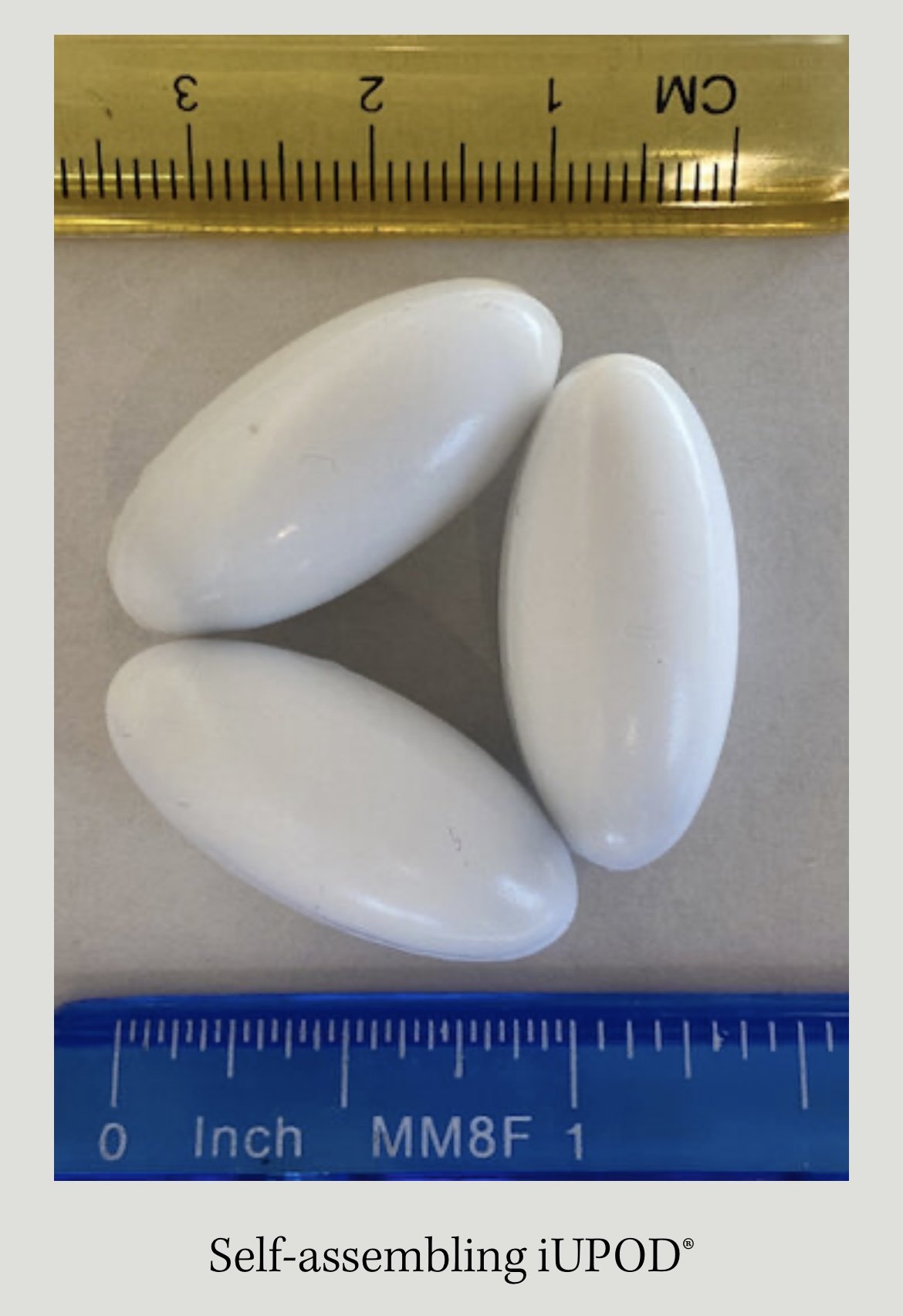
IUDs have been historically utilised to mimic early pregnancy in the mare with varying success. These require an ovulation to act upon to extend the life of the corpus luteum by blocking the hormonal release that normally brings them back into season. Therefore, they are only useful once the mare is already cycling.
Glass marbles have been the most used IUD historically; however, these are no longer recommended due to multiple evidenced side effects including risk of glass fragmentation in the uterus. The use of PMMA spheres or magnetic devices such as the iUPOD would be a preferable and safer alternative if an IUD was going to be used.
Interestingly, in the author’s experience speaking with clinicians who have administered these devices, there is surprisingly positive client satisfaction despite the inconsistent and variable evidence of the success of these devices in the literature.
Oxytocin
Administration of the hormone, oxytocin, at specific time points when the mare is in diestrus can extend diestrus by up to 60-90 days. This technique is evidenced by multiple studies. For optimal success, reproductive ultrasound would be used to identify ovulation and carefully plan the timing of injectable administration.
However, some studies have also evidenced successful extension of the diestrus phase without known timing of ovulation. The major downside of this technique is the need for administration of multiple injections/multiple reproductive examinations to time ovulation.
Immunological approach
Gonadotrophin releasing hormone (GnRH) is a hormone produced by the brain that is responsible for stimulating follicle growth in the ovaries and activation of a hormonal cascade to bring the mare into oestrus.

GnRH vaccinations generate an immune response against GnRH, suppressing the hormonal cascade and ovarian activity and therefore, oestrus behaviour. An equine licensed product has previously been available in Australia. However, this is no longer in production. We have the option of a swine formulation, Improvac®, which has commonly been used in equids off licence.
Major drawbacks for the use of this are common adverse injection site reactions, risk of anaphylaxis and concern over extended length of ovarian suppression. Therefore, this option would not be recommended in mares with a future breeding potential.
Surgical approach
Ovariectomy is a treatment option for hormonal behaviour in mares. The ovary is the only supply of progesterone in the horse but is not the only supply of oestrogen.
Ovariectomy has been associated with good client satisfaction in many cases to resolve unwanted hormonal behaviour. However, in some mares, whilst removal of the ovaries would prevent cyclicity, it can occasionally result in persistent oestrus behaviour in the absence of progesterone produced by the ovaries. This is also a permanent option that will remove breeding potential.
The techniques discussed so far are not exhaustive and there are many other methods that have been used to affect cyclicity or hormonal behaviour including pregnancy, induction of diestrus ovulation, GnRH analogue medication and infusion of intrauterine medical grade plant oils.
Colts/stallions
There are a few medicated options for hormonal manipulation in males. Progestagen administration e.g. oral altrenogest administration can quieten stallion like behaviour in males but is banned for use in racing and training.
Immunisation with off licence GnRH vaccines such as Improvac®, suppresses pituitary-gonadal hormone production aiming to cause a ‘chemical castration.’ However, results can be variable, particularly in mature stallions. As mentioned previously with mares, the downside of these vaccines are injection site reactions, risk of anaphylaxis and risk of prolonged sterility in future breeding animals.
Occasionally nutritional supplements have been used with effect in stallions such as L-tryptophan, a precursor of the neurotransmitter serotonin. This has induced calm and fatigue-like behaviour in a number of species.
Synthetic preparations of calming pheromones based on an equine appeasing pheromone produced in perimammary gland secretions of lactating females have also been used with such success. Of course, the use of these to calm behaviour vs the desire to generate an athletic performance animal is a consideration and results are likely to have wide individual variation.

Probiotics as an alternative to antibiotics to reduce resistance in the gut
Article by Kerrie Kavanagh
The leading causes of horse mortality can be attributed to gastrointestinal diseases. Therefore, maintaining the balance of the gut microbiota and avoiding a shift in microbial populations can contribute to improved health status. The gut microbiota, however, can be influenced by countless dynamic events: diet, exercise, stress, illness, helminth infections, aging, environment and notably, antimicrobial therapy (antibiotics). These events can lead to gut dysbiosis—a fluctuation or disturbance in the population of microorganisms of the gut, which can contribute to a wide range of disease. The use of antibiotics in horses is thought to have one of the most notable effects on the gut microbiota (gut dysbiosis), which can lead to diseases such as colitis, colic and laminitis.
Antibiotics, which are antimicrobial agents active against bacteria, are important to equine medicine; and bacterial infections can be resolved quite successfully using antibiotics for antimicrobial therapy, but there are consequences to their use. An antimicrobial agent can be defined as a natural or synthetic substance that kills or inhibits the growth of microorganisms such as bacteria, fungi and algae. One of the consequences of antibiotic use is that of antibiotic-associated diarrhoea, which can contribute to poor performance in the horse and even mortality. In antimicrobial therapy, the target organism is not the only organism affected by the antimicrobial agent but also the commensal microbiota too (the normal flora of the equine gut). Antibiotics can promote fungal infections and resistant organisms and impede or even eliminate the more sensitive organisms; and they can have both short and long-term consequences on the gut microbiota composition and function.

Research has indicated that antibiotic treatment may adversely affect metabolic function in the gut by decreasing protein expression responsible for biochemical pathways such as glycolysis, iron uptake, glutamate hydrolysis and possibly even more metabolic functions. The use of antimicrobial drugs directly impacts and possibly contributes to the most notable effect on the gut microbiota of the host, leading to gut dysbiosis; and certain antibiotics can have further-reaching consequences on the microbiota than others. The type of antibiotic and mode of action (bacteriostatic versus bactericidal) will differ in their influences on the gut microbiota composition, e.g., clindamycin operates a bacteriostatic mode of action by inhibiting protein synthesis and exerts a larger impact on the gut microbiota compared to other antimicrobials. These influential consequences that are imparted by the antimicrobial agent are relatively yet to be elucidated and may result in the manifestation of illness or conditions later in life. For example, the development of asthma in humans has been linked to antibiotic treatment in early childhood as a result of bacterial infections. It may yield interesting results if researchers were to examine the gut microbiome of horses suffering from chronic obstructive pulmonary disease (COPD) and other chronic respiratory illnesses and to establish if there is indeed a link with antibiotic therapy used in horses from an early age.
In comparison to the vast wealth of human studies conducted so far, the volume of equine studies falls disappointingly far behind, but that is changing as researchers focus their interest on developing and filling this gap of knowledge. One such study which examined the effect of antibiotic use on the equine gastrointestinal tract, demonstrated a significant reduction in culturable cellulolytic bacteria (>99%) from equine faeces during the administration period of trimethoprim sulfadiazine and ceftiofur in a study comparing responses to antibiotic challenge. That reduction was still evident at the end of the withdrawal period when compared to the control group. In other words, there was a significant reduction in the ‘normal’ bacteria of the gut. The ability of antibiotics to modulate the gut microbiota was evidenced by the proliferation of pathogenic Salmonella and Clostridia difficile (commonly associated with diarrhoea in horses) in the antibiotic challenged horses. This trend of reduction in cellulolytic bacteria associated with antibiotic use was also mirrored in a relatively recent study conducted in 2019, where a short-term reduction in culturable cellulolytic bacteria was combined with a progressive increase in amylolytic bacteria. The heavy reliance on cellulolytic bacteria in the role of equine digestion (without these types of bacteria the horse cannot break down their food) may, therefore, adversely affect the dietary energy available from forage during antimicrobial therapy and may therefore impact performance.
Another study that compared the effect of penicillin, ceftiofur and trimethoprim sulfadiazine (TMS) on the gut microbiota in horses using next-generation sequencing showed that TMS had the most profound impact on the microbiota, in particular the phylum Verrucomicrobia. This same study also reported a significant decrease in bacterial richness and diversity of the faecal microbiota. A reduction in bacterial diversity is certainly a trend that is commonly seen in gastrointestinal disease in horses. The restoration of the normal gut microbiota after completion of antibiotic treatment can take up to 40 days, but the organisational structure of the bacterial populations can take many years to re-establish the original structure map that was laid out in the gut pre-antibiotic treatment.
The equine studies certainly show similarities to the human studies, indicating the consequences of antibiotics that can be seen across more than one species. Human studies have reported long-term consequences of antibiotic treatment on the human microbiota. One such human study investigated a 7-day clindamycin treatment and monitored the patients for two years. The impact on the human microbiota remained evident two years post-treatment, where a reduction in bacterial diversity and detection of high-resistance to clindamycin were detected.
Interestingly, no resistant clones were detected in the control group over the two-year sampling period. Another study focusing on the effects of antibiotic treatment for Helicobacter pylori showed findings mirrored in similar studies of that field. The findings demonstrated the rapidly reducing bacterial diversity (one week) after antibiotic treatment and found that disturbances in the microbiota and high levels of macrolide resistance were evident four years post-treatment. Human studies may predict that equine studies will find similar trends with equine antimicrobial therapy. These studies highlight the impact of antibiotic use and the long-term persistence of antibiotic resistance remaining in the intestinal microbiome, which is a concern for both humans and animals.
Antibiotics can lead to the selectivity and proliferation of resistant bacteria, which is evidenced by the long-term effects observed on the gut microbiota harbouring drug-resistant encoded genes. Horizontal gene transfer (HGT) commonly occurs in the gut (can be up to 25 times more likely to occur in the gut than in other environments). HGT can be attributed to the close proximity of the microbiota in the gut, allowing the transfer of genetic material via routes such as plasmids and conjugation; in other words, the bacteria in the gut have developed a pathway to transfer antibiotic resistant genes from one generation to another. Resistance to antibiotics is now a global issue for the treatment of many diseases.
With the unfavourable association tied to Clostridium difficile infections (CDI) and the onset of colitis particularly in mature horses treated with β-lactam antibiotics (commonly used for equine infections), the incidences in which antimicrobial therapy is considered should be minimised and only used if entirely necessary. The use of broad-spectrum antibiotics in recurrent presentations of symptoms of disease such as urinary tract infections in humans or diarrhoea as a result of CDI in both humans and horses is promoting drug resistance.
The antibiotics, by disrupting the gut microbiota (which act as a defence against the establishment and proliferation of such pathogenic bacteria) are allowing the opportunity of growth for these multi-resistant microorganisms such as C. difficile, vancomycin-resistant enterococci (VRE), and multi-resistant Staphylococcus aureus (MRSA). The organism C. difficile and its antibiotic resistance has been demonstrated in the treatment of CDI for both humans and animals. The introduction of vancomycin (a glycopeptide antibiotic) in 1959 for the control of CDI remained effective until the 1990s when a more virulent form of C. difficile emerged. This new form of C. difficile with reported broad-spectrum antibiotic resistance resulted in chronic conditions and increased human mortality. C. difficile is most noted with human hospital-acquired infections. C. difficile BI/NAP1/027 has been shown to have resistance to fluoroquinolone antibiotics, moxifloxacin and gatifloxacin, which was not seen in historical genotypes. As C. difficile infections are found to cause gastrointestinal disease in horses as well as humans, this is certainly of concern.
Alternative therapies to antibiotic therapy to restore or modulate the gut microbiome after a gut dysbiosis event could be considered in certain circumstances where antibiotics are no longer effective (e.g., CDI), nor may they not be the best course (presence of Extended-spectrum -β-lactamase producing (ESBL) organisms) nor essential for example, when the diagnosis of the bacterial cause is uncertain. The rationale to using probiotic treatment along with antimicrobial treatment is that the antibiotic will target the pathogenic bacteria (e.g., C. difficile) and also the commensal microbiota of the gut, but the probiotic bacteria will help to re-establish the intestinal microbiota and in-turn prevent the re-growth of the pathogenic bacteria in the case or residual spores of C. difficile surviving the antibiotic treatment. Alternative therapies such as faecal microbiome transplant (FMT) or probiotic solutions can reduce the risk of proliferation of antibiotic-resistant bacteria and also have fewer implications on the gut microbiome as evidenced by antibiotic use.
Probiotics have been defined by the Food and Agricultural Organisation (FAO) and the World Health Organisation (WHO) as “live non-pathogenic microorganisms that, when administered in adequate amounts, confer a health benefit on the host”. The word ‘probiotic’ is Greek in origin, meaning, ‘for life’; and the term was coined by Ferdinand Vergin in 1954. While the mechanisms of action of probiotics are complex and require a deeper knowledge of the modulations of the gastrointestinal microbiota, and the health benefits due to their use are the subject of some debate, there is no doubt that probiotics are considered by many as a vital resource to human and animal health.
The use of probiotics in animal production, particularly in intensive swine and poultry production, has increased in recent years, primarily as an alternative to the use of antimicrobials in the prevention of disease. The problem of antibiotic-resistance and antimicrobial residues in food-producing animals (the horse is considered a food-producing animal), as a result of historical antibiotic use with the corresponding reduction in antibiotic efficacy in humans, leads to having to look at more sustainable options such as probiotic use to combat disease. Probiotics in horses are predominantly used as a treatment modality in the gastrointestinal microbial populations to combat illnesses such as diarrhoea—to prevent diarrhoea (particularly in foals) or help improve digestibility. Shifts or fluctuations in the microbial populations of the equine gastrointestinal tract have been associated with diseases such as laminitis and colic.
Gut dysbiosis, as mentioned previously is, a fluctuation or disturbance in the population of microorganisms of the gut is now being recognised as a cause of a wide range of gastrointestinal diseases; and in horses, it is one of the leading causes of mortality. The ability of probiotics in conferring health benefits to the host can occur via several different mechanisms: 1) inhibiting pathogen colonisation in the gut by producing antimicrobial metabolites or by competitive exclusion by adhering to the intestinal mucosa preventing pathogenic bacteria attachment by improving the function and structure; 2) protecting or restabilising the commensal gut microbiota; 3) protecting the intestinal epithelial barrier; 4) by inducing an immune response.
It is known that there is a wealth of factors that will adversely affect the gut microbiome, antibiotics, disease, diet, stress, age and environment are some of these compounding contributors. To mirror one researcher’s words echoing from an era where antibiotics were used as growth promoters in the animal industry, “The use of probiotic supplements seeks to repair these deficiencies. It is, therefore, not creating anything that would not be present under natural conditions, but it is merely restoring the flora to its full protective capacity”. In the case of using concurrent antibiotic and probiotic treatment, this strategic tweaking of the microbiota could be used as a tool to prevent further disease consequence and perhaps help improve performance in the horse.

The benefits of probiotic use in horses have not been investigated extensively but as mentioned previously, they are now being focused upon by researchers in the equine field. The most common bacterial strains used in equine probiotic products are Lactobacillus, Bifidobacterium, Streptococcus, Enterococcus, Bacillus and yeast strains of Saccharomyces. Lactobacillus, Bifidobacterium and Enterococcus strains typically account for less than 1% of the microbiota large gastrointestinal populations.
Regulation is lacking regarding labelling of probiotic products, often not displaying content with clarification and quality control (such as confirmed viability of strain[s]) not excised with over-the-counter probiotic products. There is evidence to suggest that host-adapted strains of bacteria and fungi enjoy a fitness advantage in the gut of humans and animals. Therefore, there may be an advantage in using the individual animal’s own bacteria as potential probiotics. Probiotics and antibiotics used concurrently could be the way to minimise the introduction of antibiotic-resistant bacterial strains in the gut, and in turn, protect future antibiotic efficacy.
Small wounds leading to synovial infections

Article by Peter Milner
Most experienced trainers will know from bitter experience that a seemingly tiny wound can have a big impact if a horse is unlucky enough to sustain a penetrating injury right over a critical structure like a joint capsule or tendon sheath. Collectively, joints and tendon sheaths are called synovial structures, and synovial infection is a serious, potentially career-ending and sometimes life-threatening problem.
A team of veterinary researchers from Liverpool University Veterinary School, published a study in Equine Veterinary Journal that examined factors influencing outcome and survival. This article was first published in European Trainer (issue 50 - summer 2015) but is being republished due to popular demand.
What is synovial infection?
Infection involving a synovial cavity, such as a joint or tendon sheath, is a common and potentially serious injury for the horse. The most prevalent cause is a wound, although a smaller proportion of cases result following an injection into a joint or tendon sheath, or after elective orthopaedic surgery to the area. Additionally, infection can occur via the bloodstream, particularly in foals that have not received enough colostrum. Left untreated, the horse will remain in pain, and ongoing infection and inflammation can result in permanent damage. This can ultimately result in euthanasia on welfare grounds.
What factors are important for horse survival?

When a synovial infection occurs there is a huge inflammatory response, leading to swelling and pain. The horse usually shows severe lameness but following a good clinical examination, the cause is often quickly identified. Prompt veterinary recognition of involvement of a joint or tendon sheath and aggressive treatment (involving flushing the affected synovial cavity and the correct use of systemic and local antibiotics) will often result in a good outcome for the horse. Flushing removes inflammatory debris including destructive enzymes and free radicals, and it eliminates contaminating bacteria in most cases. This is performed most effectively by arthroscopic guidance (“keyhole” surgery) under general anaesthesia. Using a “scope” to do this is considered superior to flushing through needles because arthroscopy allows the inside of the problem area to be inspected, foreign material (for example, dirt or splinters of wood) to be removed, and any concurrent damage (such as damage to the cartilage or a cut into a tendon) to be evaluated. In addition, targeted high volume lavage is best achieved via arthroscopy.
Survival following arthroscopic treatment of synovial sepsis is good – approximately 80-90% of adult horses undergoing a flush are discharged from hospital. In foals, however, the figure is much lower, at around 55%, and this likely due to complicating factors such as concurrent sepsis involving multiple organs. Our study, recently published in Equine Veterinary Journal, investigated what factors might be involved in determining survival to hospital discharge in 214 horses undergoing arthroscopic treatment for synovial sepsis. We used statistical modelling to evaluate the interactions with different factors at three key time points during the management of the condition at Liverpool Veterinary School, one of the leading UK referral veterinary hospitals. Information collected on admission to the hospital included when the horse was last seen to be normal, the cause of the infection, the degree of lameness present, and the level of white blood cells and protein in synovial fluid collected from the infected joint or tendon sheath. These lab tests are an important method which veterinarians use to determine how severe the infection is. Additional data collected included whether the surgery was performed out-of-normal working hours, if foreign material was present, the amount of inflammation present in the area, and whether any additional cartilage or tendon damage was found at surgery. Post-operative information gathered included what the levels of white blood cells and protein were in the synovial fluid after surgery and whether the horse needed further surgical treatment.
All horses in this study were greater than six months old and the majority had sustained a wound that communicated with a joint or tendon sheath. Eighty-six per cent of the 214 horses admitted to the hospital survived to hospital discharge. Of the 31 horses that did not survive, 27 were euthanised due to persistent infection or lameness.
An angry, protein-soup
A high level of protein in the synovial fluid of the affected joint or tendon sheath on admission and levels that remained high after surgery were strongly associated with a poor outcome and loss of the horse. Protein concentrations are normally fairly low in a normal joint or tendon sheath, but protein leaks into the synovial cavity from surrounding blood vessels when inflamed. Protein is also produced by cells in the synovial cavity when they are activated in response to a severe insult such as infection. Protein clots trap bacteria in the joint, making it harder to remove infection. The protein soup also includes lots of inflammatory mediators such as enzymes and signalling molecules, and these cause further inflammation, tissue damage, and sensitise pain receptors in the synovial cavity magnifying the inflammatory response and increasing the pain experienced by the horse. Unchecked, this angry, inflamed environment can result in cartilage degeneration, bone damage, and adhesion (scar) formation. This fits well with another observation from this study linking the presence of moderate or severe synovial inflammation at surgery as a negative factor for survival.
Small wounds can lead to big trouble
Interestingly, horses presenting with an obvious wound (as opposed to a small penetrating injury or no visible wound) were more likely to survive to hospital discharge. This may be due to the injury being noticed earlier and hence prompting earlier veterinary intervention. Alternatively, open wounds may allow drainage of inflammatory synovial fluid and lessen the detrimental effects of increased pressure within the joint as well as reducing ongoing exposure to inflammatory mediators. This finding highlights the fact that trainers should act promptly when faced with a wound – it is easy to underestimate just how much damage may be going on under the surface.

Horses undergoing surgical treatment of a joint or tendon sheath infection out-of-hours (for example in the middle of the night) were three times less likely to survive to hospital. Often, horses with a synovial infection arrive stressed and painful and not in an ideal state for having an anaesthetic. Early identification of an infection and appropriate management is important but stabilisation of the horse and preparation for surgery appear to outweigh any perceived benefits of undertaking immediate surgery. This is borne out by the finding that time from initial injury to treatment was not associated with outcome and is in agreement with previous findings from other researchers. It is important to reiterate that prompt recognition and treatment of a horse with an infection in a synovial cavity is essential but that surgical management within 12-24 hours of diagnosis, so that the horse is in the best condition for undergoing anaesthesia, does not affect outcome.
Do horses return to work after a synovial infection?

The big question that owners and trainers want to know is whether the horse will regain full function of the joint or tendon sheath after having an infection. Figures for return to function following surgical (arthroscopic) treatment for a synovial infection vary between 54-81%. Various factors appear to relate to outcome but when looking at a predominately thoroughbred racing population, the statistic for return to training appears to be at the higher end of this range. Factors associated with failure to return to athletic performance include the presence of thickened inflammatory tissue (known as pannus) at the time of surgery and that may relate to the development of fibrous adhesions and scar tissue within joint or tendon sheath longer-term. Some structures are particularly likely to compromise future function, and horses with an infection of the navicular bursa in the foot following a nail penetration generally do worse.
Take home message
Horses sustaining an infection to a joint or tendon sheath have a good chance of the infection clearing up and surviving the injury, with the likelihood of racing as high as around 80%. Our key message for trainers from this study is that it is essential that they recognise early when an infection involves one of these structures and have a veterinarian fully evaluate the injury. Aggressive treatment is important and involves flushing the synovial cavity using a “scope” under anaesthesia to remove as much inflammatory and infective debris as possible.
