VETERINARY EUT Webmaster VETERINARY EUT Webmaster

The importance of the Sacroiliac joint

Article by Annie Lambert

Horses that present as sore in the hindquarters can be perplexing to diagnose. Sometimes the problem is found in the last place you look – the sacroiliac joint.

Location of the sacroiliac joint on the horse

Even though the sacroiliac joint (SI) was on veterinary radars long ago, due to its location buried under layers of muscle in the equine pelvic region, the joint and surrounding ligaments were tough to diagnose and treat.

The sacroiliac joint is often a source of lower back discomfort in race and performance horses. Trainers may notice several clinical signs of a problem. These hints include sensitivity to grooming, objections to riders getting legged up, stiffness of motion, pain to manual palpation of the rump or back, resistance to being shod behind and poor performance.

Of course, those symptoms could describe other hind limb soundness issues, making the origin of the problem arduous to ascertain. A thorough physical examination with complete therapeutic options can relieve sacroiliac pain. The treatments are complicated, however, by the anatomy of the SI area. 

Location of the sacroiliac joint on the horse

The equine pelvis is composed of three fused bones: ilium, ischium and pubis. The sacrum, the lower part of the equine back, is composed of five fused vertebrae. The sacroiliac joint is located where the sacrum passes under the top of the pelvis (tubera sacrale). The dorsal, ventral and interosseous sacroiliac ligaments help strengthen the SI joint. 

The SI and surrounding ligaments provide support during weight bearing, helping to transfer propulsive forces of the hind limbs to the vertebral column—creating motion much like the thrust needed to break from the starting gate.

Sound complicated? It certainly can be.

Diagnosing Dilemmas

It wasn’t until modern medical technology advanced that the SI could be explored seriously as a cause of hind lameness.

Dr Michael Manno

“The sacroiliac is one of the areas that’s very hard to diagnose or image,” explained Dr. Michael Manno, a senior partner of San Dieguito Equine Group in San Marcos, California. “[Diagnostics] of the area probably correlated with bone scans or nuclear scintigraphy. You can’t really use radiographs because the horse is so massive and there is so much muscle, you can’t get a good image.

“About the only time you can focus on the pelvis and get a decent radiograph is if the horse is anesthetized—you have a big [x-ray] machine and could lay the horse down. But, it’s hard because with anything close to a pelvic injury, the last thing you want to do is lay them down and have them have to get back up.”  

The nuclear scintigraphs give a good image of hip, pelvis and other anatomical structures buried deep in the equine body, according to Manno, a racetrack practitioner. “Those images can show areas of inflammation that could pretty much be linked right to the SI joint.”

Ultrasound scan of the Sacroiliac joint

The other modern technological workhorse in the veterinary toolbox is the digital ultrasound machine. Manno pointed out that veterinarians improved diagnostics as they improved their ultrasounding skills and used those skills to ultrasound areas of the body they never thought about before. Using different techniques, frequencies and various heads on the machine’s probe, the results can be fairly remarkable.

“The ultrasound showed you could really image deeper areas of the body, including an image of the sacroiliac joint,” Manno said. “It can also show some ligament issues.”

Where the SI is buried under the highest point of a horse’s rump, and under heavy gluteal muscles, there are two sets of ligaments that may sustain damage and cause pain. The dorsal sacroiliac ligaments do not affect the sacroiliac joint directly, but help secure the ilium to the sacral spine. The ventral sacroiliac ligaments lie deeper, in the sacroiliac joint area, which they help stabilize. These hold the pelvis tight against its spine. The joint itself, being well secured by these ligaments, has little independent movement and therefore contains only minimal joint fluid.

Trotting up for diagnostic purposes

Diagnosing the SI can be complex because horses often travel their normal gait with no change from normal motion—no signs of soreness. Other horses, however, are sore on one leg or another to varying degrees, sometimes with a perceptible limp. 

“I don’t know that there is a specific motion,” Manno explained. “You just know that you have a hind end lameness, and I think a lot of performance horses have mildly affected SI joints. 

“The horses that are really severe become acutely lame behind, very distinct. You go through the basic diagnostics, and I think most of these horses will show you similar signs as other issues behind. We palpate along the muscles on either side of their spine and they are sore, or you palpate over their croup and you can get them to drop down—that kind of thing. Other times you do an upper limb flexion on them and they might travel weird on the opposite leg. So, it can be a little confusing.” 

In the years prior to the early 2000s, the anatomical location of the SI hindered a definite diagnosis; decisions on hind soreness were more of a shrug, “time and rest” treatment evaluation. As one old-time practitioner called it, a SWAG – “Scientific Wild Ass Guess.”

Even with modern tools, making a conclusive diagnosis can be opaque.

“The less affected horses, through exercise and with medications like Robaxin [muscle relaxer] or mild anti-inflammatories, seem to be able to continue to perform,” Manno said. “I don’t know how you can be perfectly sure of an inside joint unless you try to treat it and get results.”

“That’s why bone scans came into play and are really helpful,” Manno added. “You can image that [SI] area from different angles with the machine right over the path of the pelvis, looking down on it or an angle view into it, and then you see it from the side and the back very often. We can get an idea from the different views and angles of where the inflammation is and pinpoint the problem from that.” 

Once Manno has a generalized idea of where the problem is, he fine-tunes his hypothesis using more diagnostics with a digital ultrasound machine. 

“You can ultrasound from up above and see the joint that way,” he said. “As ultrasound has progressed, we’ve found that the rectal probes the breeding vets have used can also be tuned in to start looking for other things. If you turn them upwards, you can look at the bottom of the pelvis and the SI joint. You can see things through the rectum by just looking straight up. That is a whole new thing that we probably never thought about doing. I don’t profess to be very great at it; it’s not something I do a lot, but there are people that are just wonderful at it.” 

Treating a Theorem

But, if the diagnosis is incorrect, the prescribed treatment may be anything but helpful.

Palpation of the sacroiliac joint

“In many cases, if a horse is really sore, you need to be very careful,” cautioned Manno. “What you don’t want to do is go from a strain or some sort of soft tissue injury into a pelvic fracture by trying to keep them going. In many cases you are back in the old rest and time type of treatment.”

Manno pointed out one treatment that has advanced over many years is injecting the SI joint directly. There are a couple of techniques used when injecting the SI. With a blind injection the practitioner directs a long, straight needle into the joint by relying solely on equine anatomy. The other technique employs an ultrasound machine to guide the placement of the needle into the joint.

“Normally we are just injecting cortisone in those cases,” Manno noted. “We are trying to get the inflammatory response to settle down. Hopefully that gives the horse some relief so that they’re a bit more relaxed in their musculature. You know how it is when you get a sore back; it’s hard to keep yourself from cramping, which makes everything worse.”

A slight tweak of that technique is to use a curved needle. When you are positioning the curved needle, it follows the curve of the horse’s anatomy and helps the practitioner direct the injection into the joint.

“It curves right into position for you; it gives you a little help,” Manno confirmed of the curved needle. “Some people are really good with that technique; others still like to go to the straight needle. [The curved needle] helps you approach the site without interference from the bones in that area.”

SI joint injuries affect most performance horses, including Standardbred trotters and pacers, Western performance athletes as well as hunters, jumpers and dressage horses.

The older show horses are often diagnosed with chronic SI pain, sometimes complicated by arthritis. These chronic cases—and admittedly some racehorses—are treated with different therapies. These conservative, nonsurgical treatments have been proven effective.

In addition to stall rest and anti-inflammatories, physical training programs can be useful in tightening the equine patient’s core and developing the topline muscles toward warding off SI pain. Manno, a polo player who also treats polo ponies, believes the hard-working ponies avoid having many SI injuries due to their fitness levels. 

“I think these polo horses are similar to a cross between a racehorse and a cutting horse,” Manno opined. “They are running distances and slide stopping and turning.”

Other treatments utilized include shockwave, chiropractic, acupuncture, therapeutic laser and pulsed electromagnetic therapy.

Superior Science

With the new diagnostic tools and advanced protocols in their use, veterinarians can pinpoint the SI joint and surrounding areas much closer. This gives them an improved indication that there definitely is an issue with the sacroiliac. 

When there is a question about what is causing hind end lameness, most practitioners begin with blocking from the ground up.

Flexion test for diagnostic purposes

“In many cases with hind end lameness that we can’t figure out, we block the lower leg; if it doesn’t block out down low, we conclude the problem is up high,” Manno said. “Once you get up to the hock you’re out of options of what you can figure out. You start shooting some x-rays, but by the time you get to the stifle, you’re limited. Bone scans and ultrasounds have certainly helped us with diagnosing.”

Manno doesn’t see a lot of SI joint injuries in his practice, but he noted there were cases every now and again. He also opined that there were probably other cases that come up in racehorses on a short-term basis. He also noted that, although it may not be a real prominent injury, that’s not to say it has not gone undiagnosed.

“I think we realize, in many of the horses we treat, that the SI joint is something that may have been overlooked in the past,” Manno concluded. “We just didn’t have the ability to get any firm diagnosis in that area.”

Read More
VETERINARY EUT Webmaster VETERINARY EUT Webmaster

EIPH - could there be links to sudden death and pulmonary haemorrhage?

Dr Peter W. Physick-Sheard, BVSc, FRCVS, explores preliminary research and hypotheses, being conducted by the University of Guelph, to see if there is a possibility that these conditions are linked and what this could mean for future management and training of thoroughbreds. 

EIPH / bleeders in Thoroughbreds.jpg

"World's Your Oyster,” a three-year-old thoroughbred mare, presented at the veterinary hospital for clinical examination. She won her maiden start as a two-year-old and placed once in two subsequent starts. After training well as a three-year-old, she failed to finish her first start, easing at the top of the stretch, and was observed to fade abruptly during training. Some irregularity was suspected in heart rhythm after exercise. Thorough clinical examination, blood work, ultrasound of the heart and an ECG during rest and workout revealed nothing unusual. 

Returning to training, Oyster placed in six of her subsequent eight starts, winning the last two. She subsequently died suddenly during early training as a four-year-old. At post-mortem, diagnoses of pulmonary haemorrhage and exercise-induced pulmonary haemorrhage were established—a very frustrating and unfortunate outcome.

Across the racing world, a case like this probably occurs daily. Anything that can limit a horse's ability to express its genetic potential is a major source of anxiety when training. The possibility of injury and lameness is the greatest concern, but a close second is respiratory disease, with bleeding  from the lungs (most often referred to as exercise induced pulmonary [lung] haemorrhage or EIPH) being high on the list. 

EIPH is thought to occur in as many as 85 percent of racehorses, and may initially be very mild without obvious clinical consequences. In some cases it can be associated with haemorrhage of sufficient severity for blood to appear at the nostrils, even at first occurrence. In many racing jurisdictions this is a potentially career-ending problem. In these horses, an impact on performance is unquestionable. Bleeding from the lungs is the reason for the existence of ‘Lasix programs,’ involving pre-race administration of a medication considered to reduce haemorrhage. Such programs are controversial—the justifications for their existence ranging from addressing welfare concerns for the horse to dealing with the performance impacts. 

Much less frequently encountered is heavy exercise-associated bleeding from the nostrils (referred to as epistaxis), which can sometimes be accompanied by sudden death, during or shortly after exercise. Some horses bleed heavily internally and die without blood appearing at the nostrils. Haemorrhage may only become obvious when the horse is lying on its side, or not until post-mortem. Affected animals do not necessarily have any history of EIPH, either clinically or sub-clinically. There is an additional group of rare cases in which a horse simply dies suddenly, most often very soon after work and even after a winning performance, and in which little to nothing clearly explains the cause on post-mortem. This is despite the fact most racing jurisdictions study sudden death cases very closely.

EIPH is diagnosed most often by bronchoscopy—passing an endoscope into the lung after work and taking a look. In suspected but mild cases, there may not be sufficient haemorrhage to be visible, and a procedure called a bronchoalveolar lavage is performed. The airways are rinsed and fluid is collected and examined microscopically to identify signs of bleeding. Scoping to confirm diagnosis is usually a minimum requirement before a horse can be placed on a Lasix program. 


Are EIPH, severe pulmonary haemorrhage and sudden death related? Are they the same or different conditions? 

Thoroughbredlungs being scoped.jpg

At the University of Guelph, we are working on the hypothesis that most often they are not different—that it’s degrees of the same condition, or closely related conditions perhaps with a common underlying cause. We see varying clinical signs as being essentially a reflection of severity and speed of onset of underlying problems. 

Causes in individual cases may reflect multiple factors, so coming at the issues from several different directions, as is the case with the range of ongoing studies, is a good way to go so long as study subjects and cases are comparable and thoroughly documented. However, starting from the hypothesis that these may all represent basically the same clinical condition, we are approaching the problem from a clinical perspective, which is that cardiac dysfunction is the common cause. 

Numerous cardiac disorders and cellular mechanisms have the potential to contribute to transient or complete pump (heart) failure. However, identifying them as potential disease candidates does not specifically identify the role they may have played, if any, in a case of heart failure and in lung haemorrhage; it only means that they are potential primary underlying triggers. It isn't possible for us to be right there when a haemorrhage event occurs, so almost invariably we are left looking at the outcome—the event of interest has passed. These concerns influence the approach we are taking.


Background

The superlative performance ability of a horse depends on many physical factors:

  • Huge ventilatory (ability to move air) and gas exchange capacity

  • Body structure including limb length and design - allows it to cover ground rapidly with a long stride

  • Metabolic adaptations - supports a high rate of energy production by burning oxygen, tolerance of severe metabolic disruptions toward the end of race-intensity effort

  • High cardiovascular capacity - allows the average horse to pump roughly a brimming bathtub of blood every minute

At race intensity effort, these mechanisms, and more, have to work in coordination to support performance. There is likely not much reserve left—two furlongs (400m) from the winning post—even in the best of horses. There are many wild cards, from how the horse is feeling on race day to how the race plays out; and in all horses there will be a ceiling to performance. That ceiling—the factor limiting performance—may differ from horse to horse and even from day to day. There’s no guarantee that in any particular competition circumstances will allow the horse to perform within its own limitations. One of these factors involves the left side of the heart, from which blood is driven around the body to the muscles.


A weak link - filling the left ventricle

The cardiovascular system of the horse exhibits features that help sustain a high cardiac output at peak effort. The feature of concern here is the high exercise pressure in the circulation from the right  ventricle, through the lungs to the left ventricle. At intense effort and high heart rates, there is very little time available to fill the left ventricle—sometimes as little as 1/10 of a second; and if the chamber cannot fill properly, it cannot empty properly and cardiac output will fall. The circumstances required to achieve adequate filling include the readiness of the chamber to relax to accept blood—its ‘stiffness.’ Chamber stiffness increases greatly at exercise, and this stiffened chamber must relax rapidly in order to fill. That relaxation seems not to be sufficient on its own in the horse at high heart rates. Increased filling pressure from the circulation draining the lungs is also required. But there is a weak point: the pulmonary capillaries.

These are tiny vessels conducting blood across the lungs from the pulmonary artery to the pulmonary veins. During this transit, all the gas exchange needed to support exercise takes place. The physiology of other species tells us that the trained lung circulation achieves maximum flow (equivalent to cardiac output) by reducing resistance in those small vessels. This process effectively increases lung blood flow reserve by, among other things, dilating small vessels. Effectively, resistance to the flow of blood through the lungs is minimised. We know this occurs in horses as it does in other species; yet in the horse, blood pressure in the lungs still increases dramatically at exercise. 

If this increase is not the result of resistance in the small vessels, it must reflect something else, and that appears to be resistance to flow into the left chamber. This means the entire lung circulation is exposed to the same pressures, including the thin-walled capillaries. Capillaries normally work at quite low pressure, but in the exercising horse, they must tolerate very high pressures. They have thin walls and little between them, and the air exchange sacs in the lung. This makes them vulnerable. It's not surprising they sometimes rupture, resulting in lung haemorrhage.

Recent studies identified changes in the structure of small veins through which the blood flows from the capillaries and on toward the left chamber. This was suspected to be a pathology and part of the long-term consequences of EIPH, or perhaps even part of the cause as the changes were first identified in EIPH cases. It could be, however, that remodelling is a normal response to the very high blood flow through the lungs—a way of increasing lung flow reserve, which is an important determinant of maximum rate of aerobic working. 

The more lung flow reserve, the more cardiac output and the more aerobic work an animal can perform. The same vein changes have been observed in non-racing horses and horses without any history or signs of bleeding. They may even be an indication that everything is proceeding as required and a predictable consequence of intense aerobic training. On the other hand, they may be an indication in some horses that the rate of exercise blood flow through their lungs is a little more than they can tolerate, necessitating some restructuring. We have lots to learn on this point.

If the capacity to accommodate blood flow through the lungs is critical, and limiting, then anything that further compromises this process is likely to be of major importance. It starts to sound very much as though the horse has a design problem, but we shouldn't rush to judgement. Horses were probably not designed for the very intense and sustained effort we ask of them in a race. Real-world situations that would have driven their evolution would have required a sprint performance (to avoid ambush predators such as lions) or a prolonged slower-paced performance to evade predators such as wolves, with only the unlucky victim being pushed to the limit and not the entire herd. 

Thoroughbred lungs.jpg

Lung blood flow and pulmonary oedema

There is another important element to this story. High pressures in the capillaries in the lung will be associated with significant movement of fluid from the capillaries into lung tissue spaces. This movement in fact happens continuously at all levels of effort and throughout the body—it's a normal process. It's the reason the skin on your ankles ‘sticks’ to the underlying structures when you are standing for a long time. So long as you keep moving a little, the lymphatic system will draw away the fluid. 

In a diseased lung, tissue fluid accumulation is referred to as pulmonary oedema, and its presence or absence has often been used to help characterise lung pathologies. The lung lymphatic system can be overwhelmed when tissue fluid is produced very rapidly. When a horse experiences sudden heart failure, such as when the supporting structures of a critical valve fail, one result is massive overproduction of lung tissue fluid and appearance of copious amounts of bloody fluid from the nostrils. 

The increase in capillary pressure under these conditions is as great as at exercise, but the horse is at rest. So why is there no bloody fluid in the average, normal horse after a race? It’s because this system operates very efficiently at the high respiratory rates found during work: tissue fluid is pumped back into the circulation, and fluid does not accumulate. The fluid is pumped out as quickly as it is formed. An animal’s level of physical activity at the time problems develop can therefore make a profound difference to the clinical signs seen and to the pathology.

Usual events with unusual consequences 

If filling the left ventricle and the ability of the lungs to accommodate high flow at exercise are limiting factors, surely this affects all horses. So why do we see such a wide range of clinical pictures, from normal to subclinical haemorrhage to sudden death? 

Variation in contributing factors such as type of horse, type and intensity of work, sudden and unanticipated changes in work intensity, level of training in relation to work and the presence of disease states are all variables that could influence when and how clinical signs are seen, but there are other considerations.

Although we talk about heart rate as a fairly stable event, there is in fact quite a lot of variation from beat to beat. This is often referred to as heart rate variability. There has been a lot of work performed on the magnitude of this variability at rest and in response to various short-term disturbances and at light exercise in the horse, but not a lot at maximal exercise. Sustained heart rate can be very high in a strenuously working horse, with beats seeming to follow each other in a very consistent manner, but there is in fact still variation. 

Some of this variation is normal and reflects the influence of factors such as respiration. However, other variations in rate can reflect changes in heart rhythm. Still other variations may not seem to change rhythm at all but may instead reflect the way electrical signals are being conducted through the heart. 

Thoroughbred lungs inflated.jpg

These may be evident from the ECG but would not appear abnormal on a heart rate monitor or when listening. These variations, whether physiologic (normal) or a reflection of abnormal function, will have a presently, poorly understood influence on blood flow through the lungs and heart—and on cardiac filling. Influences may be minimal at low rates, but what happens at a heart rate over 200 and in an animal working at the limits of its capacity.

Normal electrical activation of the heart follows a pattern that results in an orderly sequence of heart muscle contraction, and that provides optimal emptying of the ventricles. Chamber relaxation complements this process. 

An abnormal beat or abnormal interval can compromise filling and/or emptying of the left ventricle, leaving more blood to be discharged in the next cycle and back up through the lungs, raising pulmonary venous pressure. A sequence of abnormal beats can lead to a progressive backup of blood, and there may not be the capacity to hold it—even for one quarter of a second, a whole cardiac cycle at 240 beats per minute. 

For a horse that has a history of bleeding and happens to be already functioning at a very marginal level, even minor disturbances in heart rhythm might therefore have an impact. Horses with airway disease or upper airway obstructions, such as roarers, might find themselves in a similar position. An animal that has not bled previously might bleed a little, one that has a history of bleeding may start again, or a chronic bleeder may worsen. 

Relatively minor disturbances in cardiac function, therefore, might contribute to or even cause EIPH. If a horse is in relatively tough company or runs a hard race, this may also contribute to the onset or worsening of problems. Simply put, it's never a level playing field if you are running on the edge.

Severe bleeding

It has been suspected for many years that cases of horses dying suddenly at exercise represent sudden-onset cardiac dysfunction—most likely a rhythm disturbance. If the rhythm is disturbed, the closely linked and carefully orchestrated sequence of events that leads to filling of the left ventricle is also disturbed. A disturbance in cardiac electrical conduction would have a similar effect, such as one causing the two sides of the heart to fall out of step, even though the rhythm of the heart may seem normal. 

The cases of horses that bleed profusely at exercise and even those that die suddenly without any post-mortem findings can be seen to follow naturally from this chain of events. If the changes in heart rhythm or conduction are sufficient, in some cases to cause massive pulmonary haemorrhage, they may be sufficient in other cases to cause collapse and death even before the horse has time to exhibit epistaxis or even clear evidence of bleeding into the lungs. 

EIPH and dying suddenly

If these events are (sometimes) related, why is it that some horses that die of pulmonary haemorrhage with epistaxis do not show evidence of chronic EIPH? This is one of those $40,000 questions. It could be that young horses have had limited opportunity to develop chronic EIPH; it may be that we are wrong and the conditions are entirely unrelated. But it seems more likely that in these cases, the rhythm or conduction disturbance was sufficiently severe and/or rapid in onset to cause a precipitous fall in blood pressure with the animal passing out and dying rapidly. 

In this interpretation of events, the missing link is the heart. There is no finite cutoff at which a case ceases to be EIPH and becomes pulmonary haemorrhage. Similarly, there is no distinct point at which any case ceases to be severe EIPH and becomes EAFPH (exercise-associated fatal pulmonary haemorrhage). In truth, there may simply be gradation obscured somewhat by variable definitions and examination protocols and interpretations.

The timing of death

It seems from the above that death should most likely take place during work, and it often does, but not always. It may occur at rest, after exercise. Death ought to occur more often in racing, but it doesn't. 

The intensity of effort is only one factor in this hypothesis of acute cardiac or pump failure. We also have to consider factors such as when rhythm disturbances are most likely to occur (during recovery is a favourite time) and death during training is more often a problem than during a race. 

A somewhat hidden ingredient in this equation is possibly the animal's level of emotional arousal, which is known to be a risk factor in humans for similar disturbances. There is evidence that emotions/psychological factors might be much more important in horses than previously considered. Going out for a workout might be more stimulating for a racehorse than a race because before a race, there is much more buildup and the horse has more time to adequately warm up psychologically. And then, of course, temperament also needs to be considered. These are yet further reasons that we have a great deal to learn.

Our strategy at the University of Guelph

Heart rate being taken.jpg

These problems are something we cannot afford to tolerate, for numerous reasons—from perspectives of welfare and public perception to rider safety and economics. Our aim is to increase our understanding of cardiac contributions by identifying sensitive markers that will enable us to say with confidence whether cardiac dysfunction—basically transient or complete heart failure—has played a role in acute events. 

We are also looking for evidence of compromised cardiac function in all horses, from those that appear normal and perform well, through those that experience haemorrhage, to those that die suddenly without apparent cause. Our hope is that we can not only identify horses at risk, but also focus further work on the role of the heart as well as the significance of specific mechanisms. And we hope to better understand possible cardiac contributions to EIPH in the process. This will involve digging deeply into some aspects of cellular function in the heart muscle, the myocardium of the horse, as well as studying ECG features that may provide insight and direction. 

Fundraising is underway to generate seed money for matching fund proposals, and grant applications are in preparation for specific, targeted investigations. Our studies complement those being carried out in numerous, different centres around the world and hopefully will fill in further pieces of the puzzle. This is, indeed, a huge jigsaw, but we are proceeding on the basis that you can eat an elephant if you're prepared to process one bite at a time.

How can you help? Funding is an eternal issue. For all the money that is invested in horses there is a surprisingly limited contribution made to research and development—something that is a mainstay of virtually every other industry; and this is an industry. 

Look carefully at the opportunities for you to make a contribution to research in your area. Consider supporting studies by making your experience, expertise and horses available for data collection and minimally invasive procedures such as blood sampling. 

Connect with the researchers in your area and find out how you can help. Watch your horses closely and contemplate what they might be telling you—it's easy to start believing in ourselves and to stop asking questions. Keep meticulous records of events involving horses in your care— you never know when you may come across something highly significant. And work with researchers (which often includes track practitioners) to make your data available for study. 

Remember that veterinarians and university faculty are bound by rules of confidentiality, which means what you tell them should never be ascribed to you or your horses and will only be used without any attribution, anonymously. And when researchers reach out to you to tell you what they have found and to get your reactions, consider actually attending the sessions and participating in the discussion; we can all benefit—especially the ultimate beneficiary which should be the horse. We all have lots to learn from each other, and finding answers to our many challenges is going to have to be a joint venture.  

Finally, this article has been written for anybody involved in racing to understand, but covering material such as this for a broad audience is challenging. So, if there are still pieces that you find obscure, reach out for help in interpretation. The answers may be closer than you think!

Oyster

And what about Oyster? Her career was short. Perhaps, had we known precisely what was going on, we might have been able to treat her, or at least withdraw her from racing and avoid a death during work with all the associated dangers—especially to the rider and the associated welfare concerns. 

Had we had the tools, we might have been able to confirm that whatever the underlying cause, she had cardiac problems and was perhaps predisposed to an early death during work. With all the other studies going on, and knowing the issue was cardiac, we might have been able to target her assessment to identify specific issues known to predispose. 

In the future, greater insight and understanding might allow us to breed away from these issues and to better understand how we might accommodate individual variation among horses in our approaches to selection, preparation and competition. There might be a lot of Oysters out there!


For further information about the work being undertaken by the University of Guelph

Contact - Peter W. Physick-Sheard, BVSc, FRCVS.

Professor Emeritus, Ontario Veterinary College, University of Guelph - pphysick@uoguelph.ca

Research collaborators - Dr Glen Pyle, Professor, Department of Biomedical Sciences, University of Guelph - gpyle@uoguelph.ca

Dr Amanda Avison, PhD Candidate, Department of Biomedical Sciences, University of Guelph. ajowett@uoguelph.ca


References

Caswell, J.I. and Williams K.J. (2015), Respiratory System, In ed. Maxie, M. Grant, 3 vols., 6th edn., Jubb, Kennedy and Palmer’s Pathology of Domestic Animals, 2; London: Elsevier Health Sciences, 490-91.

Hinchcliff, KW, et al. (2015), Exercise induced pulmonary hemorrhage in horses: American College of Veterinary Internal Medicine consensus statement, J Vet Intern Med, 29 (3), 743-58.

Rocchigiani, G, et al. (2022), Pulmonary bleeding in racehorses: A gross, histologic, and ultrastructural comparison of exercise-induced pulmonary hemorrhage and exercise-associated fatal pulmonary hemorrhage, Vet Pathol, 16:3009858221117859. doi: 10.1177/03009858221117859. Online ahead of print.

Manohar, M. and T. E. Goetz (1999), Pulmonary vascular resistance of horses decreases with moderate exercise and remains unchanged as workload is increased to maximal exercise, Equine Vet. J., (Suppl.30), 117-21.

Vitalie, Faoro (2019), Pulmonary Vascular Reserve and Aerobic Exercise Capacity, in Interventional Pulmonology and Pulmonary Hypertension, Kevin, Forton (ed.), (Rijeka: IntechOpen), Ch. 5, 59-69.

Manohar, M. and T. E. Goetz (1999), Pulmonary vascular resistance of horses decreases with moderate exercise and remains unchanged as workload is increased to maximal exercise, Equine Vet. J., (Suppl.30), 117-21.

Read More
TRAINING EUT Web Master TRAINING EUT Web Master

How technology can quantify the impact saddles have on performance

By Dr. Russell Mackechnie-Guire

Thanks to advances in technology, it is getting easier for scientists to study horses in a training environment. This, combined with recent saddlery developments in other disciplines, is leading to significant progress in the design and fit of exercise saddles.

Back pain, muscle tension and atrophy are common issues in yards. Although there are many contributory factors, the saddle is often blamed as a potential cause. Unlike other equestrian sports, where the effect of tack and equipment on the horse has been investigated, until now there has been little evidence quantifying the influence of exercise saddles.

New era

The technological advances used in sport horse research are sparking a new era in racing, enhancing our understanding of the physiological and biomechanical demands on the horse, and helping improve longevity and welfare. For the trainer this translates into evidence-based knowledge that will result in marginal or, in some cases, major gains in terms of a horse’s ability to race and achieve results. Race research has always been problematic, not least due to the speed at which the horse travels. Studies have previously been carried out in gait laboratories on treadmills, but this is not representative of normal terrain or movement. Thanks to new measuring techniques, we can now study the horse in motion on the gallops. Evidence of this new era arises from a recent study published in the Journal of Equine Veterinary Science. It found areas of high pressures under commonly used exercise saddles which had a negative influence on back function, affecting the horse’s gallop and consequently performance. 

The pressure’s on

Researchers used a combination of pressure mapping and gait analysis (see Technology in focus panel) to investigate three designs of commonly used exercise saddles: full tree, half tree and three-quarter tree. The aim was to identify pressure magnitude and distribution under each of the saddles then to establish whether the gait (gallop) was improved in a fourth saddle designed to remove these pressures. 

Areas of high pressure were found in the region of the 10th-13th thoracic vertebrae (T10-T13). Contrary to popular belief, none of the race exercise saddles tested in this study produced peak pressure on or around the scapula. The pressures around T10-T13 at gallop in the half, three-quarter and full tree were in excess of those detected during jumping or dressage in sport horses. They were also higher than pressures reported to be associated with clinical signs of back pain. Therefore, it is widely accepted that high pressures caused by the saddle could be a contributory factor to back pain in horses in training.  

Three most commonly used saddle-tree lengths, plus the new design (purple 40cm)

Three most commonly used saddle-tree lengths, plus the new design (purple 40cm)

Half tree: High peak pressures in the region of T10-T14 were consistent with the end of the tree.Three-quarter tree: Peak pressure was localised on one side of the back at a time, depending on the horse’s gallop lead.Full tree: Peak pressure was fur…

Half tree: High peak pressures in the region of T10-T14 were consistent with the end of the tree.

Three-quarter tree: Peak pressure was localised on one side of the back at a time, depending on the horse’s gallop lead.

Full tree: Peak pressure was further back and, although not high, gait analysis demonstrated a reduction in the extent to which the hindlimb comes under the horse, reducing the power in the stride.

New design: A more uniform pressure distribution, recording the lowest peak pressures at each location.

Lower pressure leads to longer strides

When looking at propulsion, there are two important measurements: the angle of the femur relative to the vertical and hip flexion. When pressures were reduced beneath the saddle, researchers saw an increased femur-to-vertical angle in the hindlimb and a smaller hip flexion angle (denoting the hip is more flexed).

A greater femur-to-vertical angle indicates that the hindlimb is being brought forward more as the horse gallops.

A greater femur-to-vertical angle indicates that the hindlimb is being brought forward more as the horse gallops.

A smaller hip flexion angle denotes the hip is more flexed, allowing the horse to bring his quarters further under him and generate increased power.

A smaller hip flexion angle denotes the hip is more flexed, allowing the horse to bring his quarters further under him and generate increased power.

Improved hip flexion was recorded in the new saddle design (A) compared to a commonly used saddle (B).

Improved hip flexion was recorded in the new saddle design (A) compared to a commonly used saddle (B).

When pressure is reduced in the region of T13, the hindlimb is allowed to come more horizontally under the horse at this point in the stride, leading to an increase in stride length. Researchers speculate that this could be due to the fact that the thorax is better able to flex when pressure is reduced.

Perhaps surprisingly, the study found that reducing saddle pressures did not result in any significant alteration in the forelimb at gallop. The major differences were recorded in hindlimb function. This could be explained anatomically; the forelimb is viewed as a passive strut during locomotion, whereas the hindlimbs are responsible for force production.

This is consistent with findings in the sport horse world, where extensive research investigating pressures in the region of the 10th-13th thoracic vertebrae has shown that reducing saddle pressure is associated with improved gait features in both dressage and jumping. 

Speed matters…

BUY THIS ISSUE IN PRINT OR DOWNLOAD

WHY NOT SUBSCRIBE?

DON'T MISS OUT AND SUBSCRIBE TO RECEIVE THE NEXT FOUR ISSUES!

Read More
PRODUCT FOCUS EUT Webmaster PRODUCT FOCUS EUT Webmaster

equinITy - the workout tracking technology as used for Mubtaahij

Fine Equinity Ltd was established in 2011 by racehorse owners Keith Hanson and Steve Catchpole to develop a product that would satisfy their need for a more interactive ownership experience. They felt that by collecting data relating to their horses’ fitness, they would have an insight without physically being present on the gallops each morning.

equinITy has evolved over the last two years and now provides a powerful, state of the art equine training and welfare tool, designed to be simple to use whilst gaining as much important data as possible. The bespoke GPS device is held in a pocket on a lightweight girth sleeve which also incorporates an equine specific heart rate monitor. Speed, heart rate, stride, split times and location are all live streamed to any web enabled device anywhere in the world either through the equinITy website or via equinITy’s App onto smartphones or tablets.

As well as real time delivery, the data collected is also available for retrospective analysis through equinITy’s web based software. This processes and graphically illustrates the data producing highly informative performance reports of speed vs heart rate and stride, split times, distance and recovery rates enabling valuable comparison analysis.

equinITy has already been used by trainers as far afield as Australia, USA and Dubai as well as all over Europe, providing themselves and their owners with a method of accurately assessing ability and fitness in advance of running. equinITy has also proved a good indicator of impending health deterioration saving on expensive and disappointing trips to the races.

Mike de Kock says of equinITy:  “Having used the equinITy system for a number of years I am delighted that the stride monitoring and live streaming components are now available. Access to accurate speed, heart rate and stride data via equinITy allows me to make decisions based on scientific facts as well as my instinct.

I was thrilled with Mubtaahij’s win in the UAE Derby on Dubai World Cup night. We used the new equinITy device on him in the build up to the Derby, which was a great help. Not only did he look amazing physically, but his data backed up exactly what I was seeing in front of me. His work showed that he could cruise at a high speed with a long stride length allowing him to conserve energy and thus quicken off a strong pace, which is exactly what he did on the big night.”

Click here for further information about equinITy

Read More
TRAINING EUT Webmaster TRAINING EUT Webmaster

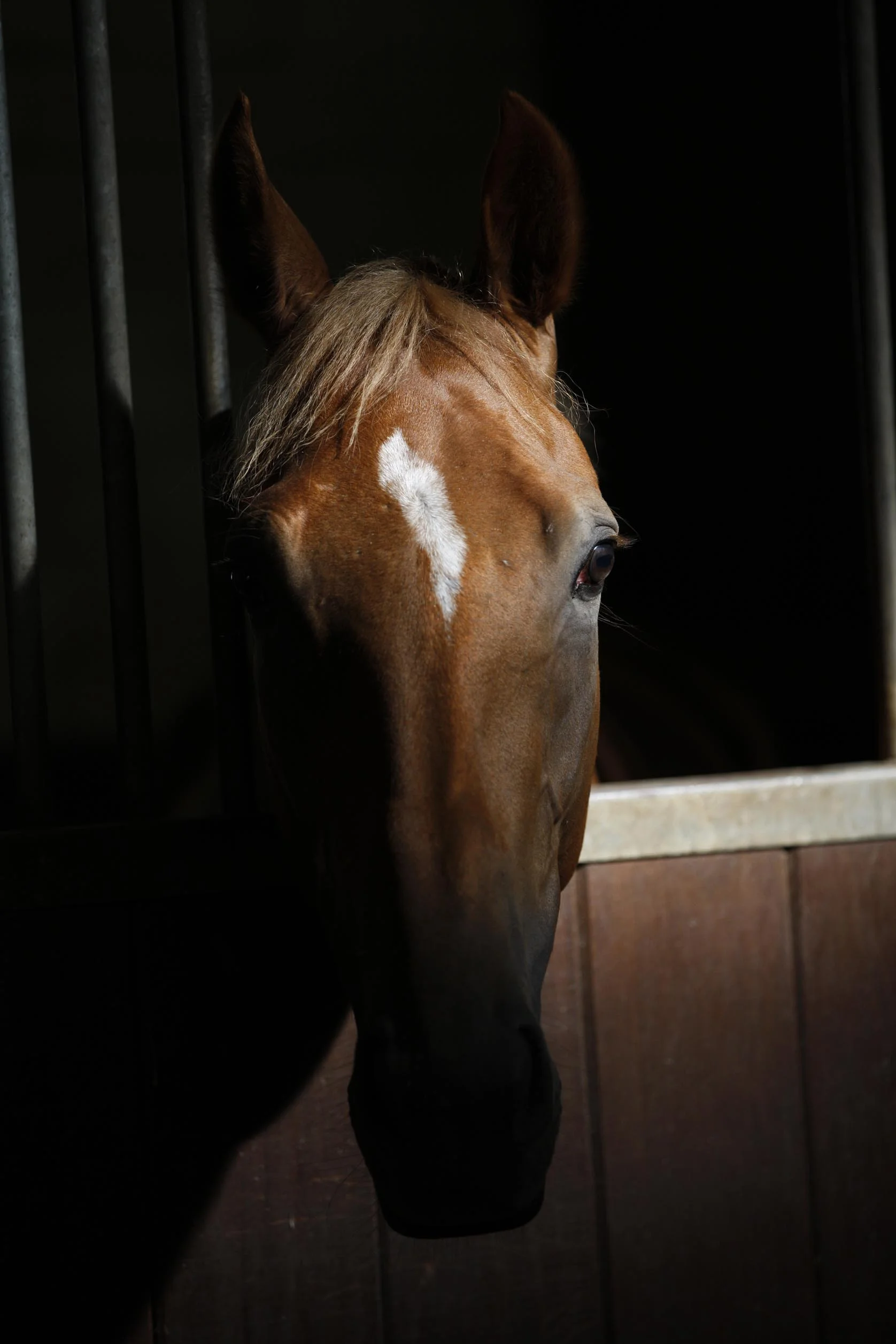
Let there be light - how daylight affects performance and safety

Recipient of multiple awards, including the Saratoga Trainer’s Title and the Eclipse Award for Outstanding Trainer, Bill Mott is no stranger to travelling with his horses. For example, Mott was trainer and chaperone of Cigar, winner of the inaugural Dubai World Cup in 1996. How do Mott and other elite trainers consider the impact of jet lag, light-dark cycles, and other factors associated with shipping across times zones on their horses’ performance?

CLICK IMAGE TO READ FULL ARTICLE

Read More
VETERINARY EUT Webmaster VETERINARY EUT Webmaster

Overtraining the racehorse

thumb_277.jpg

 Almost all trainers will have experienced a problem with individual horses, groups of horses, or sometimes even a whole yard where performance drops off for no immediately apparent reason. In human medicine we talk about chronic fatigue syndrome (CFS) which can affect both athletes and non-athletes, but in athletes we may be more likely to talk about overtraining.

In people, chronic fatigue syndrome is well recognised but often poorly understood. It typically affects young to middle aged adults with women being more commonly affected than men. It is estimated that somewhere between 150,000-250,000 people in the UK alone are affected by CFS. CFS is also referred to as simply chronic fatigue, post-viral fatigue syndrome, or myalgic encephalomyeltis (ME). The latter term describes muscle pain and central nervous system inflammation but that is not always apparent in chronic fatigue patients and so the term CFS has become more commonly used.

Dr David Marlin (European Trainer - issue 32 - Winter 2010)

Read More
VETERINARY EUT, NUTRITION EUT Webmaster VETERINARY EUT, NUTRITION EUT Webmaster

Racing power - supporting muscular effort through nutrition

The powerhouse for a horse in training is found in its large muscle mass. Whilst genetic makeup within the Thoroughbred breed has a large impact on a horse’s innate racing ability, dietary factors will also influence subsequent performance.

Dr Catherine Dunnett (European Trainer - issue 19 - Autumn 2007)

The powerhouse for a horse in training is found in its large muscle mass. Whilst genetic makeup within the Thoroughbred breed has a large impact on a horse’s innate racing ability, dietary factors will also influence subsequent performance. There are many elements found in a racehorse’s diet that will help to support muscle function. Hydrolysable carbohydrate (sugar and starch), assisted by fermentable fibre, will help to maintain important muscle stores of glycogen (a carbohydrate fuel).

Dietary electrolytes, which are integrally involved in muscle contraction, are essential to offset electrolyte loss in sweat. Key dietary antioxidants such as vitamins E and C and also antioxidant co-factors, such as copper, manganese, zinc and selenium, are also important as part of the body’s antioxidant team which strives to reduce the formation of free radicals or reactive oxygen species, and to limit their damaging effects on the body. Free radical damage has previously been implicated in the process of exercise induced muscle damage.

GLYCOGEN STORES MUST BE REPLENISHED FOLLOWING EXERCISE

One of the most important functions of the diet is to replenish the horse’s energy stores in muscle on an ongoing basis. A racing ration needs to support the synthesis of glycogen to maintain the store of this important fuel, which is used in increasing amounts during exercise. Glycogen, which consists of a large branched chain of glucose units, is stored in both skeletal muscle and the liver and it represents one of the largest potential energy stores in the body. Horses being natural athletes, have a relatively large muscle glycogen store when compared to other species. As the glycogen content of horse muscle is influenced by the proportion of different muscle fibre types present, this means that there is a genetic influence on the overall glycogen content. Fast twitch fibres (Type IIb), which are found in increased numbers in talented sprinting horses, store relatively more glycogen than the slower type I and type IIa fibres. However, both diet and training can influence the level of glycogen stored in muscle. Exercise training for example has been reported to increase muscle glycogen content by 30-60% in horses. Logically, diet should have a significant effect on the storage of muscle glycogen as it provides the building blocks for glycogen synthesis. Glycogen can be synthesised efficiently from dietary starch, which is another polymer of glucose found in cereals. Glycogen can also be produced from certain glycogenic amino acids, released from the protein content of feed. In addition, propionic acid, which is a significant volatile fatty acid produced in the horse’s hindgut during the fermentation process, can also ultimately be converted to muscle glycogen.

In terms of the day to day diet, starch is by far the most direct and most efficient precursor for glycogen and so it is therefore not surprising that cereals, which are high in starch, have been the mainstay of racing diets for many years. In recent years we have seen the introduction of racing feeds that are lower in starch and sugar than traditional racing rations, with a greater emphasis being placed on digestible fibre and oil as energy sources. Whilst there are many health benefits attributable to this type of diet, the effect of changing the level of starch in the diet on muscle glycogen should always be considered.

MUSCLE GLYCOGEN - AN IMPORTANT FUEL BUT NOT THE KEY FACTOR IN FATIGUE

Muscle glycogen is a major source of energy (ATP) to working muscle during intense exercise, which is characteristic of racing. The amount of muscle glycogen used during training or racing will depend on its rate of utilisation, which in turn is affected by the speed and duration of the exercise undertaken. In general terms, the higher the speed, the faster muscle glycogen is broken down and used.

The duration of fast exercise is normally curtailed, which limits the overall amount of glycogen used. During slower work, although the rate of glycogen utilisation is much lower, exercise can usually be continued for a much longer time allowing more glycogen to be utilised overall (see figure 1). Total muscle glycogen content can be reduced by about 30% during a single bout of maximal exercise in horses. However, as muscle is a mix of different fibre types, the depletion of glycogen in individual fibres may be greater than this depending on the pattern of fibre recruitment during the exercise. Studies, however, have shown that even the IIB muscle fibres, which use glycogen at the fastest rate, are not totally depleted of glycogen following racing.

This supports the notion that although glycogen is an important fuel source for racehorses, glycogen depletion is not the most important factor in fatigue. However, exercise studies do suggest that power output and exercise performance can be decreased in horses where muscle glycogen has failed to be adequately replaced following a previous race or piece of hard work. This was the conclusion drawn by Lacombe and co-workers (2001) who reported that horses with replete muscle glycogen stores were able to run for longer periods during a maximal exercise test compared to horses whose muscle glycogen level remained low following a previous exercise bout. Whilst there are always horses that will buck the trend, this research emphasises the need to allow a suitable period of time between races, but also between bouts of fast work and subsequent racing to allow muscle glycogen stores to be replenished.

In contrast to human athletes, muscle glycogen replenishment in horses is relatively slow. Following racing or a hard work, research suggests that muscle glycogen can take up to 72 hours to return to pre-exercise levels when a traditional high cereal racing ration is fed. Certainly research carried out in the past 3 years would suggest that a high glycemic racing ration would be better placed to support glycogen replenishment more quickly following racing or hard work. There are many factors that affect the glycemic response to feed, which in simple terms describes the relative rise in blood glucose following feeding.

The starch and sugar content of a feed, however, is one of the most significant factors affecting glycemic response. Feeds that are high in starch and sugar e.g. a high cereal-containing mix produce a greater glycemic response compared with feeds that are very low in starch and sugar e.g. a forage only ration. Rate of glycogen synthesis following a glycogen depleting exercise bout was significantly higher in horses fed a high glycemic diet compared to those fed a very low glycemic control diet (Lacombe et al 2004, Lacombe et al 2006). In addition, absolute glycogen concentration in muscle was significantly higher both 48 and 72 hours following exercise in the high glycemic group compared to the control horses and muscle glycogen concentration had returned to pre-exercise levels following 72 hours. The benefit of a high glycemic diet for glycogen repletion does, however, appear to be time dependent. Jose-Cunelleras (et al 2006) reported a minimal difference in glycogen repletion in the first 24 hours following a glycogen depleting exercise bout between horses that were fed a high glycemic feed compared with a group where feed was withheld for 8 hours and another group of horses where only hay was fed.

A recent study also concluded that the route of administration of carbohydrate given post-exercise significantly affects the rate of glycogen replenishment. Horses that were given an intravenous infusion of glucose following exercise exhibited significantly greater glycogen storage rates and glycogen concentration in the first 6 hours following exercise compared to horses fed a similar quantity of glucose orally. In fact, the repletion of glycogen in response to oral glucose was minimal over this time period compared to the unsupplemented control horses (Geor et al 2007). Whilst it is difficult to draw direct comparisons with feeding practices used in racing, it is worth appreciating the possible differences in the rate of glycogen repletion when very high glycemic feeds are fed compared to very low glycemic feeds. The reality in many training yards I would suspect lies somewhere between these two extremes.

LOW GLYCEMIC DIETS CAN OFFER RACEHORSES MANY BENEFITS

There are many health-related benefits to feeding a ration that is lower in starch and sugar. However, one should be mindful of muscle glycogen when considering horses that are consistently fed a low glycemic diet. Specifically horses may be fed this type of ration because they are behaviourally more manageable, or because a specific condition such as the muscular disease recurrent exertional rhabdomyolysis (tying up) (RER) is present. A low starch diet is actively encouraged for horses that suffer from RER. McKenzie (et al 2003) reported that plasma creatine kinase activity (CK), elevations of which can indicate muscle damage, was significantly reduced following exercise in RER horses fed a low starch high fat diet versus a high starch low fat diet. In addition, lower resting heart rates have also been reported in horses fed a low starch high fat diet compared to the reverse.

A lower resting heart rate may be beneficial especially in RER horses where it reflects a calmer horse as stress has been implicated as a trigger factor for the condition. The current thinking on feed for horses with RER continues to be a low starch and sugar diet supplemented with oil. It is also important that the diet is well balanced, especially with respect to calcium and phosphorus. Adequate electrolyte provision is equally important, as is the intake of antioxidants such as vitamin E and other related trace minerals such as selenium. Any potential individual limitation in mineral or electrolyte absorption and retention should be investigated further with veterinary assistance in order that individualised adjustments can be made to the diet.

A SUPPORTING ROLE FOR PROTEIN IN MUSCLE RECOVERY

Whilst we are all no doubt aware that the amino acids that make up protein are important for muscle development and repair, protein and its constituent amino acids have received very little attention in horses in terms of their potential to limit exercise induced muscle damage and aid muscle recovery. In human athletes, co-consumption of a protein and carbohydrate drink during and after exercise appears to limit exercise induced muscle damage, ultimately allowing faster recovery (Baty et al 2007; Saunders et al 2004). Recent introduction of ingredients containing partially hydrolysed protein may improve absorption of these amino acids and peptides possibly offering further benefit. Finally, some nutraceutical ingredients including carnitine and creatine have been hailed as being beneficial to muscle function and recovery in human athletes. Creatine, which has been studied in the horse, has failed to offer any great advantage, largely due to its poor absorption. Likewise, carnitine has been reported to improve muscle blood flow during exercise in humans, helping to reduce muscle damage. However, this aspect has not as yet been investigated in horses and previous dietary studies with carnitine were not unequivocal about the ability of oral carnitine to increase muscle carnitine content.


Read More
VETERINARY EUT Webmaster VETERINARY EUT Webmaster

Nasal Strips - increasing performance, reducing EIPH

Nasal strips’ future in Thoroughbred racing seemed limitless in the fall of 1999. Just two weeks after longshot Burrito won a race at Keeneland wearing one, 29 of the 101 horses competing in the 1999 Breeders’ Cup at Gulfstream Park November 6th had the 4-by-6-inch strip affixed 1.5 inches above their nostrils. 

Bill Heller (European Trainer - issue 18 - Summer 2007)


Nasal strips’ future in Thoroughbred racing seemed limitless in the fall of 1999. Just two weeks after longshot Burrito won a race at Keeneland wearing one, 29 of the 101 horses competing in the 1999 Breeders’ Cup at Gulfstream Park November 6th had the 4-by-6-inch strip affixed 1.5 inches above their nostrils. More importantly, three of the eight winners wore them, including Cat Thief, who captured the $4 million Classic at odds of 19-1 under Pat Day, who was sporting a human equivalent, himself. The image of both Cat Thief and Day posing in the winner’s circle with nasal strips was a powerful one. Cat Thief’s victory was the second that day for Hall of Fame trainer D. Wayne Lukas, who earlier saddled 32-1 longshot Cash Run to win the $1 million Breeder’s Cup Two-Year-Old Juvenile Fillies.

She, too, wore the non-invasive strip designed to reduce an exercising horse’s airway resistance and decrease exercise-induced, pulmonary hemorrhaging (EIPH). The nasal strips received enormous national publicity after the Breeders’ Cup. Wouldn’t almost everyone in North America emulate Lukas? Stan Bergstein, the executive vice-president of Harness Tracks of America and a columnist for the Daily Racing Form, postulated long ago that if a horse wearing a blue balloon tied to his tail won a race, you’d see dozens of horses with blue balloons tied to their tails in the paddock the next day. Lukas, however, preached caution regarding the role of nasal strips in Cash Run and Cat Thief’s surprise Breeders’ Cup victories.

Regardless, Lukas and trainer Bob Baffert spoke at a meeting of the California Horse Racing Board Medication Committee meeting, January 12th, 2000, in support of nasal strips. According to a CHRB press release, CHRB Commissioner Marie Moretti expressed hope that using the strips could lead to the decreased use of bleeder medication for some racehorses. That never happened, as Lukas proved prophetic. He saddled three horses in the 2000 Kentucky Derby, two with nasal strips, and none of them finished higher than 12th.

According to Equibase, between October 23rd, 1999, and April 24th, 2000, 8,402 Thoroughbreds wore the strip and 1,077 won, nearly 13 percent. Apparently that wasn’t high enough. Less and less trainers used them, though Lukas still does. By the end of 2000, there was a story on the Internet site www.suite101.com entitled “The Demise of Nasal Strips.” Published December 12th, 2000, the article began, “The rise and fall of nasal strips was short and sweet.” Noting that the Daily Racing Form had originally listed the nasal strip in past performance lines for all tracks and that by mid-June was only listing them at Hollywood Park, the story concluded, “As quick as they appeared in the spotlight, they vanished.” The obituary was more than a bit premature. Miesque’s Approval won the 2006 Breeders’ Cup Mile at Churchill Downs wearing a nasal strip for trainer Marty Wolfson, who uses them on all of his 30 horses. “I’ve been using them on all my horses for two years,” Wolfson said in mid-March. “I use them on myself. I run and they help me when I run. I breathe easier. The only time I couldn’t use one was when Pomeroy was in the 2006 Forego Handicap at Saratoga.” Pomeroy won that stakes. He was denied the nasal strip at Saratoga because the New York Racing Association mysteriously banned nasal strips, a day after the New York State Racing and Wagering Board approved them for both Thoroughbred and harness racing. Currently, New Jersey is the only other state which doesn’t allow them, while Pennsylvania allows them for Thoroughbreds but not for Standardbreds. According to nasal strip co-inventor and president of Flair Nasal Strips Jim Chiapetta, some 15,000 nasal strips are sold world-wide each year: 9,000 in the United States, 3,500 in Europe, 2,000 in Australia and New Zealand and 500 in Dubai. He said they were used mostly on horses in eventing, then on Thoroughbreds, Standardbreds and Quarter Horses. Should they be used more often? Are they a realistic alternative to the powerful diuretic Lasix, which is now used by roughly 95 percent of all

Thoroughbreds in the U.S., though the rest of the horse racing world bans Lasix and all other race-day medications? Lasix, which is used ostensibly to reduce EIPH, can improve a horse’s performance dramatically the first and/or second time it is used, if for no other reason that its diuretic properties. Horses can lose 10 to 20 pounds through urination after Lasix is injected. That alone improves most horses’ performance. Think about it. If there is an apprentice jockey with even a modicum of ability, trainers scramble for his services just to decrease the weight his horse is carrying by five pounds. The efficacy of nasal strips can be judged in comparison to Lasix or by itself. “Lasix and nasal strips work in very similar ways,” said David Marlin, a consultant who worked for the Animal Health Trust in Newmarket, England, and co-authored Equine Exercise

Physiology. “From scientific studies, they seem to be equally effective in reducing bleeding.” Breathe Right strips were invented in 1987 by Bruce Johnson, who suffered from allergies. By the early 1990’s, they were being used for colds, allergies, snoring and athletic performance. They work by reducing the partial collapse of the soft tissues of the nose when it is under pressure because of the vacuum caused by the lungs during exercise. The mechanical, spring device maintains optimum air flow. Humans have an option for breathing: nose or mouth. Horses do not. They breathe only through their nostrils. Could nasal strips benefit horses? That’s a question Jim Chiapetta and his partner Ed Blach decided to explore. They had become friends at the Littleton Large Animal Clinic in Littleton, Colorado. Chiapetta, 48, returned to his clinic in Shakopee, Minnesota, to finish law school at William Mitchell College of Law. Blach, a former veterinarian who is now an animal products consultant, called Chiapetta in 1996 to discuss a possible equine version of a nasal strip. “We talked to a bunch of people and they said it wouldn’t work for horses, but I told Ed I think it could,” Chiapetta said. “We went ahead and made some prototypes.” Then they consulted Monty Roberts, the horse whisperer. “Ed used to be Monty’s resident veterinarian,” Chiapetta explained. Roberts was interested enough to have them test the strip at a track at Roberts’ farm north of Santa Barbara in California. “We didn’t have the adhesive done right,” Chiapetta said. “The riders were coming back and saying, `This horse felt better, more relaxed.’ So we figured there was something there.” Having breakfast one morning with Roberts, Chiapetta and Bloch came up with a name. “I was thinking about flaring nostrils, then I was thinking about air, and we came up with the name Flair,” Chiapetta said. Next, they consulted with CNS, the Minnesota company which manufactured Breathe Right. “They agreed to license it if it showed it reduces bleeding,” Chiapetta said. “They funded a study at Kansas State University.” That study and a majority, but not all, of a handful of subsequent studies - all involving a standard small sample of horses - showed positive results from nasal strips. “The nasal strips seem to help,” Dr. Howard Erickson of Kansas State University, a co-author of one of the studies, said last February. “We’ve done studies here.

There have been studies in Kentucky, California and Florida. In most of the studies, it decreases the bleeding by 50 percent and it also decreases the airway resistance.” He believes that most horses would benefit from both, because he believes almost all horses suffer from EIPH: “I think it’s nearly 100 percent that have some degree of bleeding for the movement of fluid from the capillaries to the airway. For some, it may be negligible. Quarter Horses will respond the same way. Standardbreds, too. You see it in rodeo horses and barrel horses.” That sentiment is shared by David Marlin, who has worked with researchers at Kansas State. “The bottom line is that all horses will break blood vessels in a race,” he said. “It happens with camels; it happens with humans, it happens with greyhounds.” Marlin also believes that nasal strips may be a more preferable treatment than Lasix. “It’s less complicated and you can’t build up tolerance,” he said. “If you think about a diabetic who uses insulin, he develops tolerance and needs more of it.

Do horses develop tolerance of Lasix? Generally, when you use drugs repeatedly, there’s a chance of adaptation to it. The nasal strip is different because it’s a mechanical device.” Then why aren’t trainers around the world, and especially in the United States, using them? Ironically, Chiapetta believes that the success of Cash Run and Cat Thief in the 1999 Breeders’ Cup is a major reason why. “It was the worst possible thing that could have happened,” he said. “We were on the front page of the New York Times Sports Section, the Wall Street Journal and Sports Illustrated. I think horsemen said, `Hey, this will make us win.’ So they strapped them on. And when they didn’t win, they took them off.” Some, not all. “They’re expensive ($7.95 per strip),” Wolfson said. “Some people don’t want to spend the money, but I think it’s worth it.” Day, the retired Hall of Fame jockey, knew they worked on him. “I found them to be quite helpful when I was riding a number of races back to back,” he said. “It seemed that I was less fatigued because I believed I was getting much more air into my lungs. I would have thought that would be more helpful to horses than riders. Horses only breathe through their noses. They cannot or will not breathe through their mouths.

If you can open up the nasal passages, open the airways, you would think it would be beneficial to the horses.” At the Havemeyer Foundation Workshop investigating EIPH, March 9th-12th, 2006, in Vancouver, Canada, Dr. Frederick Derksen, of the Department of Large Animal Clinical Sciences at Michigan State University, spoke about the role of airways in EIPH. He said, “A series of studies demonstrated that the use of a nasal strip decreases the number of red cells in bronchoalveolar laverage fluid after exercise. In horses, the majority of inspiratory resistance to airflow is located in the upper airway. The nasal valge region, located just cranial to the nasoincisive notch is a high resistance region, not supported by bone or cartilage.

These characteristics make this region particularly susceptible to collapse during inhalation. Application of the nasal strip in this region prevents nasal collapse and decreases upper airway resistance during exercise. This in turn is expected to reduce negative alveolar pressure during inhalation and decrease transmural capillary pressures.” The nasal strips are certainly a hit in New Zealand, especially with harness horses. After reading about the use of nasal strips in the 1999 Breeders’ Cup, Brian McMath, a committee member of the New Zealand Standardbred Breeders Association, imported a few samples. After the strips were approved by Harness Racing New Zealand, several trainers began using them and many had success, including Jim and Susan Wakefield’s Glacier Bay, who won the $105,000 PGG Sales Series Final at Alexandria Park in April, 2000, for trainer Cran Daigety. Eventually, Thoroughbred trainers began using the strip, too.

By the end of 2004, more than 700 winners in both harness and Thoroughbred racing won wearing the strip. “I have a technology background in chemistry and engineering, and what convinced me the strips work was basic physics,” McMath said. “It’s all about windpipe pressures and how a simple mechanical device like the springs in the nasal strip can beneficially alter these pressures.” The reception in Europe, at least for Thoroughbreds, was decidedly cooler. In an April 11th, 2000, letter, Peter Webbon, the Chief Veterinary Adviser to the British Jockey Club, noted that the senior veterinary surgeons from the European Horserace Scientific Liaison Committee (Britain, France, Italy, Germany) considered the question of nasal strips and decided to recommend to their racing authorities that their use should be banned for the following reasons: 1 “Other `gadgets’, such as tongue ties, which are allowed, are intended to address a specific clinical entity. Nasal strips are seen by trainers as a non-specific way of improving performances. 2 “If they improve performance, they should be banned, in line with performance enhancing medication. 3 “If they are ineffective, they should be banned because they give the impression that we condone practices that are intended to improve performance. 4 The manufacturers claim that they reduce the frequency/severity of EIPH.

The EHSLC veterinarians felt very strongly, for the sake of the breed, that horses should run on their merits. What would be the effect on the Thoroughbred in the long term if a horse won the Derby, wearing a nasal strip,that without the strip was unable to win a selling race?” To this day, they are banned throughout Europe for racing but allowed for training. Two years ago, Chiapetta met with Webbon and his assistant in Newmarket. “He said, `It reduces fatigue, which improves performance,’” Chiapetta related. “I said, `If you shoe them, do they run better? If you feed them, do they run better? If you train them, do they perform better? Where do you draw the line?’” Event horses are allowed to use them throughout the world because they were approved by the International Federation for Equine Sports (FEI).

On June 26th, 2006, Horse & Hound wrote that nasal strips “are becoming commonplace on the noses of top event horses,” and noted that Andrew Hoy’s Moon Fleet won the Badminton, a premier cross-country event in England. “I started using them two years ago,” Andrew Hoy said. “I’d seen them being used on horses and humans, and discussed their use with a vet. I had used a human one myself when I had a cold, and it seemed to help. I now use them on my horses at top events to give them every opportunity.” The story said that another eventer, Francis Whittington, uses them on his “advanced” horse Spin Doctor. “I tried the human version and noted the difference,” he said. “I believe it makes it easier for him to breathe so he can last the distance.” That’s the whole point. “Some people may think that more oxygen makes them run faster,” co-inventor Blach said. “That’s not the case. Rather, horses perform at their optimum level for a longer time so they can do what they’re made to do over the long haul. Maybe it’s too simple. It’s based on very simple physics that if you maintain the size of an opening, you’re going to maximize what goes through it, in this case air.” Asked if nasal strips help horses, Blach said, “Absolutely.” Perhaps the most confounding question about nasal strips is that even the single negative clinical study about them said that they do not reduce EIPH, but offered no tangible downside to their usage. Asked if there is a downside, Marlin said, “I think, as far as anyone knows from a scientific point of view, there is no evidence that there is.” Referring to that study, Chiapetta said it showed that horses using them “certainly weren’t less healthier. I don’t think there’s any downside to it.” Dr. Ted Hill, the New York Racing Association steward for the Jockey Club, said on April 11th, “Our only downside was how to regulate it. If a horse comes to the paddock and it falls off, what do we do? Do we treat it as equipment? We can’t put it back on. The significant problem we had originally was it possibly being an aid to bleeders, and relaying that to the public. That came up in an international meeting at a round table in Tokyo last October. It did not receive wide acceptance because it has some efficacy.”

So Japan does not allow them. Australia allows them for Standardbreds, but not for thoroughbreds. Yet, nasal strips are allowed for Thoroughbreds in Dubai and Singapore, as well as New Zealand. “It’s probably been embraced more in other countries than here, but in Thoroughbred racing here, furosemide (Lasix) is so embedded,” Kansas State’s Erickson said. “Furosemide reduces weight. It certainly reduces bleeding. But maybe we have to look for something better.” Maybe something better has been out there for eight years.


Read More
NUTRITION EUT Webmaster NUTRITION EUT Webmaster

Forage - so much more than just a filler

Too often thought of as just a ‘filler’, or occupational therapy to while away the time between hard feeds, forage is worth so much more than that. Simply feeding an inadequate quantity of forage, or choosing forage that has an inappropriate nutrient profile, or is of poor quality can have a negative impact both on health and performance in racehorses.

Dr Catherine Dunnett (European Trainer - Issue 18 - Summer 2007)

Too often thought of as just a ‘filler’, or occupational therapy to while away the time between hard feeds, forage is worth so much more than that. Simply feeding an inadequate quantity of forage, or choosing forage that has an inappropriate nutrient profile, or is of poor quality can have a negative impact both on health and performance in racehorses. Inappropriate choice of forage and its feeding can easily lead trainers down the slippery slope towards loose droppings and loss of condition.

Forage can also have a significant impact on the incidence and severity of both gastric ulcers and respiratory disease, including inflammatory airway disease (IAD) and recurrent airway obstruction (RAO).

When choosing forage the main elements to consider are

• Good palatability to ensure adequate intake • Adequate digestibility to reduce gut fill

• Fitness to feed to maintain respiratory health

• A profile of nutrients to complement concentrate feeds

FORAGE CAN ONLY BE GOOD WHEN PALATABLE

Palatability is a key issue, as even the best forage from a quality and nutritional standpoint is rendered useless if the horses do not eat sufficient quantities on a daily basis. Palatability is a somewhat neglected area of equine research and so we largely have to draw on practical experience to tell us what our horses like and what they don’t. Some horses appear to prefer softer types of hay, whilst others prefer more coarse stemmy material. Many horses readily consume Haylage, whilst some trainers report that other horses prefer traditional hay. Apart from the physical characteristics, the sugar content of hay or haylage may affect its palatability. Forage made from high sugar yielding Ryegrass is likely to have a higher residual sugar content compared with that made from more fibrous and mature Timothy grass. Some interesting research carried out a few years ago by Thorne et al (2005), provided some practical insight into how forage intake could be increased in the reluctant equine consumer.

This work reported that the amount of time spent foraging (which will increase saliva production), was increased when multiple forms of forage were offered to horses at the same time. From a practical viewpoint this can be easily applied in a training yard and it should help to increase the amount of forage consumed. For example, good clean hay could be offered together with some haylage, and a suitable container of alfalfa based chaff or dried grass all at the same time.

A Healthy Intake Racehorses in training often eat below what would be considered to be the bare minimum amount of forage to maintain gastrointestinal health. Whilst sometimes this is due to the amount of forage offered being restricted, in other instances it is because the horses are limiting their own intake. This may be due to either their being over faced with concentrate feed, or due to unpalatable forage being fed. Establishing a good daily intake of forage during the early stages of training and then maintaining the level through the season is important. Typically the absolute minimum amount of forage fed should be about 1% or 1.2-1.5% of bodyweight for hay or haylage, respectively.

This equates to 5kg of hay or a rounded 7kg of haylage for an average sized horse (500kg). The weight of haylage fed needs to be greater than that of hay due to the higher water content of the latter. Intake of haylage needed to achieve a similar dry matter intake to 5kg of hay Moisture Dry Matter Weight of forage % Increase above hay Hay (Average) 15% 85% 5kg Haylage 1 30% 70% 6kg 20% Haylage 2 45% 55% 7.5kg 50% The dry matter of haylage needs to be consistent to allow a regular intake of fibre and reduce the likelihood of digestive disturbance or loose droppings.

Ideally trainers should be aware of any significant change in dry matter, so that they can adjust the intake accordingly. Forage intake is restricted in racehorses to firstly ensure that a horse consumes adequate concentrate feed to meet their energy needs and requirement for vitamins and minerals within the limit of their appetite. Secondly, the amount of forage fed is restricted in order to minimise ‘gut fill’ or weight of fibre and associated water in the hindgut, as this will restrict their speed on the racetrack. BUT… inadequate amounts of forage in a horses’ diet has such a negative effect on health that the minimum amount fed must be kept above recognised ‘safe limits’.

Choosing an early cut forage that is less mature and with more digestible fibre means that the ‘gut fill’ effect is lessened. In addition, horses can always be fed more forage during training with the daily quantity being reduced (within the safe limits) in the few days before racing where this is practical.

FITNESS TO FEED

Quality of forage, in terms of its mould, yeast and mycotoxin load, can have a major impact on respiratory health. A recent Australian report (Malikides and Hodgson 2003) highlighted the cost of inflammatory airway disease (IAD) in horses in training, in terms of loss of training time and of potential earnings, together with the associated cost of veterinary treatment. They estimated from their study group that in Australian racing up to 33% of horses in training can have lower airway inflammation, yet show no overt clinical signs. Type and therefore quality of forage, as well as the quality of ventilation were singled out as the most significant risk factors in the development of IAD.

Forage is potentially a concentrated source of bacteria, mould spores and even harvest mites. Hay that has heated during storage, or that has been bailed with a high moisture content is likely to provide a greater load of these undesirable agents that can harbour substances that promote airway inflammation, such as endotoxin. Purchasing good quality and clean forage from a respiratory perspective will certainly reduce the pressure placed on young racehorses’ respiratory systems.

However, how does one achieve this?

• Microbiological Analysis – the price paid for a microbiological analysis of a prospective batch of hay is a worthwhile cost when the consequences of poor hay are considered.

Assuming the analysis is favourable, purchasing a larger batch for storage gives further peace of mind and spreads the cost further, providing of course that the storage conditions are appropriate. Interpretation of the microbiology results as CFU/g (colony forming units/gram) for moulds, yeasts and Thermophillic actinomycetes is not difficult. As a rule of thumb the lower the CFU count the better. Whilst a very low mould or yeast count (<10-100) should not usually cause concern, more consideration of the merits of a batch of forage should be triggered by a CFU count that reaches 1000-10,000. Certainly if any Aspergillis species of mould are identified the alarm bells should be ringing.

Aspergillis Fumagatus has particular association with respiratory disease including ‘Farmers Lung’ in humans.

• Storage –A suitably sized storage area will allow storage of a good-sized batch of your chosen forage giving consistency through the season. It makes financial sense for the welfare of racehorses to make adequate provision for a good-sized storage area. Third party storage is also sometimes an option where this is not available on site.

• Forage merchant or farmer - A good working relationship with one or more farmers or forage merchants is essential to be able to consistently buy good hay. They need to know what you want to buy and you need to be able to rely on them to provide a high quality product through the season. Newmarket based forage merchant Robert Durrant stands by the principle that “A good forage merchant should be able to supply a trainer with the same high standard of hay for much if not all of the season”. He adds that in his opinion “American hay English hay or haylage are all good options when they have been made well and the quality is high, but the quality of the American hays are consistently more reliable.”

PRO’S AND CON’S

Hay from colder climates e.g. UK, Ireland commonly used quality can be variable usually palatable economical Haylage. Usually clean dry matter can be variable Fermentation inhibits mould growth Need to feed more than hay Feed value often higher May need to adjust hard feed Usually palatable Beware of punctured bales Newmarket trainer James Eustace has used big bale haylage for many years he says “I found it increasingly difficult to reliably source good clean English hay. I am very happy with the haylage, as it is pretty consistent and it provides the dust free option that I wanted.”

Hay from warmer climates e.g. USA / Canada usually very clean May need to adjust hard feed Feed value often higher Premium price Usually palatable Newmarket trainer Ed Dunlop appreciates the advantages of using more than one forage source he says, "American hay gives us the consistent good quality that we need and the horses eat it well. Feeding it alongside other forage gives us the flexibility needed for different horses throughout the season." Alfalfa (High temperature dried or sun dried)

Good adjunct to forage (e.g 1-2kg) High intakes can oversupply protein and calcium Can be used as chaff Leaf fragments can add to dust High feed value & digestibility Less gut fill Many of Forage merchant Robert Durrants clients choose sun dried alfalfa as an extra treat for the horses he says “the horses get a large double handful daily as a treat and they love it.”

NUTRITIONAL CONSIDERATIONS

The nutritional contribution made by forage should complement that made by the concentrate feed. Most racing rations are high in energy, high in protein and low in fibre. Therefore a suitable forage needs to be contrastingly high in digestible fibre with a limited level of energy and protein. However, where you have sourced early cut hay or haylage that is more digestible and higher in energy and protein, the concentrate feed intake should be adjusted to account for this. This will help to avoid the issue of over feeding of energy or protein. An excess of energy can result in undesired weight gain or over exuberance, whilst an excessive intake of protein at the very least increases the excretion of ammonia, which is a respiratory irritant.

Whilst it is important to know the calcium and phosphorus content of forage, the trace mineral content is less significant as the concentrate feed will meet the majority of the horse’s requirement. The exception to this, however is where a batch of forage is identified as having a severe excess of one particular element, e.g. Iron which can reduce the absorption of copper. Much emphasis is placed on finding an optimum concentrate feed and associated supplements, to enhance the diet of horses in training. The same emphasis should ideally be placed on a trainer’s choice of forage. Forage can so easily make or break the best thought out feeding plan.


Read More
VETERINARY EUT Webmaster VETERINARY EUT Webmaster

Inhalation therapy - treating airway problems in the racehorse

Physiologically speaking, one of the major limiting factors to racehorse performance is how efficiently the lungs can exchange gasses. Clearly any threat to the efficiency of the lungs will result in poor performance.

Paul Peacock (European Trainer - issue 14 - Summer 2006)

Physiologically speaking, one of the major limiting factors to racehorse performance is how efficiently the lungs can exchange gasses. Training maximises the potential of any athlete, equine or human, to continue functioning at full throttle while the metabolism changes to deal with an oxygen debt in the muscle tissues. Clearly any threat to the efficiency of the lungs will result in poor performance. Horses are subject to a wide range of respiratory diseases; heaves, lung bleeding or exercise- induced pulmonary haemorrhage (EIPH), and exercise induced Airway Inflammatory Disease (IAD) among them. Another description of the same basic problem is Chronic Obstructive Pulmonary Disease or (COPD). Like all mammals, horses suffer from allergic reactions as well as viral and bacterial infections. The epithelial linings of the airways and the lungs are sensitive to infection or foreign bodies of any size, and the result is usually a mucosal discharge which blocks the airway.

Whichever route an animal has airway problems, by infection or allergy, the result is almost the same; mucous builds up, coughing and irritation, frequent nose bleeding and considerably reduced performance. Moreover, some animals with tendencies towards heaves can show little signs of respiratory stress at times, but can be triggered later, more frequently by pollen and dust, when a change in regime occurs. The treatment options can be quite different for horses with a zootic infection to those with an allergy. Treatment of IAD involves the use of bronchial dilators and steroids, which have treatment implications of their own. Some of the drugs used can cause the gut to become sluggish, and can lead to colic.

Many of them induce tachycardia, the speeding of the heart rate, and still others make the animal skittish and nervous. Similarly, the use of corticosteroids in cases of allergic response, can affect the immune system, lead to numbers of other opportunistic infections, particularly in the mouth and have been implicated in laminitis. Bronchodilators include substances well known to human medicine and their function is to cause the dilation of the airways, thus allowing more air in and out of the lung. When irritated, the airways constrict and then produce mucous, which is then countered by the drug. There are two types of drug used for dilation of the airways, and they work very differently in the horse.

The Salbutamol type inhalation works on receptors on the epithelial cells of the airway, relaxing the muscle, thus causing dilation. They work at best for around an hour. A second class of drug, anticholinergics, work on various parts of the larger airway. Consequently, a mixture of the two types of drug is frequently used. Nigel Haizelden of the Ledston Equine Clinic in Castleford, West Yorkshire has been using this therapy for over 12 years and states that all kinds of drugs are administered using this system. Using a nebuliser, antibiotics, corticosteroids and bronchodilators are regularly applied. He points out that “the nebuliser is used to get the specific particle size which is required to reach a certain part of the lung – this is critical to the treatment.” Another important aspect of the bronchodilator is that the easier breathing allows the animal to relax under exercise, something which tends to promote further airway dilation. However, they do nothing for inflammation. Treatment should be associated with a regime which removes the animal from possible irritants. Trainer magazine has dealt with varying aspects in recent issues, from dust-free bedding to pollen allergy; particularly that produced by Oil Seed Rape.

One of the problems of treatment has included the fact that in order to get the drugs into the animal, the whole horse has to be treated. Injecting a horse with drugs means providing a high enough concentration in the animal’s blood which, when diluted by the circulatory system and metabolised by the liver, there is enough at the site of operation to do its work. Consequently a much higher concentration of drug is used than would be required if it could somehow be administered solely where it is needed and nowhere else. Inhalation therapy has been used in humans for a long time, from the vapour baths of Victorian days to modern viral carrier gene manipulation therapy proposed for such disorders as cystic fibrosis.

There are a number of benefits. Firstly the lung is an excellent way of getting a balanced concentration of drug into the blood stream. It works very quickly. In the case of airway disease, the drug is being used directly at the point that it is needed, and consequently the amount of drug required to be effective is greatly reduced. This improves treatment options by reducing the possibility of side effects. There are a couple of products on the market that allow this type of therapy. The Aeromask and the Equine-haler. Both are available via the vet and come from the United States. Their use has become increasingly widespread across Europe, particularly in France and Germany, where there have been particular links with American racing practices. IN the UK they have been used for at least fifteen years and the treatment regimes have developed accordingly. The Aeromask is strapped onto the head and the drug is held in a reservoir called the spacer. The Equine-haler is a cone which has to be held over the nose of the animal while the drugs are placed in a compartment at the bottom. This allows for a metered dose aerosol to deliver a dose to the spacer which is then inhaled by the horse. It only works on one nostril, and a puff of medicine is released into the nose.

The Equine-haler need not be held in position all the time, it allows for a puff of medicine to be fired into a spacer which then can be applied to the horse when it breathes in next time. Between the two it should be possible to find a regime which will ideally suit any animal, those shy of the head bag of the Aeromask could easily treated by the Equine-haler and visa versa according to the treatment required. It is important that only a measured amount of drug is administered, under veterinary control, so that overdoses do not occur. Similarly, the equipment should not be used to administer anything other than prescribed medicines. One yard on the continent was reported to have used their own remedies in association with the mask, which consequently caused some blistering to the horse’s mouth.

There are some risks associated with the use of inhalation therapy. One is associated with the drugs themselves. These drugs are particularly effective on the metabolism of the animal. It produces dilation of blood vessels, particularly in the liver, and it also promotes the production of insulin. In America at least, where there are different rules in various states regards doping, trainers are advised to take advice before racing. However, this method of treatment has meant that withdrawal periods for horses under treatment are considerably reduced in comparison to former treatments. Another possible problem is associated with the effect of the drug on the mouth, where fungal infections have been associated in humans with constant use.


Read More
VETERINARY EUT Webmaster VETERINARY EUT Webmaster

Do horses suffer from jet-lag?

The consequences of jet lag for the equine athlete have become more relevant in recent times due to increased travel of performance horses across multiple time zones for international competition.

Barbara Murphy (European Trainer - issue 7 - Spring 2004)


The consequences of jet lag for the equine athlete have become more relevant in recent times due to increased travel of performance horses across multiple time zones for international competition. The effects of jet lag are significantly more detrimental for the professional athlete hoping to perform optimally in a new time zone. Before defining the implications of jet lag for the horse, it is first necessary to understand the effects of light on any mammalian system. Most all life on earth is influenced by the daily cycles of light and dark brought about by the presence of the sun and the continuous rotation of our planet around its own axis.

From the simplest algae to mammals, nearly all organisms have adapted their lifestyle in such a way that they organize their activities into 24-hour cycles determined by sunrise and sunset. For this reason, many aspects of physiology and behaviour are temporally organized into circadian rhythms driven by a biological clock. Thus, biological clocks have evolved that are sensitive to light and so enable physiological anticipation of periods of activity. Light is the primary cue serving to synchronize biological rhythms and allows organisms to optimise survival and adapt to their environment. An example of this environmental adaptation is clearly evident in the mare’s natural breeding season. As the number of hours of light gradually increases in the early spring, the mare’s reproductive system reawakens and within weeks is ready for conception.

With an 11 month, one week gestation period, horses have evolved to produce their young when the days are long and warm and the grass is green – the ideal environment for a growing animal and for a lactating mare with increased nutritional needs. However, we have interfered with nature’s design. With the creation of a universal birthday for Thoroughbred horses of January 1st and the economic demands to produce early foals for sale as mature yearlings, we have succeeded in altering the mare’s natural breeding season through use of artificial lighting programmes. A 200-watt light-bulb, in a 12-foot by 12-foot stall, switched on from dusk until roughly 11:00 pm nightly and beginning December 1st, is sufficient to advance the onset of the mares reproductive activity such that she should be ready to be bred by February 15th, the official start of the breeding season. From this, it is clear that light can control seasonal rhythms. What is more important in relation to jet lag is that light also closely regulates daily, or circadian rhythms. These circadian rhythms include changes in body temperature, hormone secretion, sleep/wake cycles, alertness and metabolism. A disruption of these rhythms results in jet lag.

It can be defined as a conflict between the new cycle of light and dark and the previously entrained programme of the internal clock. The first step to understanding jet lag is to examine the workings of the biological clock and the extent to which the daily cycles of light and dark can control physiological processes. All mammals possess a “master” circadian clock that resides in a specific area of the hypothalamic region of the brain. This area of the brain is responsible for regulating diverse physiological processes such as blood pressure, heart rate, wakefulness, hormone secretion, metabolism and body temperature. Each of these processes is in turn affected by time of day.

During daylight hours, the eye perceives light and light energy is transmitted via a network of nerve fibres to the brain. Here, the light signal activates a number of important genes and these “clock” genes are responsible for relaying signals conveying the time of day information to the rest of the body. Recent advances in the study of circadian rhythms and clock genes have shown that a molecular clock functions in almost all tissues and that the activities of possibly every cell in a given tissue are subject to the control of a clockwork mechanism. The role of the “master” clock in the brain is to communicate the light information to the clocks in the peripheral tissues, so that each tissue can use this information for its own purpose.

Thus, as day breaks and eyes perceive morning light, hormones are produced to help us to wake up, enzymes are activated in our digestive systems in anticipation of breakfast, heart rates increase, muscles prepare for exercise and many more circadian rhythms are initiated. This master clock in the brain, that controls so many bodily functions, must be reset on a daily basis by the photoperiod, whether it is sunrise and sunset or lights on and lights off, in order for an organism to be in harmony with its external environment. Jet lag occurs due to an abrupt change in the light-dark cycle and results from travel across multiple time zones, which in turn causes de-synchronization between an organism’s physiological processes and the environment.

Coupled to this is the fact that the circadian clock can only adapt to a new lighting schedule gradually and while the brain receives the light information directly, there is a further lag period involved in transmitting the time of day message to peripheral tissues. As a consequence, behavioural and physiological adaptation to changes in local time is delayed. This means that following a transmeridian journey, travellers are forced to rest at an incorrect phase of their circadian cycle, when they are physiologically entrained to be active and more importantly for the athlete, they are expected to perform when they are physiologically set to rest. As mammals, horses also suffer from the effects of jet lag.

Research is needed to understand the extent of physiological disruption caused by a transmeridian journey, the time period of the disruption and the overall effect it has on equine performance. Until now, no studies have been undertaken to investigate the physiological effects of jet lag in the horse. Studies in human athletes have demonstrated the detrimental effects of translocation on exercise capacity and performance. One early study examined human subjects following intercontinental flights consisting of eastward or westward journeys across multiple time zones (1). Results clearly demonstrated significant disturbances in heart rate, respiratory rate, body temperature, evaporative water loss and psychological function. Interestingly, these disturbances were found to be more profound following the eastward flight.

A more recent study conducted using top athletes from the German Olympic team investigated the effects of time-zone displacement on heart rate and blood pressure profiles (2). In athletes, blood pressure and oxygen supply to the organs are of utmost importance for optimal performance and successful competition. Rhythm disturbances in the 24-hour profiles of heart rate and blood pressure were found to be present up to day 11 after time-zone transition. The athletes were involved in intensive training programmes throughout the study and underwent frequent bouts of strenuous exercise.

Regular exercise at a set time in the 24-hour clock can strengthen circadian rhythms that are integral to physiological processes and can act as a timing cue secondary to light. It is also thought to aid in resynchronisation to a new time-zone. However, exercise was not found to improve the jet lag effects in this study, an observation that has relevance for the athletic horse in intensive training. The investigators concluded that following a flight across six time zones, athletes should arrive for their competition at least two weeks in advance in order to overcome the jet lag effects before competing.

Another study using fit human subjects examined performance times before and after an eastward journey across 6 time zones (3). Performance times for a 270m sprint were slower for the first 4 days following translocation as were times for a 2.8km run on the second and third days. This can be explained by the fact that the athletes’ internal body rhythms, including several neuromuscular, cardiovascular and metabolic variables and indices of aerobic capacity are out of synchrony with the environmental light-dark cycle following a transmeridian journey. Small mammals such as rats and mice have historically been used to study human circadian disorders such as jet lag. Current research being conducted at the Gluck Equine Research Center at the University of Kentucky has resulted in successful isolation of a number of ‘clock’ genes.

A comparison of these equine specific genes with their human counterparts has revealed an unusually high similarity between these two species at the DNA level, closer than the similarity observed between small mammals and humans. Unlike humans and horses, rats and mice are nocturnal animals and have yet to be proven as elite athletes. Further research is underway to investigate in detail the effects of jet lag on equine performance that will eventually lead to the development of measures to counteract these effects. Until then, information on the effects of transmeridian travel derived from studies on human performance can be used to provide guidelines to horse trainers, especially based on the similarity between the species in question. The severity of the jet lag effects can depend on a number of factors. These include the ability to preset the bodily rhythms prior to flying (4), the number of time zones crossed, the direction of the flight and individual variability.

Just as set exercise times can affect circadian rhythms in many physiological processes, feeding schedules also play an important role in entraining biological clocks, particularly within the digestive system. Horses anticipate feeding times. Banging of hooves on doors and rattling of empty feed buckets are common sounds that greet those responsible for feeding a yard of hungry horses. Therefore, it is important to change both feeding times and exercise schedules to mimic the new time zone prior to travel, in order to shorten the amount of time required for resynchronisation of digestive function and performance capacity upon arrival. Lighting is also of paramount importance. Exposing animals to early morning bright light for several days prior to an eastward journey across multiple time zones, or, extended hours of evening light prior to a westward journey, will help synchronize circadian rhythms to the new time zone prior to travel.

A recent study that tested a combination of approaches to hasten the resynchronisation of a group of elite sports competitors and their coaches to a westerly transmeridian flight, demonstrated the usefulness of combining melatonin treatment, an appropriate environmental light schedule and timely applied physical exercise to help the athletes overcome the consequences of jet lag (5). Melatonin, a hormone secreted by the pineal gland of the brain during the hours of darkness, is thought to help synchronize sleep-wake cycles and resynchronisation to a new time zone, but its suitability for these purposes has yet to be tested in the horse. Of course, these procedures to preset bodily rhythms need not be implemented if it is possible to arrive at the destination in sufficient time to allow natural re-entrainment to the new light-dark cycle.

For financial reasons, this is not necessarily feasible for the equine athlete. Two other important factors that determine the severity of jet lag effects are the number of time zones crossed and the direction of the flight. As one would expect, the greater the number of time zones traversed, the more severe the physiological disruption. For example, a flight from Europe to the East Coast of the United States, across six time-zones, would require a significantly greater resynchronisation time than a flight from the East Coast to the West Coast (three time zones), within the continental U.S. Any transmeridian journey in an eastward direction will result in a more profound disruption of circadian rhythms than a similar journey in a westward direction. The reason for this is a molecular one and involves the individual characteristics of certain clock genes. Suffice to say that clock genes react more rapidly to light than to darkness. When travelling in a westward direction i.e. from Europe to the United States, travellers enter an environment consisting of extended hours of evening light. The light continues to stimulate clock genes in the brain and adaptation to the new time zone occurs more rapidly. To some extent this may explain the success experienced by European horses at U.S. racetracks, even when they arrive three to four days prior to a race.

Knowing exactly how long it takes for the equine athlete to overcome any travel effects that may impinge on performance following such a flight, should provide valuable information to European trainers. In contrast, an eastward journey results in a shortened day length at the destination and requires a phase advance of the circadian clock. Travellers experience earlier nightfall and as the clock genes cannot respond well to darkness, an extended duration of jet lag. To emphasize, it will take an animal longer to adapt to the new light-dark cycle following an eastward flight and consequently longer to reach optimal performance levels following transit.

Pharmacokinetics deals with absorption, distribution, metabolism and elimination of drugs and these steps are influenced by physiological function of the body, which we now know to be influenced in turn by time of day. The implications of this for the athletic horse following transmeridian travel is worth highlighting, as it underlines the importance of knowing approximate physiological resynchronisation time to a new time zone. For example, terbutaline, a bronchodilator similar to clenbuterol commonly used by equine practitioners, has a significantly longer half-life when administered in the morning than in the evening (6). This implies that drug clearance times can be affected by transmeridian travel. In addition to the disruption of circadian rhythms, travel stress can also be a significant factor in further compounding the effects of jet lag following the transportation of horses across multiple time-zones.

Major complications associated with long-distance travel include pleuropneumonia, otherwise known as ‘shipping fever’, dehydration and colic. Even in cool conditions, horses will often lose 2-5 pounds of body weight for every hour they travel, as they do not like to drink while travelling (7). Care of horses during long-distance transportation is an extensive topic that requires separate attention. At the Gluck Equine Research Center, preparations are underway to conduct several experiments that will simulate phase advances and delays in the lighting schedule of groups of horses, thus mimicking eastward and westward journeys, so that the molecular and physiological effects of jet lag and the time duration of these effects can be investigated. The goal of this research is ultimately to provide practical guidelines to trainers in order that measures can be taken to counteract the detrimental effects of jet lag on performance, therefore leveling the playing field for horses competing away from home.


Read More