Young racehorse development through the lens of biotensegrity and fascia science
The debate surrounding the appropriate age to commence racehorse training remains a contentious topic. Advocates of traditional biomechanical models argue that training at 18 months is premature, as a horse's skeletal system does not reach full maturity for several more years. However, skeletal development alone presents a limited perspective. I would like to introduce another perspective from a rising research field. Through the lens of biotensegrity and fascia science, a more comprehensive approach emerges—one that considers the interconnectedness of a horse’s entire physiological system. Well-structured training at a relatively young age can support the holistic development of the racehorse, fostering both physical and psychological adaptability.
The Influence of gravity and early adaptation
From the moment a foal is born, it must quickly adapt to the force of gravity. Passage through the birth canal initiates its structural alignment, and within hours, the foal is standing and moving independently. Foals are born with predominantly fast muscle fibres (2X). The ability to travel up to seven kilometers daily alongside its dam is a testament to the foal’s inherent adaptability. This early exposure to movement and environmental stimuli plays a crucial role in its physiological and neurological development.
Challenging traditional views on skeletal maturity
This article seeks to introduce an alternative perspective on how the horse interacts with gravity, incorporating the principles of biotensegrity and fascia. It is important to note that most research is done on corpses and the fascia dries out almost directly after the circulation stops. Traditionally, equine skeletal maturation has been the primary concern regarding the timing of racehorse training. However, a singular focus on bone development overlooks the adaptability of connective tissues and the overall structural integrity of the horse. All young horses as an adaptation to their environment are in a critical phase of learning and adaptation—both physically and mentally—which must be accounted for in any training approach.
Veterinary discourse on skeletal maturity presents conflicting perspectives. Veterinarian Chris Rogers asserts that the skeleton of a two-year-old Thoroughbred is sufficiently developed for training, drawing parallels to human child development. Conversely, Dr. Deb Bennett posits that full skeletal maturity does not occur until six to eight years of age regardless of breed. All horses go through almost the same skeletal development phases, although thoroughbreds are extremely adapted through breeding to grow much quicker. While Bennett’s perspective has been widely accepted, Rogers’ viewpoint aligns with the practical realities of racehorse development, supporting the industry’s traditional training timelines.
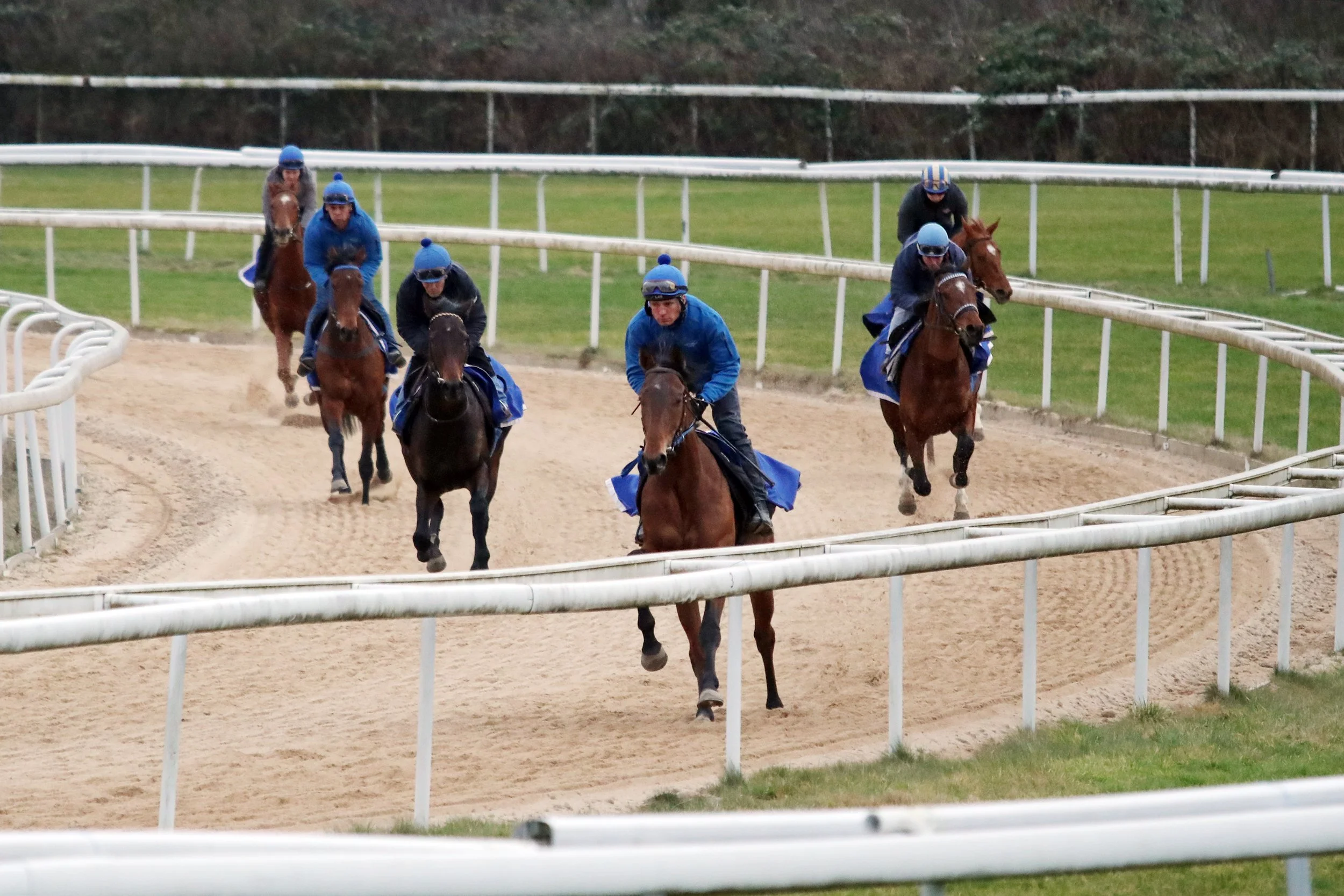
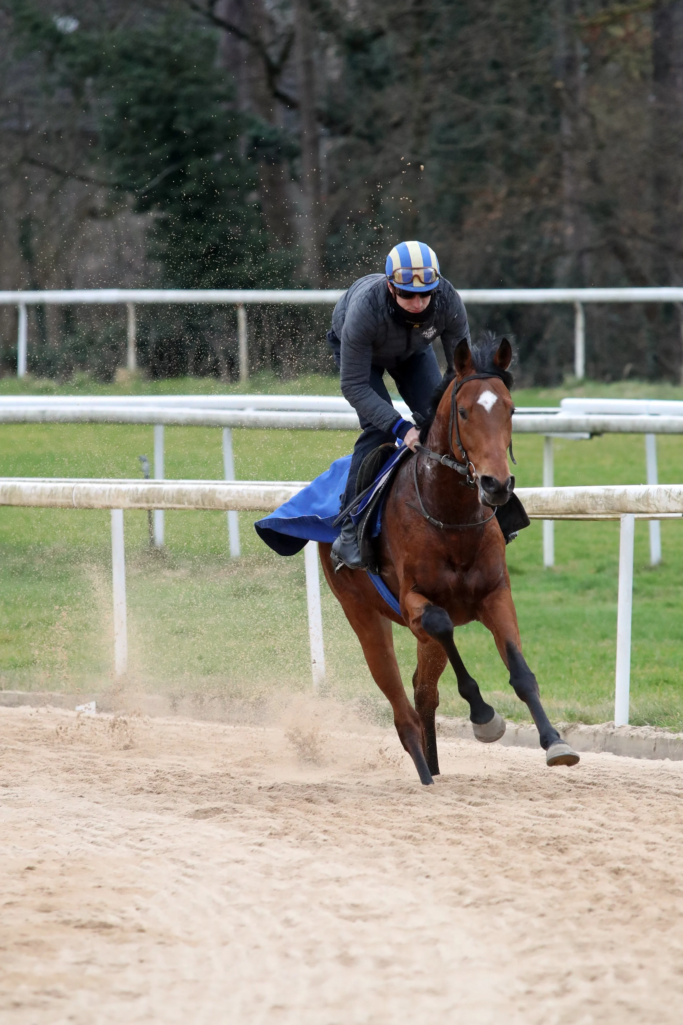
Flat racehorses typically begin training at around 18 months of age. At this stage, their skeletal and connective tissues are still developing, as research consistently shows. Cartilage, bones, muscles, and ligaments undergo intensive growth and adaptation. Every experience the young horse encounters contributes to its physiological and neurological development, shaping its ability to perform the tasks expected of a racehorse. Training at a young age offers several advantages, as young horses are highly receptive and adaptable. As explored later in this article, their connective tissues develop in response to the challenges they are exposed to, reinforcing their structural integrity over time.
Racehorse training inherently involves a selection process. Horses that do not meet performance expectations within the first few seasons are often retired from racing by the age of three or four, making way for new yearlings. Those that demonstrate both speed and durability may continue competing well into their later years. Those are often geldings. Mares and stallions that show promise may transition into breeding programs. The rest, if their foundational training has been well-structured, can adapt successfully to second careers as riding horses, often becoming ideal partners for young equestrians at the start of their horsemanship journey.
Tensegrity principles
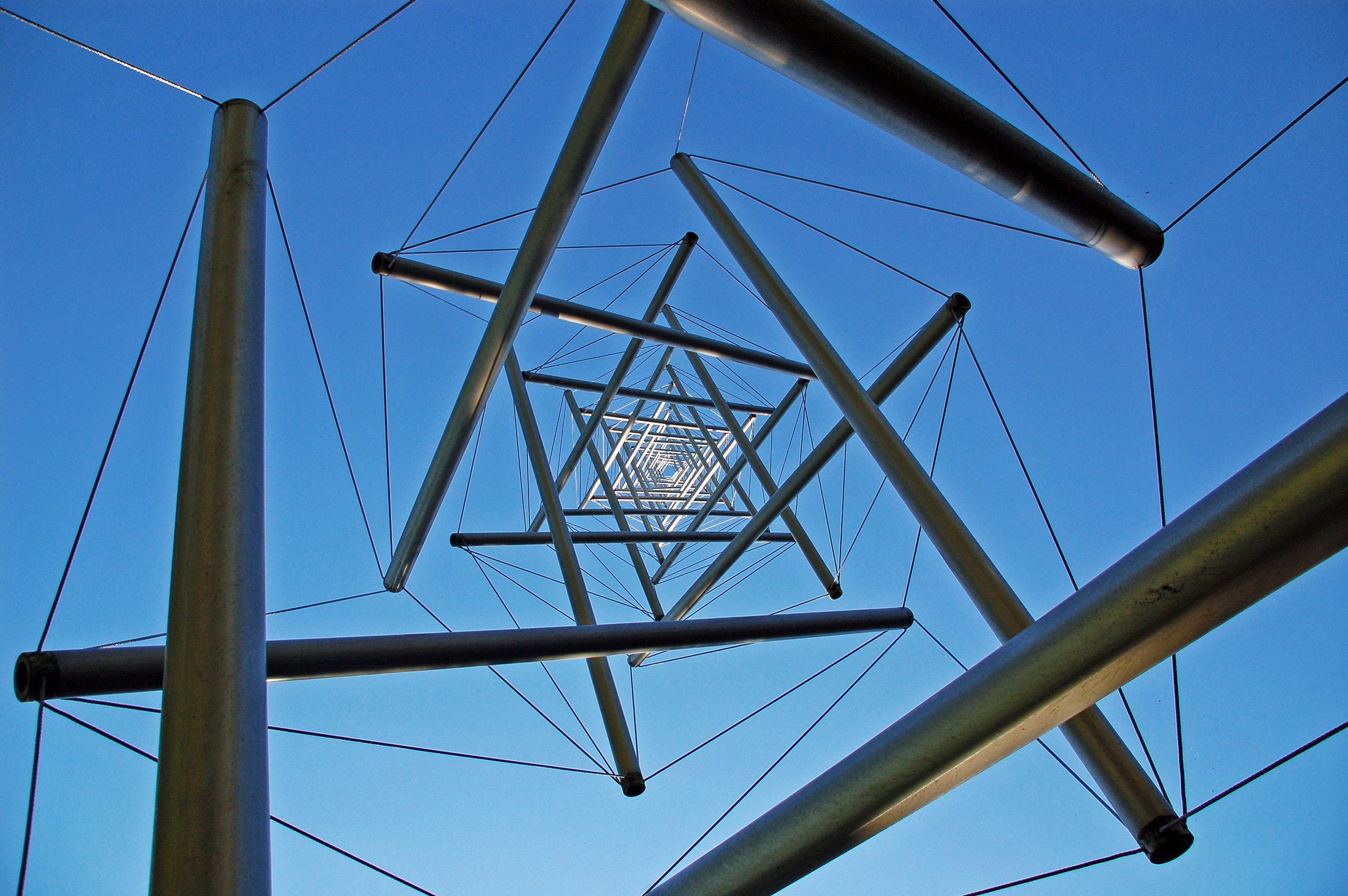
Tensegrity (tensional integrity) is a structural principle that explains how forces of tension and compression interact to create stability in a system. Originally coined by architect and engineer Buckminster Fuller, tensegrity has been widely applied in biological systems, including human and equine anatomy.
The role of fascia and biotensegrity in equine development
A traditional biomechanical view perceives the horse's skeleton like a rigid brick wall—if one part weakens, the entire structure becomes vulnerable to collapse. In contrast, a tensegrity-based perspective views the horse as a dynamic suspension bridge, where forces are distributed across an interconnected network of fascia, tendons, and ligaments. In this model, the skeleton is not a rigid load-bearing framework but rather ‘floats’ within the fascial system, allowing for adaptability, resilience, and efficient force distribution.
Biotensegrity highlights the balance between tension and compression within the body. In equine anatomy, the skeleton functions as a stabilizing framework, while fascia, tendons, and ligaments manage dynamic forces. Fascia, composed predominantly of collagen, exists in various densities, from loose connective tissue that facilitates muscle glide to the more rigid structures forming tendons and bones. This complex, fluid-filled network plays a crucial role in maintaining stability, distributing forces, and mitigating the impact of training.
Training influences the structural adaptation of connective tissues. Properly executed, it can enhance durability and resilience, reinforcing ligaments and tendons much like steel cables under controlled tension. Understanding the dynamic interplay between muscle, fascia, and skeletal development allows for training methods that optimise long-term soundness and performance.
Fascia
One of the most abundant proteins in the body is collagen, which forms connective tissue in all its various forms—from loose fascia, which separates muscles, to denser collagen structures that align with the direction of force and develop into tendons, ligaments, or bone. One of the key functions of loose fascia is to allow muscles to glide smoothly against one another without friction when one muscle contracts and another stretches.
Loose fascia consists of a collagen network, with its spaces primarily filled with water and hyaluronic acid. It is a highly hydrated structure—young horses are composed of approximately 70% water. Imagine a water-filled balloon, where the skin acts as the boundary between the internal and external environments. During fetal development, collagen structures form first, providing the framework within which the organs develop. Collagen, a semiconductive protein, relies on water to function optimally.
The water-rich environment surrounding fascia transforms it into an extraordinarily intelligent communication network. Its function is highly responsive to the body's pH levels, adapting moment by moment to internal conditions. As the central hub for force transfer and energy recycling, fascia provides immediate balance and support—often operating beyond the constraints of the nervous system.
Fascia's remarkable adaptability is rooted in its multifaceted properties. It is nociceptive, meaning it is capable of detecting pain and harmful stimuli, alerting the body to potential injury or strain. It is also proprioceptive, enabling the body to sense its position and movement in space, which aids in maintaining coordination and balance. Additionally, fascia exhibits thixotropic properties—allowing it to shift between a gel-like state and a fluid-like state depending on movement, which enhances flexibility and responsiveness.
Finally, fascia demonstrates piezoelectric properties, generating electrical charges in response to mechanical stress, playing a crucial role in cellular signaling and tissue remodeling. These combined characteristics enable fascia to dynamically adjust to both mechanical and biochemical stimuli, ensuring optimal function in response to ever-changing internal and external conditions."
Fascia has no clear beginning or end; it distributes pressure and counteracts the force of gravity.
The biotensegrity of equine locomotion and how horses rest while standing
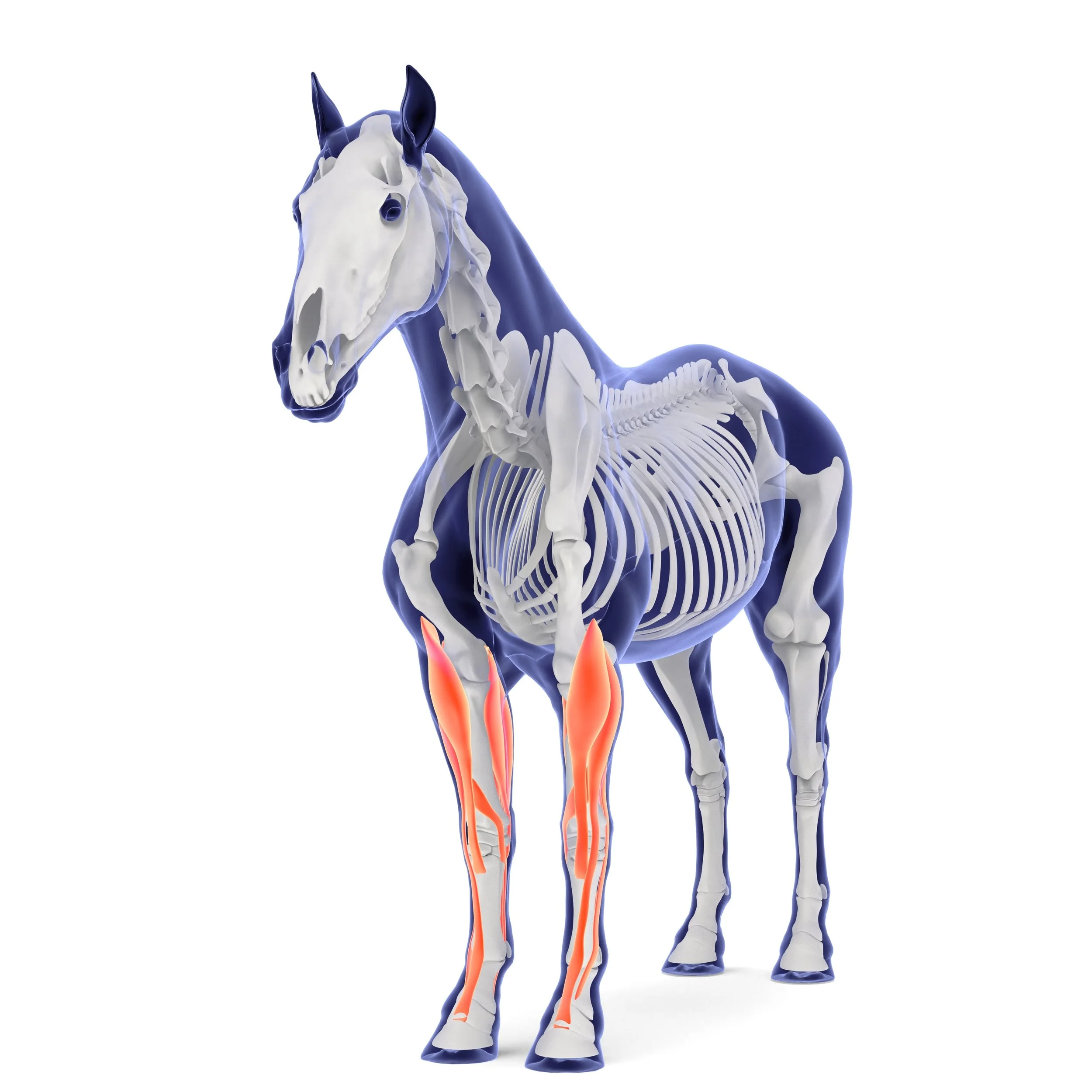
Horses possess a remarkable evolutionary adaptation that allows them to rest while standing, a capability underpinned by the principles of biotensegrity. This structural efficiency is achieved through an intricate network of tendinous and ligamentous locking mechanisms working in harmony with the skeleton.
In the forelimbs, the extensor and flexor tendons engage to stabilise the skeletal structure, minimizing muscular effort. Meanwhile, in the hind limbs, a specialised locking mechanism is activated when the patella (kneecap) is positioned against a flat section on the femur just above the stifle, further contributing to this passive support system.
This adaptation allows horses to conserve energy while remaining poised for rapid movement. In the event of sudden danger, they can instantly transition from rest to flight, ensuring their survival—an essential trait for both wild and athletic performance. The efficiency of this natural support system exemplifies the principles of biotensegrity, where tension and compression forces work in balance to maintain structural integrity with minimal effort.
The head and neck function as critical balancing structures, comprising approximately 10% of the horse's total body weight. The forelimbs bear roughly 60% of the body’s weight, but true structural support originates from above the elbow joint. The spine, a central element of equine biomechanics, acts as a suspension system. The primary function of the equine spine is to support the internal organs, a role that also enables the horse to carry a rider.
This structural foundation ensures both stability and balance, allowing for efficient movement and performance under saddle. The propulsion generated by the hind legs is efficiently transferred to the forehand through the back muscles, which are reinforced with robust connective fascia plates, ensuring optimal movement and stability. This structural complexity underscores the need for a training regimen that respects the developmental timing of multiple interrelated systems beyond just the skeletal framework.
Risks and adaptations in young racehorses
While early training offers advantages in developing resilience in young racehorses ( they have a high percentage muscle fiber 2X), it also presents risks. The spinal column, particularly the lumbar-sacral junction, endures significant forces during high-speed galloping. Without appropriate conditioning, the vulnerability of these structures can lead to pathologies such as kissing spines or pelvic instability. Their growth plates remain open, making them more susceptible to the impact of high-speed forces, compensatory adaptations to early training stress may manifest as different maladaptive adaptations in connective and skeletal tissues, potentially diminishing long-term performance capabilities.
However, when managed correctly, the high adaptability of collagen structures in young horses allows for positive adaptation. Training introduces controlled tensile and compressive forces, fostering the development of strong, functional connective tissues. The challenge lies in striking the right balance between stimulus and recovery to optimise long-term soundness and athletic potential.
The value of tacit knowledge in training practices
Experienced trainers have an intuitive understanding of the complex relationships between tissues and biomechanics, a knowledge that is often honed through years of careful observation and practical experience. This tacit expertise is fundamental in shaping training strategies that take into account the horse’s overall development. A comprehensive training program should not only focus on the maturation of the horse’s bones but also prioritise the adaptive growth of fascia, ligaments, and muscles. By doing so, trainers ensure that young racehorses develop in a way that is aligned with their evolving physiological capabilities, promoting balanced growth and minimizing the risk of injury. This holistic approach allows for the optimal performance and longevity of the racehorse, fostering a more sustainable path toward peak athleticism.
Conclusion: a holistic perspective on racehorse development
The evolution of equine training methodologies has greatly benefited from recent advancements in scientific understanding, offering a more refined approach to racehorse development. By incorporating biotensegrity principles into training programs, a more comprehensive view of the horse’s physical structure and function emerges, shifting the focus from skeletal maturity alone to a broader understanding of the interconnected roles of fascia, connective tissues, and adaptive biomechanics. This shift in perspective allows for the cultivation of healthier, more resilient athletes who can perform at their peak while minimizing the risk of injury.
With equine welfare at the forefront, adopting a holistic approach to racehorse development—one that blends cutting-edge biomechanics, physiological insights, and traditional training wisdom—will pave the way for more sustainable, ethical practices within the industry. Such an approach not only enhances performance in the short term but also ensures the longevity and well-being of racehorses throughout their careers. Ultimately, by embracing this integrated perspective, the racing industry can promote a future where both the performance and welfare of horses are prioritised, leading to a more ethical and effective standard of training.
---
Extra Reading References
- Levin, S. (Biotensegrity: www.biotensegrity.com)
- Clayton, H. M. (1991). *Conditioning Sport Horses*. Sport Horses Publications.
- Adstrup, S. (2021). *The Living Wetsuit*. Indie Experts, P/L Austrasia.
- Schultz, R. M., Due, T., & Elbrond, V. S. (2021). *Equine Myofascial Kinetic Lines*.
- Bennett, D. (2008). *Timing and Rate of Skeletal Maturation in Horses*.
- Rogers, C. W., Gee, E. K., & Dittmer, K. E. (2021). *Growth and Bone Development in Horses*.
- Ruddock, I. (2023). *Equine Anatomy in Layers*.
- Myers, T. W. (2009). *Anatomy Trains (2nd Edition)*. Churchill Livingstone.
- Diehl, M. (2018). *Biotensegrity*.
- Kuhn, T. S. (1962). *The Structure of Scientific Revolutions*. University of Chicago Press.
- Tami Elkyayam Equine Bodywork
The importance of good hoof balance to improve performance
The equine foot is a unique structure and a remarkable feat of natural engineering that follows the laws of biomechanics in order to efficiently and effectively disperse concussional forces that occur during the locomotion of the horse. Hoof balance has been a term used by veterinarians and farriers to describe the ideal conformation, size and shape of the hoof relative to the limb.
Before horses were domesticated, they evolved and adapted to survive without any human intervention. With respect to their hoof maintenance, excess hoof growth was worn away due to the varied terrain in their habitat. No trimming and shoeing were required as the hoof was kept at a healthy length.
With the domestication of the horse and our continued breeding to achieve satisfactory performance and temperament, the need to manage the horse’s hoof became essential in order to ensure soundness and performance. The horse’s foot has evolved to ensure the health and soundness of the horse; therefore, every structure of the foot has an essential role and purpose. A strong working knowledge of the biology and biomechanics of the horse’s foot is essential for the veterinarian and farrier to implement appropriate farriery. It was soon concluded that a well-balanced foot, which entails symmetry in shape and size, is essential to achieve a sound and healthy horse.
Anatomy and function of the foot
The equine foot is extremely complex and consists of many parts that work simultaneously allowing the horse to be sound and cope with the various terrains and disciplines. Considering the size and weight of the horse relative to the size of the hoof, it is remarkable what nature has engineered. Being a small structure, the hooves can support so much weight and endure a great deal of force. At walk, the horse places ½ of its body weight through its limbs and 2 ½ its weight when galloping. The structure of the equine foot provides protection, weight bearing, traction, and concussional absorption. Well-balanced feet efficiently and effectively use all of the structures of the foot to disperse the forces of locomotion. In order to keep those feet healthy for a sound horse, understanding the anatomy is paramount.
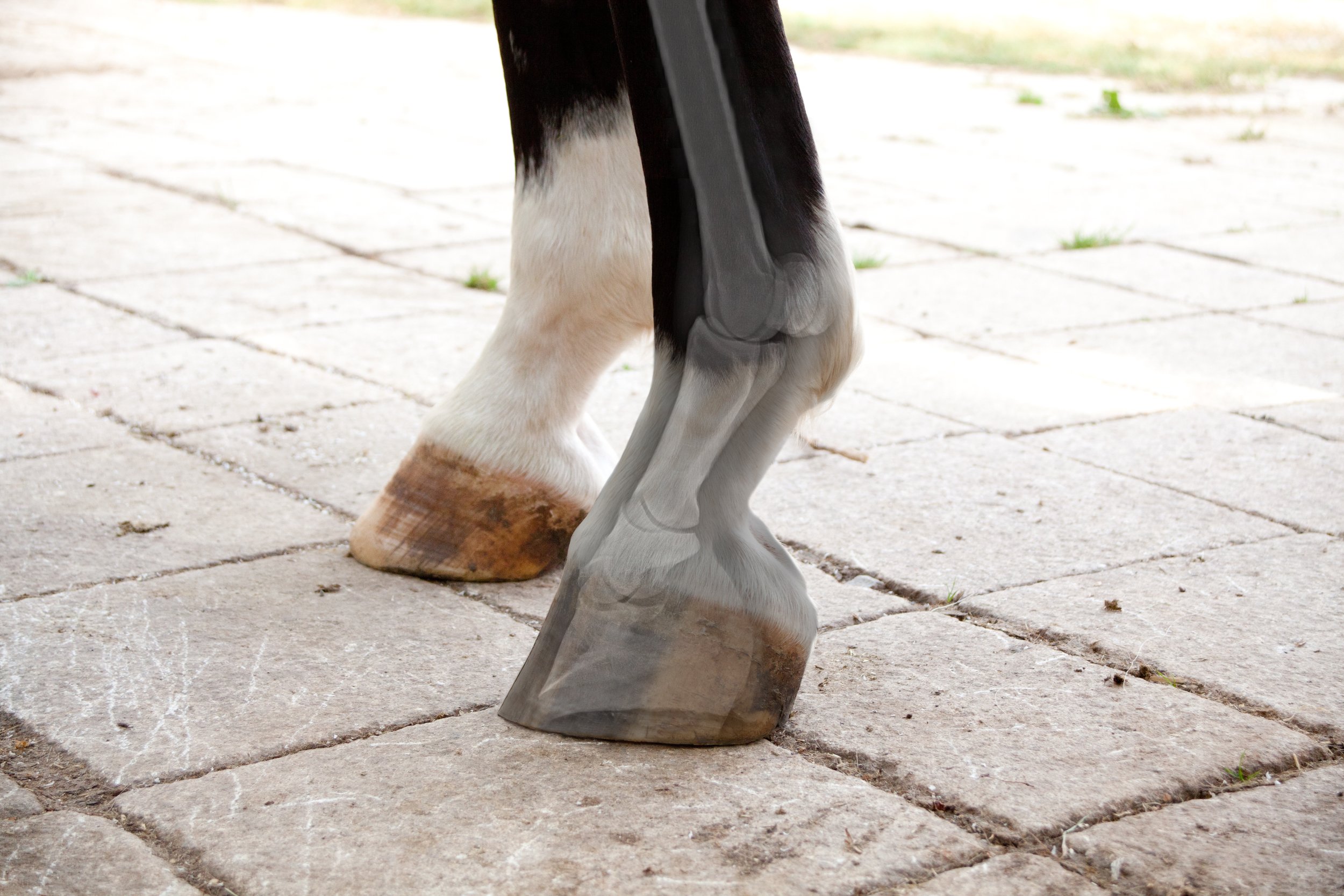
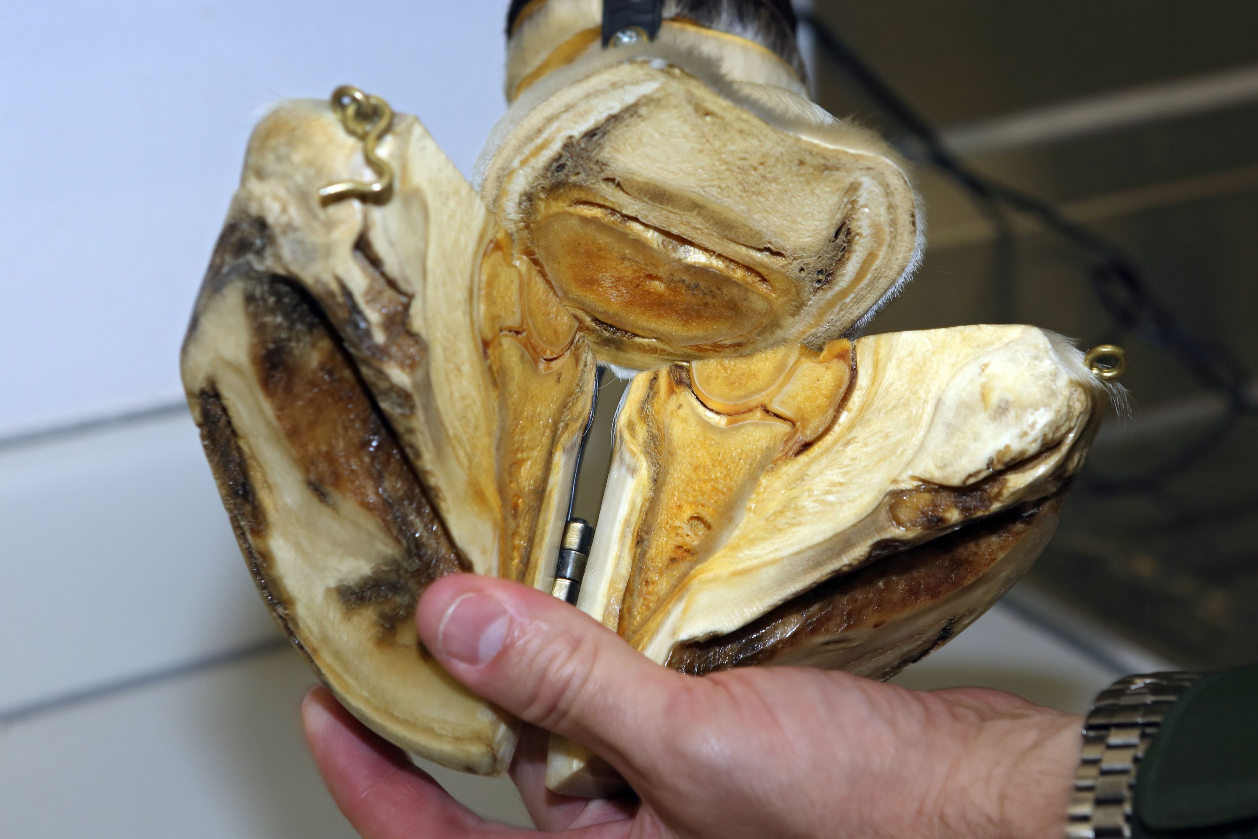
The foot consists of the distal end of the second phalanx (short pastern), the distal phalanx (pedal bone, coffin bone) and the navicular bone. The distal interphalangeal joint (coffin joint) is found between the pedal and short pastern bone and includes the navicular bone with the deep digital flexor tendon supporting this joint. This coffin joint is the centre of articulation over which the entire limb rotates. The navicular bone and bursa sits behind the coffin bone and is stabilised by multiple small ligaments. The navicular bone allows the deep digital flexor tendon to run smoothly and change direction in order to insert into the coffin bone. The navicular bursa is a fluid-filled sac that sits between the navicular bone and the deep digital flexor tendon.
The hoof complex can be divided into the epidermal weight-bearing structures that include the sole, frog, heel, bulbs, bars, and hoof wall and the anti-concussive structures that include the digital cushion, lamina, deep digital flexor tendons, and ungual (lateral) cartilage. The hoof wall encloses the dermal structures with its thickest part at the toe that decreases in thickness as it approaches the heel. The hoof wall is composed of viscoelastic material that allows it to deform and return its shape in order to absorb concussional forces of movement. There is enough deformation to diminish the force from the impact and load of the foot while preventing any damage to the internal structures of the foot and limb. As load is placed on the foot, there is deformation that consists of:
Expansion of the heels
Sinking of the heels
Inward movement of the dorsal wall
Biaxial compression of the dorsal wall
Depression of the coronary band
Flattening of the sole
The hoof wall, bars and their association with the sole form the heel base with the purposes of providing traction, bearing the horse’s weight while allowing the stability and flexibility for the expansion of the hoof capsule that dissipates concussional forces on foot fall. The sole is a highly keratinised structure like the hoof wall but made up of nearly 33% water so it is softer than the hoof wall and should be concave to allow the flattening of the sole on load application. The frog and heel bulbs serve a variety of special functions ranging from traction, protection, coordination, proprioception, shock absorption and the circulation of blood.
When the foot lands on the ground, the elastic, blood-filled frog helps disperse some of the force away from the bones and joints, thus, acting as a shock absorber. The venous plexus above the frog is involved in pumping blood from the foot back to the heart when the foot is loaded. In addition, there is shielding of the deep digital flexor tendon and the sensitive digital cushion (soft tissue beneath the sole that separates the frog and the heel bulb from the underlying tendons and bones). Like the heel bulbs, the frog has many sensory nerve endings allowing the horse to be aware of where his body and feet are and allows the horse to alter landing according to the condition of the ground (proprioception and coordination).
The soft tissue structures comprise and form the palmar/plantar aspect of the foot. The digital cushion lies between the lateral cartilages and above the frog and bars of the horse’s hoof. This structure is composed of collagen, fibrocartilage, adipose tissue and elastic fibre bundles. The digital cushion plays a role in shock absorption when the foot is loaded as well as a blood pumping mechanism. Interestingly, it has been found that the digital cushion composition varies across and within breeds. It is thought the variation of the composition of the digital cushion is partially dictated by a genetic predisposition. In addition, the composition of the digital cushion changes with age. As the horse ages the composition alters from elastic, fat and isolated collagen bundles to a stronger fibrocartilage. Finally, the digital cushion and connective tissue within the foot have the ability to adapt to various external stimuli such as ground contact or body weight. The lateral cartilage is a flexible sheet of fibrocartilage that suspends the pedal bone as well as acting as a spring to store and release energy. The lamina is a highly critical structure for hoof health. The lamina lies between the hoof wall and the coffin bone. There are two types of lamina known as the sensitive (dermal) lamina and insensitive (epidermal) lamina. The insensitive lamina coming in from the hoof wall connects to the sensitive lamina layer that is attached to the coffin bone and these two types of lamina interdigitate with each other to form a bond.
Hoof and Musculoskeletal System
The hoof and the musculoskeletal system are closely linked and this is particularly observed in the posture of the horse when resting or moving. Hoof shape and size and whether they are balanced directly affects the posture of the horse. Ultimately, this posture will also affect the loads placed on the skeletal system, which affects bone remodelling. With an imbalance, bone pathologies of the limbs, spine and pelvis may occur such as osteoarthritis. In addition, foot imbalances result in postural changes that lead to stress to the soft tissue structures that may lead to muscle injuries and/or tendon/ligament injuries.
Conformation and hoof balance
The terms balance and conformation are used frequently and used to describe the shape and size of the limb as a whole as well as the individual components of the limb and the spatial relations between them. Balance is the term often used to describe the foot and can be viewed as a subset of conformation.
Conformation should be considered when describing the static relations within the limb and excludes the foot. Balance should be considered when describing the dynamic and static relationship between the horse’s foot and the ground and limb as well as within the hoof itself.
These distinctions between conformation and balance are important to assess lameness and performance of the horse. Additionally, this allows the veterinarian and farrier to find optimal balance for any given conformation.
The term hoof balance does lack an intrinsic definition. The use of certain principles in order to define hoof balance, which in turn can be extended to have consistent evaluation of hoof balance as well as guide the trimming and shoeing regimens for each individual horse. In addition, these principles can be used to improve hoof capsule distortion, modify hoof conformation and alter landing patterns of the foot. These principles are:
Evaluate hoof-pastern axis
Evaluate centre of articulation
The need for the heels to extend to the base of the frog
Assessing the horse’s foot balance by observing both static (geometric) balance and dynamic balance is vital. Static balance is the balance of the foot as it sits on a level, clean, hard surface. Dynamic balance is assessing the foot balance as the foot is in motion. However, horses normally do not resemble the textbook examples of perfect conformation, which creates challenges for the farriers and veterinary surgeons. The veterinarian should instigate further evaluation of the foot balance and any other ailments, in order to provide information that can be used by the farrier and veterinarian in formulating a strategy to help with the horse’s foot balance. With the farrier and veterinarian working cooperatively, the assessment of the hoof balance and shoeing of the foot should deliver a harmonious relationship between the horse’s limb, the hoof and the shoe.
Dynamic Balance
The horse should be assessed in motion as one can observe the foot landing and placement. A balanced foot when in motion should land symmetrically and flat when moving on a flat surface. When viewed from the side, the heels and toe should land concurrently (flat foot landing) or even a slight heel first landing. It is undesirable to have the toe landing first and often suggests pain localised to the heel region of the foot. When observing the horse from the front and behind, both heel bulbs should land at the same time. Sometimes, horses will land first slightly on the outside or lateral heel bulb of the foot but rarely will a horse land normally on the medial (inside) of the foot. If the horse has no conformational abnormalities or pathologies the static balance will achieve the dynamic balance.
Static Balance
Hoof –pastern axis (HPA)
The hoof pastern axis (HPA) is a helpful guideline in assessing foot balance. With the horse standing square on a hard, level surface, a line drawn through the pastern and hoof should be parallel to the dorsal hoof wall and should be straight (unbroken). The heel and toe angle should be within 5 degrees of each other. An underrun heel has been defined as the angle of the heel being 5 degrees less than the toe angle. The heel wall length should be roughly 1/3 of the dorsal wall. In addition, the cannon (metacarpus/metatarsus) bone is perpendicular to the ground and when observed from the lateral side, the HPA should be a straight line. When assessing the foot from the side, the dorsal hoof wall should be aligned with the pastern. The optimal angle of the dorsal hoof wall is often cited as being 50-54°. The length of the dorsal hoof wall is variable but guidelines have been suggested according to the weight of the horse.
It is not uncommon that the hind feet are more upright compared to the fore feet at approximately 5 degrees. A broken hoof-pastern axis is the most common hoof imbalance. There are two presentations of a broken HPA known as a broken-back HPA and a broken-forward HPA. These changes in HPA are often associated with two common hoof capsule distortions that include low or underrun heels and the upright or clubfoot, respectively.
A broken-back hoof-pastern axis occurs when the angle of the dorsal hoof wall is lower than the angle of the dorsal pastern. This presentation is commonly caused by low or underrun heel foot conformation accompanied with a long toe. This foot imbalance is common and often thought to be normal with one study finding it present in 52% of the horse population. With a low hoof angle, there is an extension of the coffin and pastern joints resulting in a delayed breakover and the heels bearing more of the horse's weight, which ultimately leads to excess stress in the deep digital flexor tendon as well as the structures around the navicular region including the bone itself.
This leads to caudal foot pain so the horse lands toe first causing subsolar bruising. In addition, this foot imbalance can contribute to chronic heel pain (bruising), quarter and heel cracks, coffin joint inflammation and caudal foot pain (navicular syndrome). The cause of underrun heels is multifactorial with a possibility of a genetic predisposition where they may have or may acquire the same foot conformation as the parents. There are also environmental factors such as excessive dryness or moisture that may lead to the imbalance.
A broken-forward hoof-pastern axis occurs at a high hoof angle with the angle of the dorsal hoof wall being higher than the dorsal pastern angle. One can distinguish between a broken-forward HPA and a clubfoot with the use of radiographs. With this foot imbalance, the heels grow long, which causes the bypassing of the soft tissue structures in the palmar/plantar area of the foot and leads to greater concussional forces on the bone. This foot imbalance promotes the landing of the toe first and leads to coffin joint flexion as well as increases heel pressure. The resulting pathologies that may occur are solar bruising, increased strain of the suspensory ligaments near the navicular bone and coffin joint inflammation.
Center of articulation
When the limb is viewed laterally, the centre of articulation is determined with a vertical line drawn from the centre of the lateral condyle of the short pastern to the ground. This line should bisect the middle of the foot at the widest part of the foot and demonstrates the centre of articulation of the coffin joint. The widest part of the foot (colloquially known as “Ducketts Bridge”) is the one point on the sole that remains constant despite the shape and size of the foot. The distance and force on either side of the line drawn through the widest part of the foot should be equal, which provides biomechanical efficiency.
Heels extending to the base of the frog
With respect to hoof balance, another component of the foot to assess is that the heels of the hoof capsule extend to the base of the frog. The hoof capsule consists of the pedal bone occupying two-thirds of the space and one-third of the space is soft tissue structures. This area is involved in dissipating the concussional and loading forces and in order to ensure biomechanical efficiency both the bone and soft-tissue structures need to be enclosed in the hoof capsule in the same plane.
To achieve this goal the hoof wall at the heels must extend to the base of the frog. If the heels are allowed to migrate toward the centre of the foot or left too long then the function of the soft tissue structures have been transferred to the bones, which is undesirable. If there is a limited amount to trim in the heels or a small amount of soft tissue mass is present in the palmar foot then some form of farriery is needed to extend the base of the frog (such as an extension of the branch of a shoe).
Medio-lateral or latero-medial balance
The medio-lateral balance is assessed by viewing the foot from the front and behind as well as from above with the foot raised. To determine if the foot has medio-lateral balance, the hoof should be bisected or a line is drawn down the middle of the pastern down to the point of the toe.
You should be able to visualise the same amount of hoof on both the left and right of that midline. In addition, one should observe the same angle to the side of the hoof wall. It is important to pick up the foot and look at the bottom. Draw a line from the middle quarter (widest part of foot) on one side to the other then draw a line from the middle of the toe to the middle sulcus of the frog.
This provides four quadrants with all quadrants being relatively the same in size (Proportions between 40/60 to 60/40 have been described as acceptable for the barefoot and are dependent on the hoof slope). The frog width should be 50-60% of its length with a wide and shallow central sulcus. The frog should be thick enough to be a part of the bearing surface of the foot. The bars should be straight and not fold to the mid frog. The sole should be concave and the intersection point of both lines should be the area of optimal biomechanical efficiency.
The less concavity means the bone is nearer to the ground, thus, bearing greater concussional force. Finally, assess the lateral and medial heel length. Look down at the heel to determine the balance in the length of both heel bulbs. Each heel bulb should be the same size and height. If there are any irregularities with the heel bulbs then sheared heels may result, which is a painful condition. Medio-lateral foot imbalance results in the uneven loading of the foot that leads to an accumulation of damage to the structures of the foot ultimately causing inflammation, pain, injury and lameness. Soles vary in thickness but a uniform sole depth of 15mm is believed to be the minimum necessary for protection.
Dorso-palmar/plantar (front to back – DP) balance
Refers to the overall hoof angle and the alignment of the hoof angle with the pastern angle when the cannon bone is perpendicular to the ground surface. When assessing the foot from the side, the dorsal hoof wall should be aligned with the pastern. The optimal angle of the dorsal hoof wall is often cited as being 50-54°. The length of the dorsal hoof wall is variable but guidelines have been suggested according to the weight of the horse.
The heel and toe angle should be within 5 degrees of each other. An underrun heel has been defined as the angle of the heel being 5 degrees less than the toe angle. The heel wall length should be roughly 1/3 of the dorsal wall.
A line dropped from the first third of the coronet should bisect the base. A vertical line that bisects the 3rd metacarpal bone should intersect the ground at the palmar aspect of the heels.
Radiographs
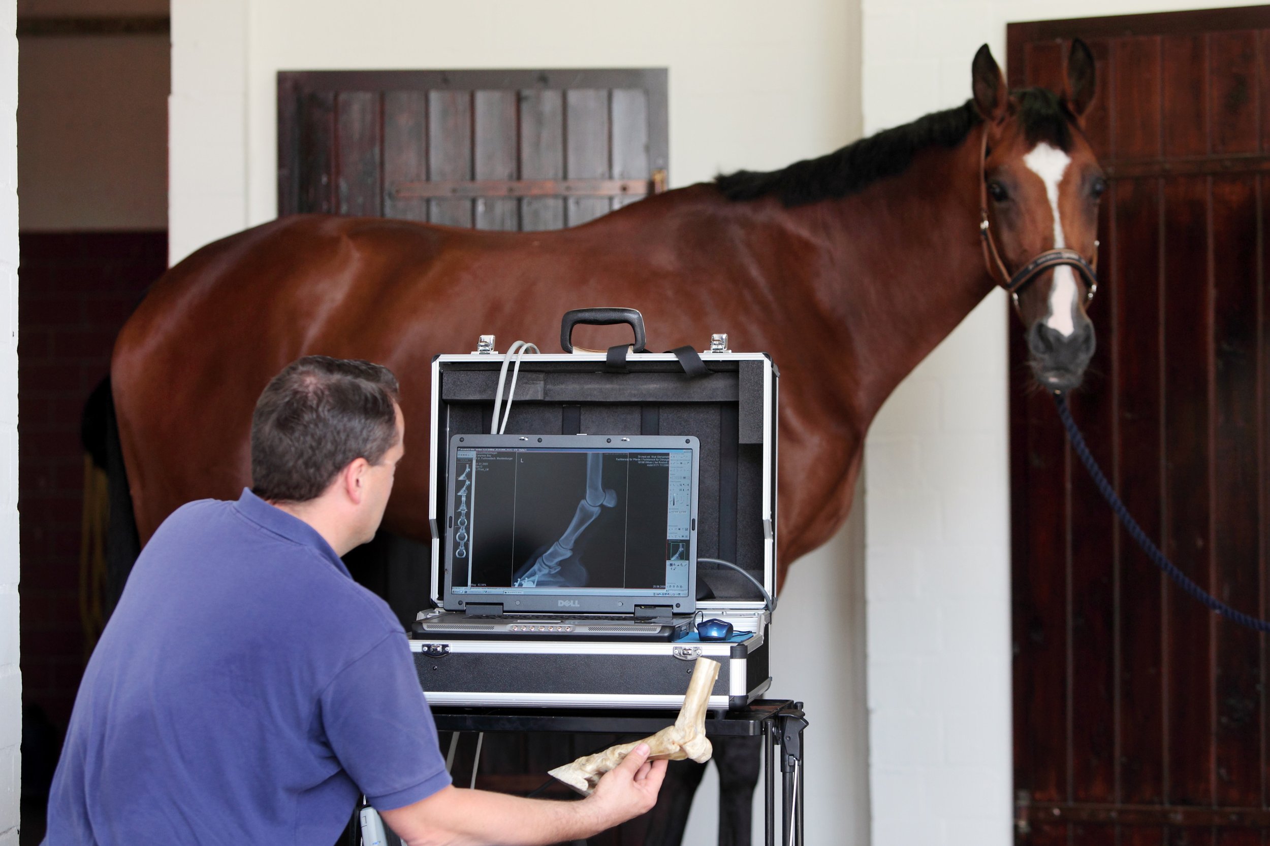
A useful way to assess trimming and foot balance is by having foot x-rays performed. Radiography is the only thorough and conclusive method that allows one to determine if the foot is not balanced and the bony column (HPA) is aligned.
Shoes should be removed and the foot cleaned before radiographs are executed. The horse is often placed on foot blocks to elevate the feet off the ground so that the foot can be centred in the cassette and x-ray beam.
Latero-medial view – The side view of the foot allows one to assess the dorsal and palmar aspects of the pedal bone as well as the navicular bone. The horse should be standing squarely on a flat, level surface. This projection is useful in determining the point of breakover and the hoof pastern axis should be parallel with the hoof wall. The lateral view will demonstrate the length of the toe and the alignment of the dorsal surface of the pedal bone with the hoof wall, which should be parallel. This view also allows one to determine the depth of the sole and inadequate solar depth is usually accompanied with excessive toe length (broken-back HPA). One may observe a clubfoot, broken forward.
One can distinguish between a clubfoot and a broken-forward HPA with radiographs. The broken-forward HPA the hoof angle of the heel is greater than the angle of the dorsal hoof wall. The clubfoot also demonstrates these steep/high hoof angles but additionally the alignment of the coffin, short and long pastern bones are broken forward.
Dorsopalmar/plantar views - this “front to back” view is also performed with the horse standing squarely on 2 positioning blocks. This projection allows the evaluation of medial to lateral balance and conformation of the foot with observation and measurement of the medial and lateral wall length and angle. Horses with satisfactory conformation present with a parallel joint surface of the pedal bone to the ground. The coffin joint should be even across its width. In addition, the lateral and medial coronet and the lateral and medial walls are of equal thickness and the distance from the lateral and medial solar margins to the ground are similar.
With foot imbalance, this author has observed that fore feet may have a higher lateral hoof wall, whereas, the hind feet may have a higher medial hoof wall. It is worth noting that the pelvis, stifle and hocks are adapted to move laterally allowing a slight rotating action as it moves. This action may cause uneven wear or poor trimming and shoeing may cause this limb movement to be out of line.
Trimming
Often, trimming and shoeing are based on empirical experience that includes theoretical assumptions and aesthetic decisions. The goals of trimming and shoeing are to facilitate breakover, ensure solar protection and provide heel support. Trimming is the most important aspect of farriery because it creates the base to which a shoe is fitted. Hoof conformation takes into account the function and shape of the foot in relation to the ground and lower limb both at rest and exercise. Each individual foot should have a conformation that provides protection and strength while maximising biomechanical efficiency often viewed as foot balance.
An important question that initially needs to be addressed is whether the horse requires shoes or not. The answer does depend on what type of work the horse performs, what is the amount of workload, the conformation of the horse (especially the limbs and foot) and are there any previous or current injuries. It must be stressed that the most important aspect, whether the horse is shod or not, is that the trim ensures an appropriately balanced foot for the horse. If there is poor trimming then this may lead to uneven and increased workload on the limb leading to an increased strain of the hoof and soft tissues (i.e. ligaments, tendons) that increase the risk of injury and developing acute and chronic lameness.
The foot can be evaluated, trimmed and/or shod in a consistent, reproducible manner that considers:
Hoof-pastern axis (HPA)
The centre of articulation
Heels extending to the base of the frog
Appropriate trimming and shoeing to ensure the base of the foot is under the lateral cartilage; therefore, maximising the use of the digital cushion, can help in creating a highly effective haemodynamic mechanism. Shoeing must be done that allows full functionality of the foot so that load and concessional forces are dissipated effectively.
To implement appropriate farriery, initially observe the horse standing square on a hard service to confirm that the HPA is parallel. If The HPA is broken forward or backward then these balances should be part of the trimming plan. To determine the location of the centre of rotation, palpate the dorsal and palmar aspect of the short pastern just above the coronary band and a line dropped vertically from the centre of that line should correlate with the widest part of the foot.
Shoeing
When the shoe is placed on the horse, the horse is no longer standing on its feet but on the shoe; therefore, shoeing is an extension of the trim. The shoe must complement the trim and must have the same biomechanical landmarks to ensure good foot balance. It is this author’s view that the shoe should be the lightest and simplest possible. The shoe must be placed central to the widest part of the foot and the distance from the breakover point to the widest part of the foot should be equal to the distance between the widest part of the foot and the heel.
It has been shown that the use of shoes that lift the sole, frog and bars can reduce the efficient workings of the caudal foot and may lead to the prevalence of weak feet. A study by Roepstorff demonstrated there was a reduced expansion and contraction of the shod foot but improved functionality of solar and frog support. With this information, appropriate shoeing should allow increased functionality of the digital cushion, frog and bars of the foot, which improves the morphology and health of the hoof and reduces the risk of exceeding the hoof elasticity.
Disease associated with hoof imbalance
Foot imbalance can lead to multiple ailments and pathologies in the horse. It must be noted that the pathologies that may result are not necessarily exclusive for the foot but may expand to other components to the horse’s musculoskeletal system. In addition, not one but multiple pathologies may result. Diseases that may result from hoof imbalance are:
Conclusion
Foot balance is essential for your horse to lead a healthy and sound life and career. With a strong understanding of the horse anatomy and how foot imbalance can lead to lameness as well as other musculoskeletal ailments, one can work to assess and alter foot balance in order to ensure optimal performance and wellbeing of the horse. It is essential that there is a team approach involving all stakeholders as well as the veterinarian and farrier in order to achieve foot balance. With focus on foot balance, one can make a good horse into a great horse.
Lower limb anatomy and how it can be conditioned for racing
Article by Adam Jackson MRCVS
Better understanding the appropriate levels of exercise and training while the horse’s body grows and develops has been a topic of research for many years. Although it has been shown that young, growing horses are well-suited to adapt to conditioning, it is vital that continued research is performed in order to develop thoughtful and strategic training methods to promote healthy, fit and sound horses with long careers and lives.
Horses’ limbs consist of dozens of muscles, bones, tendons, ligaments, and joints that allow the horse to move as well as support its body weight. The limbs function to provide thrust and movement while absorbing impact and bearing weight. Most of the horse’s weight is supported by the fore limbs, while the propulsion of the horse is provided by the hind limbs. In addition, the horse has two apparatuses referred to as the stay apparatus and suspensory apparatus. The stay apparatus allows major joints in the limbs to lock so that the horse may rest and relax while standing. The suspensory apparatus is designed to absorb shock, carry the horse’s weight, and prevent the overextension of joints. Finally, the hooves are important structures that maintain support and traction as well as provide additional shock absorption.
Since the cardiovascular system provides blood supply throughout the body, by responding to various stimuli, it can control the velocity and amount of blood carried through the vessels, thus, delivering oxygen, nutrients, hormones, and other important substances to cells and organs in the body. It plays a very important role in meeting the body’s demands during exercise, stress, and activity.
Exercise is used to increase the body’s ability to withstand repeated bouts of similar exercise with less impact. With a strong and healthy cardiovascular system, there is an improved ability of the musculoskeletal system receiving oxygen, thus, allowing muscles to better their capacity to use oxygen and energy. However, the adaptation period for each of these physiological systems do differ as the cardiovascular system adapts faster compared to the musculoskeletal system. This is often an overlooked consideration when developing training programmes for horses.
It is important to understand the various functions, structures, and adaptive processes of the horse’s musculoskeletal system such as bone, articular cartilage, tendons, and ligaments in order to develop appropriate training regimens.
Bone has many important roles that involve locomotion, the storage of minerals (especially calcium and phosphate), soft tissue and vital organ protection, and the support and containment of bone marrow. Bone is a specialised connective tissue, and together with cartilage forms the strong and rigid endoskeleton. The bone is continuously altering through two processes called bone modelling and bone remodelling, involving four cells referred to as osteoclasts, osteoblasts, osteocytes and bone lining cells.
Osteoblasts secrete bone matrix in the form of non-mineralised osteoid, which is then mineralised over a few weeks to form a bone matrix. Osteoclasts are involved in resorption of bone as this process occurs faster than the formation of bone. When the bone surfaces are not in the development or resorption phase, the bone surface is completely lined by a layer of flattened and elongated cells termed bone-lining cells. Osteocytes are derived from osteoblasts and are highly specialised to maintain the bone matrix. They are designed to survive hypoxic conditions and maintain biomineralisation of the bone matrix. Osteocytes also control osteoblastic and osteoclastic activities allowing bone remodelling.
The function of bone modelling is to alter and maintain shape during growth. As the horse grows and develops, bone modelling occurs with the acquisition and removal of bone. While the young horse grows and develops, bone modelling allows the bone to endure strains from everyday work and exercise. The adult skeleton undergoes a minimal amount of bone modelling. Due to the presence of the high frequency of bone modelling in young horses, their skeletal strength is highly influenced by strains to their bones during exercise and daily use. With this knowledge, it has been concluded and confirmed that short-term dynamic exercise of an adolescent can lead to beneficial changes to its bone morphology.
Bone remodelling is a different process, in which old and damaged bone is renewed, which enables the bone to respond and adapt to changing functional situations. Bone remodelling is usually a coordinated relationship between bone resorption and bone formation. This process occurs throughout the horse’s life with the renewal of primary, damaged or old bone. Osteoclasts absorb old and damaged bone, and the osteoblasts form new bone and lay down new bone matrix until the earlier absorbed bone is replaced. In those animals with musculoskeletal disease or damage, there is an imbalance of osteoblast and osteoclast activity. With the knowledge that osteoblast activity to make new bone takes months whilst osteoclast activity of removing old and damaged bone only takes a few days to two weeks, bone that is being repaired is at a high risk of further injury as bone removed has not been completely replaced. Multiple studies have shown that exercise while growing can provide lifelong benefits; however, it must be done with care and knowledge.
In addition, many studies have shown that exercise of a dynamic nature in moderate distances, such as that achieved in the pasture or prescribed short-distance high-speed work is beneficial to musculoskeletal development and may prevent injuries when entering race training. It has also been observed that long slow work does not increase bone strength. Below is a summary of the young horse response of the various types of exercise.
Articular cartilage is a highly specialised connective tissue found in joints with the role of providing a smooth, lubricated surface of articulation and to help transmit loads with a low amount of friction. The articular cartilage is a hyaline cartilage (flexible and strong tissue providing a smooth, slippery surface) with a dense “ExtraCellular Matrix” (ECM) consisting of specialised cells called chondrocytes, collagen and proteoglycans. These components help to retain water in the ECM that is required for the joints mechanical properties. As age increases, hydration of the matrix does decrease, resulting in stiffness. Chondrocytes are residential cells in articular cartilage that play a role in the development, maintenance, and repair of the ECM. They do respond to a variety of stimuli, including mechanical loads, growth factors, hydrostatic pressures, piezoelectric forces (formation of electric charge with force). Because of the lack of blood vessels, lymphatics, and nerves as well as being a harsh biomechanical environment, there is a limited capacity to heal and repair. In addition, chondrocytes have limited potential for replication, thus, have limited healing capacity; and chondrocytes survival depends on an optimal chemical and mechanical environment.
Maintaining joint health is vital, which requires the preservation of healthy cartilage tissue. Inactivity of joints is detrimental to articular cartilage; thus, regular movement of joints and dynamic loads is needed to provide a normal articular cartilage structure and function. Biochemical responses of the cartilage to exercise are not nearly as well known compared to bone. While the confinement of young horses stunts joint development, excessive straining of cartilage can also reduce joint development. It has been observed that pasture access was optimal for the development of joints and the confinement or excessive sprint exercise (12–32 sprints of 40 metres for 6 days a week for 5 months) causes detrimental effects on the joint and may be deemed as unnatural exercise. It is also thought that exercise is needed well before two years of age to allow cartilage thickening as well as the avoidance of confinement. It can be concluded that further studies are required with respect to level of exercise and type of exercise in order to achieve healthy cartilage tissue as there is clearly a fine line between frequency and intensity of exercise.
Tendons and ligaments are distinct but closely related tissues that have unique and important roles in musculoskeletal function and musculoskeletal disease. Tendons and ligaments are dense, fibrous connective tissues that connect muscle to bone or bone to bone, respectively. These tissues transmit mechanical forces to stabilise the skeleton and allow body movement. Tendons and ligaments consist mainly of collagen type I as well as small amounts of collagen III, IV, V, and VI. There are also various proteoglycans in tendons and ligaments that both organise and lubricate collagen fibre bundles. The elasticity of tendons and ligaments is due to the large amount of type I collagen. During locomotion, the tendon decreases energy cost to the horse by acting as a spring to store and release energy while stretching and recoiling in the stance and swing phases of each stride. Tendons and ligaments have blood vessels and nerves that allow the homeostasis and response to injury.
Tenocytes are tightly regulated by a series of growth factors and transcription factors that allow the synthesis, maintenance, and the degradation of the tendon extracellular matrix. Tendons are elastic, but tearing may occur if there is excessive loading on the tendon and the repair of collagen is a slow process. In addition, tendons have crimp morphology where the tendons buckle in a state of relaxation and act as shock absorbers. Unbuckling of the tendon occurs during loading. This crimp morphology may be disturbed if an injury occurs and also is reduced in older horses.
Due to the variation of activity of tenocytes in foals and young horses, it has been observed that both a lack of exercise and excess of exercise can impair tendon make-up and subsequent functionality. With the current data and research that has been gathered, it can be concluded that if horses take advantage of spontaneous exercise when in the paddocks (which they often do), the developing tendons may benefit and be at a lower risk of injury when racing training starts.
Conclusion
It is clear that further research is needed in order to ascertain the optimal amount and type of exercise that is needed in order to provide a strong musculoskeletal system and functional performance. However, it has been shown that prescribed exercise during the growth of the horse can increase the longevity of the horse’s health and performance. It has been observed that confinement and the lack of loading can result in weaker tissues and the loss of function of none, tendons, ligaments and articular cartilage. However, it must also be recognised that medical attempts to alleviate pain so that a horse can continue to train through an injury can greatly increase tissue damage which is detrimental to the horse’s health and career. It is far more beneficial to provide an adequate amount of time for the injury to heal, thus, putting the horse’s health and wellbeing as a top priority.
Nutritional Perspective
Bone development in yearlings from the sales ring to racing
Article by Des Cronin B.Ag.Sc, MBA
Maintaining the equine skeleton is vital to ensure optimal development of the young growing horse, minimise risk of injury in the performance horse, and promote longevity and soundness.
The skeletal development and health of a young horse begins in utero and ensuring the broodmare receives the correct intake of key nutrients will be critical to the growth of the unborn foal. Producing high-quality milk places a significant drain on the mineral reserves of the mare. Maintaining mineral intakes during peak lactation is vital to ensure the foal receives the best nutrition to support the rapid skeletal development in the early weeks and months of growth. During this time, bone formation, body size, and muscle mass greatly increase. Risk of defective bone and related tissue formation increases with one of more of the following:
Poor diet with the incorrect balance of energy and nutrients in the daily ration
Inadequate amounts of calcium (Ca) and phosphorus (P)
A reversed Ca:P ratio
Low zinc (Zn) or copper (Cu) in the diet
Low Vitamin D
Feeding a young horse for a maximum growth rate is undesirable because bone hardening lags greatly behind bone lengthening. At 12 months old, the young horse could reach about 90 to 95 per cent of its mature height but only about 75 per cent of its mature bone mineral content.
Ideally, young horses should gain weight at a rate that their developing bones can easily support. Growing bones and connective tissues don’t have the strength to support rapid weight gain from overfeeding, especially energy. Rapid weight gain can also make other skeletal anomalies worse. In these cases the risk of developmental orthopaedic disorders (DOD) and unsoundness increases.
DOD and unsoundness can also occur during uneven growth. For example, switching an underfed, slow-growing horse to a good diet that allows quick growth (compensatory growth), increases the risk of DOD. Foals between the ages of 3 and 9 months of age are at greatest risk of DOD.
Fresh forages, for example grazed grass, usually provide enough major minerals such as calcium (Ca) and phosphorus (P) for the growing horse. However, there can be significant variation in calcium and phosphorus levels in all forages but particularly preserved forages (hay and haylage). Forage analysis should always be undertaken to determine mineral composition.
For young fast-growing horses, the diet must supply the quantities of calcium and phosphorus needed for normal bone formation. In terms of Ca:P ratio, the ratio must be positive in favour of calcium. Horses are much more tolerant of high-dietary calcium than other species. For practical purposes, a good guideline would be to keep the ratio Ca:P between 1.5 to 1 and 2.5 to 1. Grains (e.g., oats) contain 10 per cent of the calcium level found in typical forages. Grains are poor sources of calcium, both in terms of the amount of calcium supplied and their effect on Ca:P ratio in the diet. Where grains are fed, supplementation will be necessary to balance the diet.
While some forages may contain adequate calcium and phosphorus, they will typically supply less than 20 per cent of the daily requirements for trace elements. Supplementation of trace elements will generally be necessary to support normal bone development.
Where concentrates are fed (especially low levels), supplementation may still be necessary to balance the overall mineral and trace element intake. Nutritional advice should be sought to ensure the horse's diet is correctly balanced.
To meet the carefully balanced requirements of key minerals, it is advisable to supplement the daily rations of growing horses and young horses entering training with an appropriate nutritional product.
Make sure that the supplement used contains the correct ratio of calcium and phosphorus, as well as other key nutrients such as vitamin D and chelated trace elements (copper, manganese, and zinc) to support normal bone development.
Supplementing branch chain amino acids in the diet ensures that growth is maintained. Lysine plays a key role when protein concentrations in the body are low. Vitamin A supports collagen formation, which is a key component of the supportive structures of joints (tendons and ligaments). Vitamin D3 is added to enhance calcium absorption.
Although growth rates slow after the age of two, they are still juvenile in their skeletal development with some growth plates, such as the shoulder and stifles, yet to completely close. Although they may look like fully grown adults, it is still important to meet nutritional requirements especially if starting training and work. With the addition of exercise and training, a young horse's nutritional needs change. The added forces from groundwork on the long bones and increased requirements of other nutrients like electrolytes need to be considered.
Finally, horses all grow and develop at different rates because of factors such as genetics. Some youngsters will need more support for longer periods of time than others, so it is important to manage accordingly.
The importance of the Sacroiliac joint
Article by Annie Lambert
Horses that present as sore in the hindquarters can be perplexing to diagnose. Sometimes the problem is found in the last place you look – the sacroiliac joint.
Even though the sacroiliac joint (SI) was on veterinary radars long ago, due to its location buried under layers of muscle in the equine pelvic region, the joint and surrounding ligaments were tough to diagnose and treat.
The sacroiliac joint is often a source of lower back discomfort in race and performance horses. Trainers may notice several clinical signs of a problem. These hints include sensitivity to grooming, objections to riders getting legged up, stiffness of motion, pain to manual palpation of the rump or back, resistance to being shod behind and poor performance.
Of course, those symptoms could describe other hind limb soundness issues, making the origin of the problem arduous to ascertain. A thorough physical examination with complete therapeutic options can relieve sacroiliac pain. The treatments are complicated, however, by the anatomy of the SI area.
The equine pelvis is composed of three fused bones: ilium, ischium and pubis. The sacrum, the lower part of the equine back, is composed of five fused vertebrae. The sacroiliac joint is located where the sacrum passes under the top of the pelvis (tubera sacrale). The dorsal, ventral and interosseous sacroiliac ligaments help strengthen the SI joint.
The SI and surrounding ligaments provide support during weight bearing, helping to transfer propulsive forces of the hind limbs to the vertebral column—creating motion much like the thrust needed to break from the starting gate.
Sound complicated? It certainly can be.
Diagnosing Dilemmas
It wasn’t until modern medical technology advanced that the SI could be explored seriously as a cause of hind lameness.
“The sacroiliac is one of the areas that’s very hard to diagnose or image,” explained Dr. Michael Manno, a senior partner of San Dieguito Equine Group in San Marcos, California. “[Diagnostics] of the area probably correlated with bone scans or nuclear scintigraphy. You can’t really use radiographs because the horse is so massive and there is so much muscle, you can’t get a good image.
“About the only time you can focus on the pelvis and get a decent radiograph is if the horse is anesthetized—you have a big [x-ray] machine and could lay the horse down. But, it’s hard because with anything close to a pelvic injury, the last thing you want to do is lay them down and have them have to get back up.”
The nuclear scintigraphs give a good image of hip, pelvis and other anatomical structures buried deep in the equine body, according to Manno, a racetrack practitioner. “Those images can show areas of inflammation that could pretty much be linked right to the SI joint.”
The other modern technological workhorse in the veterinary toolbox is the digital ultrasound machine. Manno pointed out that veterinarians improved diagnostics as they improved their ultrasounding skills and used those skills to ultrasound areas of the body they never thought about before. Using different techniques, frequencies and various heads on the machine’s probe, the results can be fairly remarkable.
“The ultrasound showed you could really image deeper areas of the body, including an image of the sacroiliac joint,” Manno said. “It can also show some ligament issues.”
Where the SI is buried under the highest point of a horse’s rump, and under heavy gluteal muscles, there are two sets of ligaments that may sustain damage and cause pain. The dorsal sacroiliac ligaments do not affect the sacroiliac joint directly, but help secure the ilium to the sacral spine. The ventral sacroiliac ligaments lie deeper, in the sacroiliac joint area, which they help stabilize. These hold the pelvis tight against its spine. The joint itself, being well secured by these ligaments, has little independent movement and therefore contains only minimal joint fluid.
Diagnosing the SI can be complex because horses often travel their normal gait with no change from normal motion—no signs of soreness. Other horses, however, are sore on one leg or another to varying degrees, sometimes with a perceptible limp.
“I don’t know that there is a specific motion,” Manno explained. “You just know that you have a hind end lameness, and I think a lot of performance horses have mildly affected SI joints.
“The horses that are really severe become acutely lame behind, very distinct. You go through the basic diagnostics, and I think most of these horses will show you similar signs as other issues behind. We palpate along the muscles on either side of their spine and they are sore, or you palpate over their croup and you can get them to drop down—that kind of thing. Other times you do an upper limb flexion on them and they might travel weird on the opposite leg. So, it can be a little confusing.”
In the years prior to the early 2000s, the anatomical location of the SI hindered a definite diagnosis; decisions on hind soreness were more of a shrug, “time and rest” treatment evaluation. As one old-time practitioner called it, a SWAG – “Scientific Wild Ass Guess.”
Even with modern tools, making a conclusive diagnosis can be opaque.
“The less affected horses, through exercise and with medications like Robaxin [muscle relaxer] or mild anti-inflammatories, seem to be able to continue to perform,” Manno said. “I don’t know how you can be perfectly sure of an inside joint unless you try to treat it and get results.”
“That’s why bone scans came into play and are really helpful,” Manno added. “You can image that [SI] area from different angles with the machine right over the path of the pelvis, looking down on it or an angle view into it, and then you see it from the side and the back very often. We can get an idea from the different views and angles of where the inflammation is and pinpoint the problem from that.”
Once Manno has a generalized idea of where the problem is, he fine-tunes his hypothesis using more diagnostics with a digital ultrasound machine.
“You can ultrasound from up above and see the joint that way,” he said. “As ultrasound has progressed, we’ve found that the rectal probes the breeding vets have used can also be tuned in to start looking for other things. If you turn them upwards, you can look at the bottom of the pelvis and the SI joint. You can see things through the rectum by just looking straight up. That is a whole new thing that we probably never thought about doing. I don’t profess to be very great at it; it’s not something I do a lot, but there are people that are just wonderful at it.”
Treating a Theorem
But, if the diagnosis is incorrect, the prescribed treatment may be anything but helpful.
“In many cases, if a horse is really sore, you need to be very careful,” cautioned Manno. “What you don’t want to do is go from a strain or some sort of soft tissue injury into a pelvic fracture by trying to keep them going. In many cases you are back in the old rest and time type of treatment.”
Manno pointed out one treatment that has advanced over many years is injecting the SI joint directly. There are a couple of techniques used when injecting the SI. With a blind injection the practitioner directs a long, straight needle into the joint by relying solely on equine anatomy. The other technique employs an ultrasound machine to guide the placement of the needle into the joint.
“Normally we are just injecting cortisone in those cases,” Manno noted. “We are trying to get the inflammatory response to settle down. Hopefully that gives the horse some relief so that they’re a bit more relaxed in their musculature. You know how it is when you get a sore back; it’s hard to keep yourself from cramping, which makes everything worse.”
A slight tweak of that technique is to use a curved needle. When you are positioning the curved needle, it follows the curve of the horse’s anatomy and helps the practitioner direct the injection into the joint.
“It curves right into position for you; it gives you a little help,” Manno confirmed of the curved needle. “Some people are really good with that technique; others still like to go to the straight needle. [The curved needle] helps you approach the site without interference from the bones in that area.”
SI joint injuries affect most performance horses, including Standardbred trotters and pacers, Western performance athletes as well as hunters, jumpers and dressage horses.
The older show horses are often diagnosed with chronic SI pain, sometimes complicated by arthritis. These chronic cases—and admittedly some racehorses—are treated with different therapies. These conservative, nonsurgical treatments have been proven effective.
In addition to stall rest and anti-inflammatories, physical training programs can be useful in tightening the equine patient’s core and developing the topline muscles toward warding off SI pain. Manno, a polo player who also treats polo ponies, believes the hard-working ponies avoid having many SI injuries due to their fitness levels.
“I think these polo horses are similar to a cross between a racehorse and a cutting horse,” Manno opined. “They are running distances and slide stopping and turning.”
Other treatments utilized include shockwave, chiropractic, acupuncture, therapeutic laser and pulsed electromagnetic therapy.
Superior Science
With the new diagnostic tools and advanced protocols in their use, veterinarians can pinpoint the SI joint and surrounding areas much closer. This gives them an improved indication that there definitely is an issue with the sacroiliac.
When there is a question about what is causing hind end lameness, most practitioners begin with blocking from the ground up.
“In many cases with hind end lameness that we can’t figure out, we block the lower leg; if it doesn’t block out down low, we conclude the problem is up high,” Manno said. “Once you get up to the hock you’re out of options of what you can figure out. You start shooting some x-rays, but by the time you get to the stifle, you’re limited. Bone scans and ultrasounds have certainly helped us with diagnosing.”
Manno doesn’t see a lot of SI joint injuries in his practice, but he noted there were cases every now and again. He also opined that there were probably other cases that come up in racehorses on a short-term basis. He also noted that, although it may not be a real prominent injury, that’s not to say it has not gone undiagnosed.
“I think we realize, in many of the horses we treat, that the SI joint is something that may have been overlooked in the past,” Manno concluded. “We just didn’t have the ability to get any firm diagnosis in that area.”


