The work being done on the prevention of serious fractures in the racehorse
Fractures: are they inevitable or preventable?

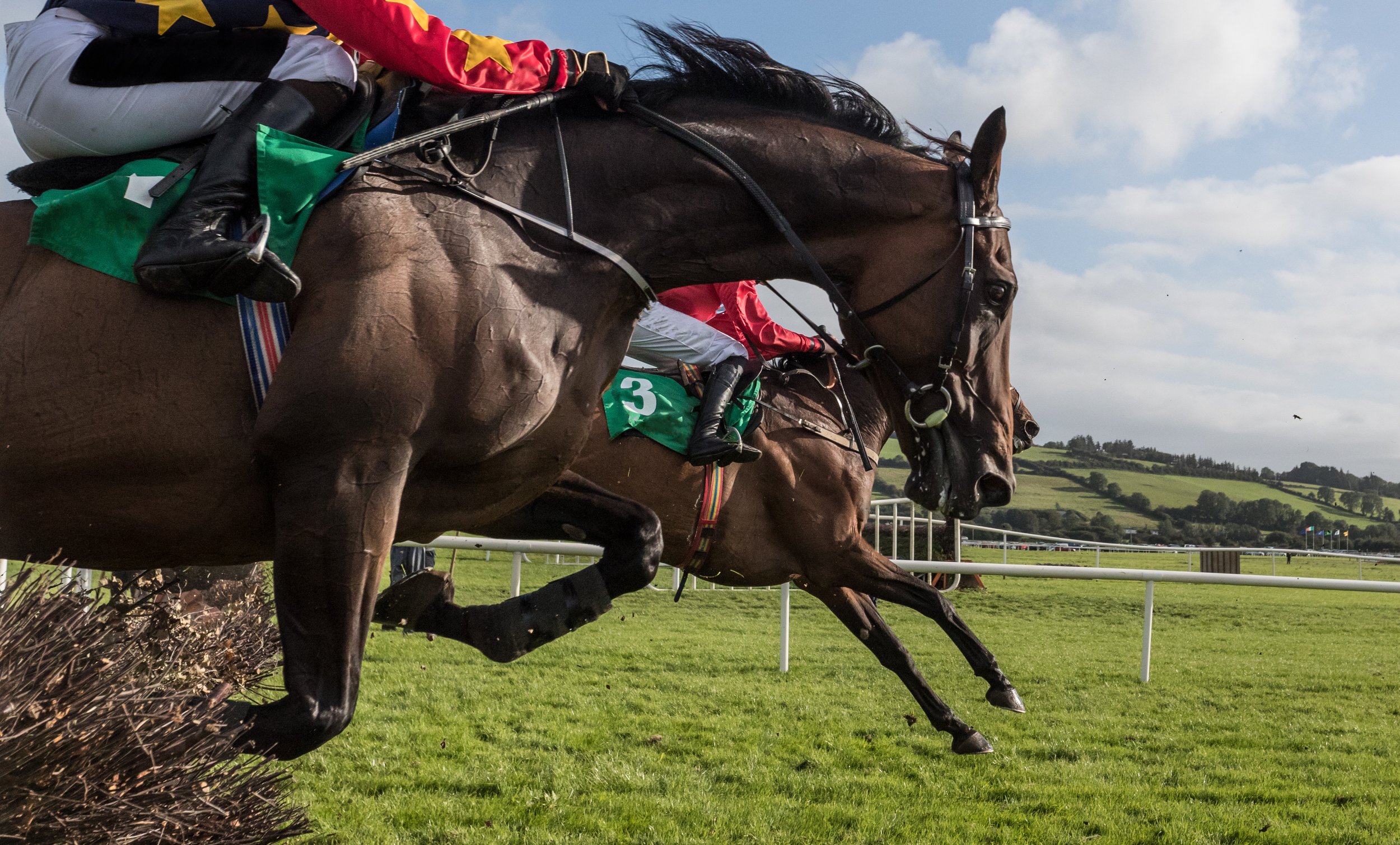
The incidence of serious fractures in horses racing on the flat is low (around one case every one to two thousand starters); but when fractures occur the consequences are often severe for the horse and, sometimes, the jockey. Both the severity and the dramatic nature of these injuries cause a significant negative impact to everyone connected with the horse, as well as spectators.
The International Federation of Horseracing Authorities (IFHA) convened the Global Summit on Equine Safety and Technology at Woodbine Racecourse, Toronto, in June 2024, to focus on how current research into causes of racehorse fatalities on the course, including fractures, can be advanced and translated into action, to better understand the factors leading to fatalities and potentially mitigate them. At the meeting there were two workshops where specialist veterinary clinicians, pathologists and researchers came together to focus on Exercise Associated Sudden Death incidents and fractures.
Why do fractures occur?
While most fractures that affect racehorses on the flat appear to arise spontaneously, while the horse is racing, and in the absence of any obvious trauma, we now know that the majority are the consequence of structural fatigue, involving a process that started weeks or even months earlier. Just as a paper clip will break if it is repeatedly bent once too often, a bone will fracture if it is loaded recurrently beyond certain limits. Microscopic damage accumulates in the bone tissue until a small fissure develops, which can extend to a severe fracture if it is not detected and the horse continues to race.
The fatigue life of bone decreases exponentially as the magnitude of load on the bone during each loading cycle increases. Loads on the skeleton are directly related to the speed of exercise, work at a fast gallop causes fatigue damage at a rate many orders of magnitude greater than when the horse is exercised at slower speeds. Unfortunately, fatigue damage, and even formation of small fissures in the bone, often proceed without the horse showing any outward signs and so, historically, affected horses have often gone undetected until it is too late.
Are there any natural biological mechanisms that protect against fracture?
An ability to run fast has been of evolutionary benefit to horses by enabling them to escape predators. Humans have refined this characteristic in specific breeds, notably the Thoroughbred, through targeted breeding and training practices. The need for a horse to undertake a fast gallop in the wild is likely to be relatively infrequent and well-within the bounds that are likely to cause fatigue damage. Conversely, society’s expectations of racehorses, and the training imposed on them, place loads on the skeleton that may be excessive.
Some readers may be surprised to learn that bone is a remarkably “smart” tissue. It is so much more than the equivalent of the “concrete structural beam” laid down to support man-made structures. While bone is composed partly of minerals, it is still very much a living tissue, packed with blood vessels, nerves and cells. Furthermore, the cells are all physically interlinked through microscopic projections, and can communicate with each other through their interconnections. This biological network allows bone tissue to detect the effects of loads applied to the bone as whole, and to initiate a cellular response to model the bone structure if those loads change.
For instance, increase in the magnitude of prevailing loads (e.g. through increase in an animal’s weight or the introduction of high-speed work) will cause greater deformation of the bone and this will stimulate a process that results in the formation of additional bone mass and, if beneficial, subtle reshaping of the bone’s geometry. The bone will be stronger as a result and deform less for the given load. Conversely, if the prevailing loads decrease (e.g. during a period of rest) bone deformation is reduced, and bone may be removed through a process of resorption. This overall mechanism of remodelling is called “adaptation”. Bone adaptation facilitates the formation and maintenance of a skeleton that is continually fit for purpose, even as that purpose changes.
Beyond this, there are biological mechanisms that detect when a bone is damaged (e.g. small cracks develop) and will initiate repair. The restoration process involves removal of microscopic packets of bone tissue, including the damaged portion, and its replacement with fresh, healthy material. This process means that fatigue-related damage of bone can be resolved and, theoretically, that a bone can tolerate infinite cycles of load. However, the process of repair has been shown to be inhibited if the bone is still regularly subjected to cycles of high load (i.e. the horse remains in training) and it can also be overwhelmed if the rate of damage accumulation is too high.
Are fractures something that racing just has to live with or can we do something about them?
Applying the knowledge we have gained from research over the past forty years has already had an impact on reducing the risk of fractures in some situations, and there are good reasons for optimism that we will be able to prevent a much greater proportion of racing fractures in the future. For example, epidemiological studies can help to identify certain management practices and design features of racecourses associated with an increased risk of fracture and these can be modified.
The introduction of strict rules governing the validity of sale of a horse in a claiming race in some jurisdictions in the USA, depending on the health status of the horse immediately after the race, has had a significant impact on reducing the incidence of fractures. Over 300 potential risk factors have been examined and those found to have a significant association with fracture, modelled in numerous studies.
Applying the findings to individual racecourses still requires much work and, ultimately, almost half of the variation in risk of fracture appears to be due to factors associated directly with the horse itself. Epidemiology has also been applied to identify characteristics of a horse’s history that may indicate that it is at higher risk of fracture. These studies depend on access to detailed and accurate data and recent work has highlighted the importance of including veterinary records. Access to these records, suitably anonymised to protect confidentiality, will have a profound impact in the development of more accurate models that can be used to predict the risk of fracture.
Studies into the genetics of Thoroughbreds have identified particular genes that are associated with a predisposition to fracture. This is a highly complex field and interaction of the environment and genetics ultimately determines the fracture status of an individual animal. However, genetic screening will help to identify horses that may benefit from closer monitoring. Clearly this is a sensitive topic, especially to breeders, although hopefully stakeholders will make choices that will be in the best interest of Thoroughbreds and racing in the long term.
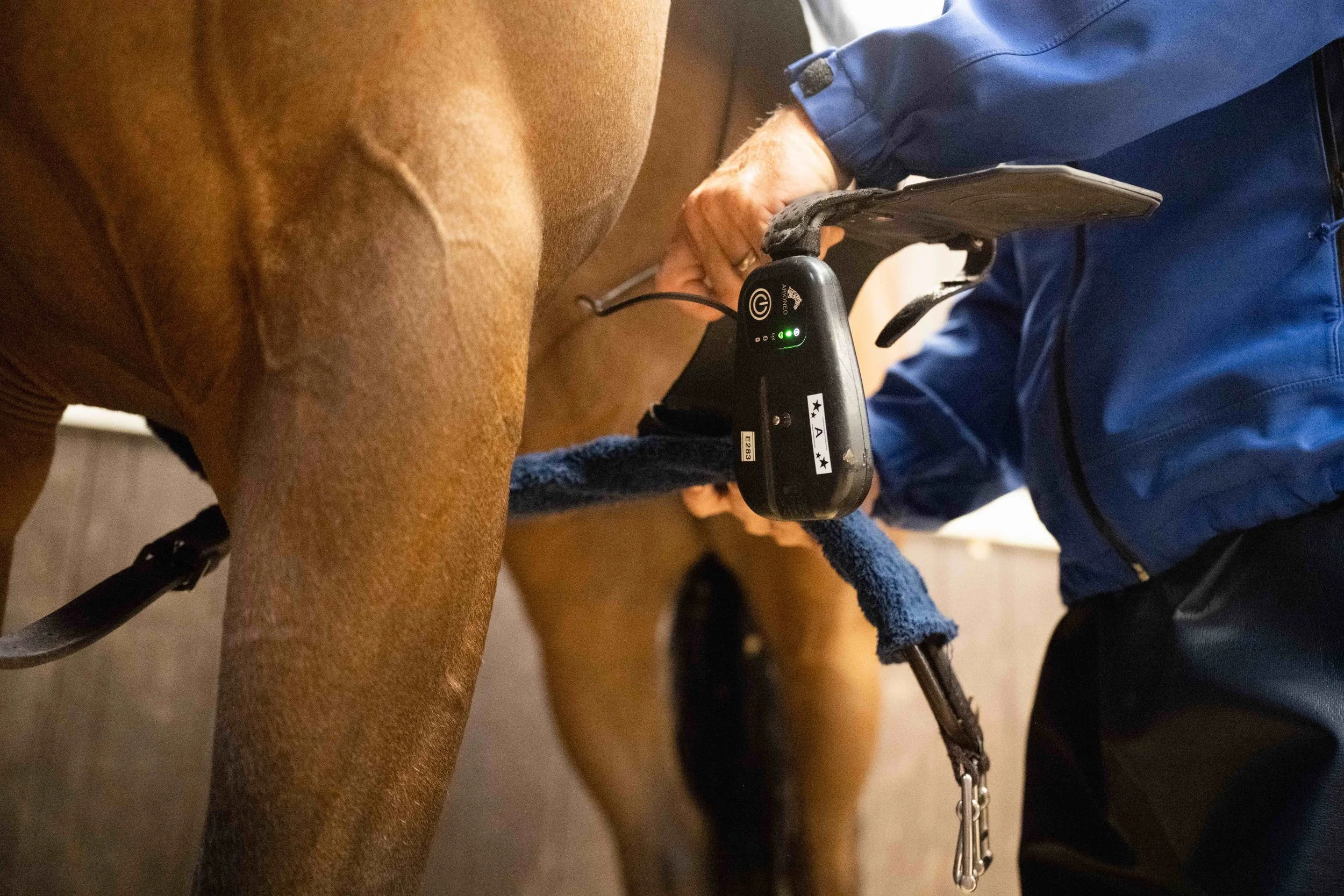
Wearable technology, carried on the horse to record cardiac and stride data shows promise in being able to identify horses at early stages of skeletal injury. Preliminary results suggest that this is possible by identifying changes in stride characteristics, such as stride length, in individual animals in races leading up to the horse sustaining an injury. There has been a lot of interest and publicity associated with wearable technology and its use to identify horses both at imminent risk of fracture as well as those with irregularities of cardiac rhythm which may lead to exercise associated sudden death, although work still needs to be undertaken to substantiate their use in both these contexts.
These devices are also proving increasingly valuable as tools to record actual workload undertaken by individual horses in training. This is important information that is needed for researchers to accurately model associations between workload and risk of fracture, and for trainers to use to apply such models in the future.

Modern clinical imaging technology, especially that which allows detailed scrutiny of areas of bones in the locations where fractures commonly originate, facilitates identification of bone damage due to fatigue much earlier than was previously possible. The increasing availability of computed tomography (CT) and positron emission tomography (PET) machines that can be used to image the lower limb of the standing horse has made their application more practical in horses that are in active training.
There is potential for this technology to be used to screen horses prior to racing, as is currently undertaken before some race meetings in the state of Victoria, Australia. The enhanced resolution of CT helps to identify subtle fissures in bone that may be overlooked by conventional x-ray procedures and in such cases its value as a screening tool is indisputable. However, work is still ongoing to decipher the importance of even more subtle changes in bone structure. Undertaking imaging studies carries expense, and is not without some risk, and the concept of regularly imaging the relevant anatomical regions of racehorses to screen for risk of fracture clearly has challenges.
A simple blood test for “biomarkers” that could identify animals at an early stage of pre-fracture pathology would be a helpful tool. Even if the accuracy of such a test was insufficient to make important decisions about whether or not to race a horse, it could at least be used to reduce the number of animals that would need to be subjected to imaging studies.
Research into changes in the patterns of arrays of many molecules in the blood (so called “omics” studies) have led to many breakthroughs in screening systems in humans and animals. Preliminary studies give hope that this technology can be used to assist in the detection of horses at increased risk of fracture, although a practical test is still likely to be several years off.
It is important to acknowledge that all screening tests carry some risk: they may falsely identify normal horses to be at increased risk (false positives) and fail to recognise horses that have a problem (false negatives). Careful development and thorough testing of screening systems is therefore essential, and this takes time and money. Furthermore, no test is ever 100% accurate, and the practical application of screening designed to prevent fractures will require racing to strike a balance between stopping some normal horses from running and failing to identify some of those likely to suffer a fracture. This will require informed and engaged discussion between researchers and all relevant industry stakeholders, and strong leadership and clear communication from racing’s regulators.
Perhaps an even greater question is whether we can recommend ways of exercising horses in training that minimise the risk of fatigue damage that leads to fracture in the first place. Research conducted in Melbourne, Australia demonstrated that trainers who worked their horses at high speed in training less frequently than others experienced a lower incidence of fracture.
A very important additional bit of detail was that, excepting the extremes, their racing results and performance outcomes were no different. Recognition that bone is like other tissues, in that it needs time to respond to changes in physical activity and requires a training programme that accounts for this, is important. We are all well aware of the need to develop muscle to improve power and endurance in training, but the concept of training bone is less familiar.
Fortunately, an adaptive response of bone has been shown to occur reasonably quickly to only a few cycles of altered load. Stimulating young, healthy racehorses to increase the strength of their bones requires only a short distance of gallop work. Therefore, introducing a very small amount of fast work at the early stages of a training programme can be sufficient to prepare the bones for more sustained work without the risk of causing substantial fatigue damage. However, if the horse is rested from high-speed work for more than a few days, the process reverses, and bone mass will start to be resorbed (in evolutionary terms, there is no point wasting energy carrying around bones that are bigger and heavier than you need them to be).
So, when a horse is brought back into work after a period of rest, the process of training the skeleton has to be considered all over again. Periods of rest in themselves are also important to allow the bone to repair because even a sensitive training programme will result in some fatigue damage, and the accumulation of this damage may eventually lead to fracture. The innate, biological mechanism of bone repair is switched off (inhibited) while horses remain in active training and a period of rest is required for this process to be reactivated and for repair of fatigue-damaged tissue to take place. Therefore, periodic periods of rest from training are required in order to give the skeleton the best opportunity to heal and “reset” itself.
In summary, a training programme that:
reduces the distance of work (especially high-speed work) that a horse undertakes in its racing career to optimal levels that promote bone health (while racing and training);
stimulates appropriate development of bones through short bursts of fast work early on in a training campaign;
builds periods of rest into a horse’s life span in training;
will all reduce the risk of accumulated fatigue damage that predisposes to fractures developing.
To move forward, the industry needs to invest in research so that metrics can be developed to help trainers to apply these concepts to their own training techniques in a practical way. There will be a range of durations and speeds of work that will reduce the risks of fracture across different populations of Thoroughbreds, and these will only be demonstrated by investing in studies measuring the effects of different training regimes in significant numbers of horses.
Scientists and Racing Working Together
Developing and applying measures that are designed to reduce the risk of fracture will require the commitment of all stakeholders. We also need to be realistic in acknowledging that, due to the athletic nature of racing, the risk of a racehorse sustaining a fracture will never be eliminated and, sadly, for some of these injuries euthanasia will be the only humane option.
When racing authorities communicate to the public that the safety of equine athletes in racing is a priority, their credibility is clearly demonstrated by investing in work to bring the risks of injury, including fractures, down to the lowest possible level.
There will inevitably be an element of “pain” associated with the work and changes required to reduce injury rates. Screening systems will disrupt normal cycles of work, there will be frustration as horses that appear fit are withdrawn from races on the basis of findings of “risks” that we might find difficult to conceptualise, there will be additional financial demands to fund studies, and a sense of exposure as records that are necessary for researchers to do their work are shared, even with promises of confidentiality and anonymity.
At the Toronto meeting, the IFHA has already committed to supporting global scientific research efforts to reduce the incidence of racehorse injuries, including fractures. I look forward to the benefits this co-ordinated approach to research can bring by both reducing injury rates in equine athletes, and at the same time, demonstrating to society the priority racing gives to equine welfare.
The Gerald Leigh Memorial Lectures 2023


The Gerald Leigh Memorial Lectures, is an annual gathering devoted to the racing industry and the health and wellbeing of the horses involved.

This year, equine veterinarians, researchers, students and industry professionals from around the world attended the event, held June 8, 2023, at the historic Tattersalls Sales in Newmarket, England.
There were insightful and informative lectures that educated the attendants but also instigated a healthy, lively debate on the health and welfare of the training and competing of horses. The underlying theme that was present during the whole event was all members of the conference had a deep passion and commitment to continuously progress and improve on managing the welfare and wellbeing of the horses in the industry, both on and off of the track.

Two very special guest speakers, Sir Mark Prescott and Luca Cumani, wonderfully illustrated these sentiments as they described their reflections on the improvement and enhancement of horse safety.

Horse racing may be regarded as an elite sport, and all activities involving horses have an element of risk. All stakeholders in the racing industry must continuously work to ensure that the risks are minimised in order to reduce the number of injuries and fatalities that may occur in training and on the racecourse. There are now well-publicised concerns regarding the acceptability of exposing horses to risk in racing. These lectures and all of the attendees embraced the values of the public will so that there can be continued acceptance of horse sports.
Reducing the incidence of fractures in racing
Christopher Riggs of The Hong Kong Jockey Club clearly outlined the various strategies to reduce the risk of fractures in racehorses. There are two principal strategies that may used to reduce the incidence of severe fractures in horses while racing and training:

Identifying extrinsic factors that increase risk and take action to minimise them.
An example would be investigating different racing surfaces in order to determine which may provide the safest racing surface. However, studies have provided limited evidence and support for subtle extrinsic factors.
2. Identifying individuals that are at increased risk and prevent them from racing or minimise that risk until the risk has subsided.
There are many research routes that are being undertaken to identify those horses that may be at a higher risk of fractures. There are investigations involving heritability and molecular studies that may provide evidence of genetic predisposition to fracture. However, Dr. Riggs explained that further understanding of the relationship between genetic, epigenetic and environmental factors is required before genetic screening is likely to be of practical use.
Pre-race screening of horses by diligent clinical examination is poor at reducing the incidence of fracture. Dr. Riggs described another strategy that may assist with a clinical examination that is the use of biomarkers in blood and urine.
Unfortunately, the precision to be of practical value has so far remained relatively unrewarding. Wearable technology that records biometric parameters, including stride characteristics, has shown some promise in identifying horses that are at increased risk of fracture; although Dr. Riggs explained that this work requires further development.
Finally, Dr. Riggs described both the use and current limitations of diagnostic imaging in identifying pre-fracture pathology in order to identify a horse at imminent risk of fracture. He conceded that further knowledge of the significance of the range of abnormalities that can be detected by imaging is incomplete.
Dr. Riggs concluded his lecture by expressing that the implementation of diagnostic imaging to screen “high-risk” horses identified through genetic, epidemiology, biomarkers and/or biometrics may be the best hope to reduce the incidence of racing fractures. This field can be advanced with further studies, especially of a longitudinal nature.

Professor Tim Parkin of Bristol Veterinary School discussed the need for further investment in welfare research and education. One avenue of investment that should be seriously considered is the analysis of data related to (fatal) injuries in Thoroughbred racing over the last 25 years.

It was expressed, with the abundance of data that has been collected, that some risk factors would be relatively simple to identify. An encouraging example in the collection and use of data to develop models in predicting and potentially preventing injury has been conducted by the Hong Kong Jockey Club funded by the Hong Kong Jockey Club Equine Welfare Research Foundation. This may provide an opportunity to pilot the use of risk profiling to contribute to decision-making about race entries. In addition, the results of the pilot study combined with other sources of data may encourage race authorities to mandate the collection of veterinary and training data in order to help in risk mitigation.
Horse racing is an international sport, and there are different governing bodies that ensure racing integrity. However, the concept of social licence equestrian sports and Thoroughbred horse racing continues to gain significant public attention. Therefore, racing governing bodies are increasingly aiming to provide societal assurances on equine welfare.

Dr. Ramzan of Rossdales Veterinary Surgeons provided an eloquent and clear message during his lecture that race yard veterinarians and trainers are instrumental in ensuring good horse health and welfare and reducing serious injury of the horse both while training or racing, which will provide sufficient trust and legitimacy from the public and society. This feasible goal can be reached with good awareness of members involved in the care and training of each individual horse and conveying this information and any concerns to their veterinarian. The veterinarian can also contribute by honing their knowledge and skills and working closely with yard staff in order to make appropriate and better targeted veterinary intervention.
In the last two decades, there has been an incredible evolution and exciting developments in diagnostic imaging in the veterinary profession. It is believed that these technologies can provide a significant contribution to helping in mitigating fracture risks to racehorses on the course and in training.

Professor Mathieu Spriet of University of California, Davis, described how these improvements in diagnostic imaging has led to the detection of early lesions as well as allowing the monitoring of the lesions’ evolution.

He continued by explaining the strengths and limitations of different imaging modalities such as computed tomography (CT), magnetic resonance imaging (MRI) and positron emission tomography (PET). Being one of the leaders in the use of PET in equine veterinary medicine, he presented further insight on how this particular modality provides high-resolution 3-D bone scans while being very sensitive to the identification of bone turn-over prior to the development of structural changes and allowing one to distinguish between active and inactive processes when structural changes are present.
He concluded his impressive lecture by providing evidence with amazing PET images that the role of imaging is not merely for diagnostic purposes to characterise clinical abnormalities, but can also be used as a screening tool in certain horse populations for fracture risk assessment or for the monitoring of lesions to provide clearance for racing.
Fractures, due to bone overloading rather than direct trauma occur commonly in Thoroughbred racehorses and are the leading cause of euthanasia on the racecourse. Despite many changes to race conditions, the number of catastrophic fractures has remained relatively static, with approximately 60 horses a year having a fatal fracture during a race in the UK.
Against this backdrop, there have been great developments in the diagnosis and treatment of fractures in the last 40 years. Prevention of racecourse and training fractures would be ideal so the development of efficacious techniques to screen horses at risk may reduce the incidence and preserve social licensing.

One technique discussed by Dr. Ian Wright of Newmarket Equine Referrals was to help mitigate the impact of racecourse fractures, which would be acute immobilisation of racecourse fractures, thus, reducing associated pain and anxiety while optimising clinical outcome and reducing on course fatality rates. Because of our increased understanding of fracture pathogenesis and their associated biomechanics, effective fracture immobilisation has been made possible. The majority of fractures that occur in flat racing and between obstacles in jump racing, are a result of stress or fatigue failure of the bone and not associated with trauma.
In addition, fractures seen on the racecourse are often found in the same specific sites (i.e., metacarpal/metatarsal condyles and the proximal sesamoid bones of the fetlock) and have repeatable configurations. With this understanding and knowledge, racecourse veterinarians can optimally immobilise a fracture in a logical and pre-planned manner.
As Dr. Wright expressed, this allows the fracture patient to have reduced pain and anxiety and enable the horse to be moved from the course comfortably so that it can be further examined. Ultimately, this allows the veterinarian and all stakeholders to make effective and judicious decisions for the sake of the horse’s welfare and wellbeing. As Dr. Wright concluded, this benefits both horses and racing.

Dr. Debbie Guest of the Royal Veterinary College discussed a different approach in mitigating the risk of fractures during training and racing by developing novel tools to reduce catastrophic fractures Thoroughbreds. Because it has been found that some horses are more inherently predisposed to fractures than other horses, Dr. Guest and her team have developed a genome-wide polygenic risk score so that one can potentially calculate an individual horse’s risk of fracturing during training or racing compared to the population as a whole.
This strategy may contribute in identifying genetically high-risk horses so that additional monitoring of the patients can be exercised during their careers and also leading to fracture risk, which are found to be the cause of approximately half of these incidents.
The system of using DNA testing to identify biological processes that may or may not be present ultimately leading to fracture risk may be a powerful tool in lowering the risk of catastrophic fracture and requires further research and application.
Cardiac events & sudden cardiac death in training and racing
In racehorses, sudden death that is associated with exercise on the racetrack or during training is a serious risk to jockeys and adversely affects horse welfare and the public perception of the sport. It is believed 75% of race day fatalities result from euthanasia following a catastrophic injury. The other 25% of fatalities is due to sudden deaths and cardiac arrhythmias are found to be the cause of approximately half of these incidents. The lectures focused on this area of concern by providing three interesting lectures on cardiac issues in the racehorse industry.

Dr. Laura Nath of the University of Adelaide, explained the difficulties in identifying horses that are at risk of sudden cardiac death. It is believed that part of the solution to this difficult issue is the further development and use of wearable devices including ECG and heart rate monitors.
With the use of these technologies, the goal would be to recognise those horses that are not progressing appropriately through their training and screen these horses for further evaluation. This course of action has been seen in human athletes that develop irregular rhythms that are known to cause sudden cardiac death with the use of computational ECG analysis, even when the ECGs appear normal on initial visual inspection.
Knowing that ECGs and particularly P-waves are used as a non-invasive electrocardiographic marker for atrial remodelling in humans, Dr. Nath recently completed a study on the analysis variations in the P-wave seen on ECGs in athletic horses and found that increases of P-waves in racehorses are associated with structural and electrical remodelling in the heart and may increase the risk of atrial fibrillation (cardiac event).

Dr. Celia Marr of Rossdales Veterinary Surgeons continued the discussion of cardiac disease in both the training and racing of horses. Unfortunately, cardiac disease knowledge does lag compared to musculoskeletal and respiratory diseases when considering the causes of poor performance in racehorses. Due to the fact that cardiac rhythm disturbances are fairly common, occurring in around 5–10% of training sessions in healthy horses in Newmarket and over 50% of horses investigated for poor performance, Dr. Marr expressed the need for further research and investigation in this area.
In addition, this research needs to determine if there is indeed a link between heart rhythm disturbances and repeated episodes of poor performance and sudden cardiac arrest. ECGs and associated technologies are helpful, but there are limitations such as the fact that rhythm disturbances do not always occur every time the horse is exercised. Therefore, it would be of great value that a robust criterion is established when evaluating ECGs in racehorses. The Horserace Betting Levy Board has provided funding for investigation by initially exploring the natural history of paroxysmal atrial fibrillation (self-correcting form) to understand risk factors and predict outcomes for affected horses.
Continuing the theme of the lectures on irregular heart rhythms and associated sudden cardiac death (SCD) in training and racing, Professor Kamalan Jeevaratnam described his exciting research in using artificial intelligence (AI) to identify horses at increased risk of developing irregular rhythms that may cause SCD.

AI is an exciting and rapidly expanding field of computer science that is beginning to be implemented in veterinary medicine. With funding by the Horserace Betting Levy Board and the Grayson Jockey Club Research Foundation, Professor Jeevaratnam of the University of Surrey, has piloted three novel algorithms that help predict horses with rhythm abnormalities through the analysis of horses’ ECGs.
It was acknowledged that further research is required to develop this technology by using data collected from multiple sources, but the initial results are promising in the development of an useful AI tool to identify horses at risk of SCD and prevent catastrophic events, thus, ensuring the welfare of the horse in racing.
Conclusion
The Gerald Leigh Memorial Lectures was a thoroughly successful and enjoyable event attended by a variety of different members of the horse racing industry. Not only did the lecturers provide interesting and valuable information but also excitement for the future of racing. It was very clear that all the lecturers and attendees were passionate and committed to the racehorse welfare and wellbeing as well as retaining the social licence for an exciting sport.
Equine Neck CT: Advancing diagnostic precision in racehorses
Article by Rachel Tucker MRCVS
Introduction
When considering neck disorders in the racehorse, we most commonly think of severe conditions such as acute neck trauma and cervical vertebral myelopathy (Wobbler Syndrome). These represent the most severe end of the scale of orthopaedic and neurologic injury to the neck; and a diagnosis, or at least prognosis, is usually clear. However, neck conditions encompass a far wider range of clinical presentations.

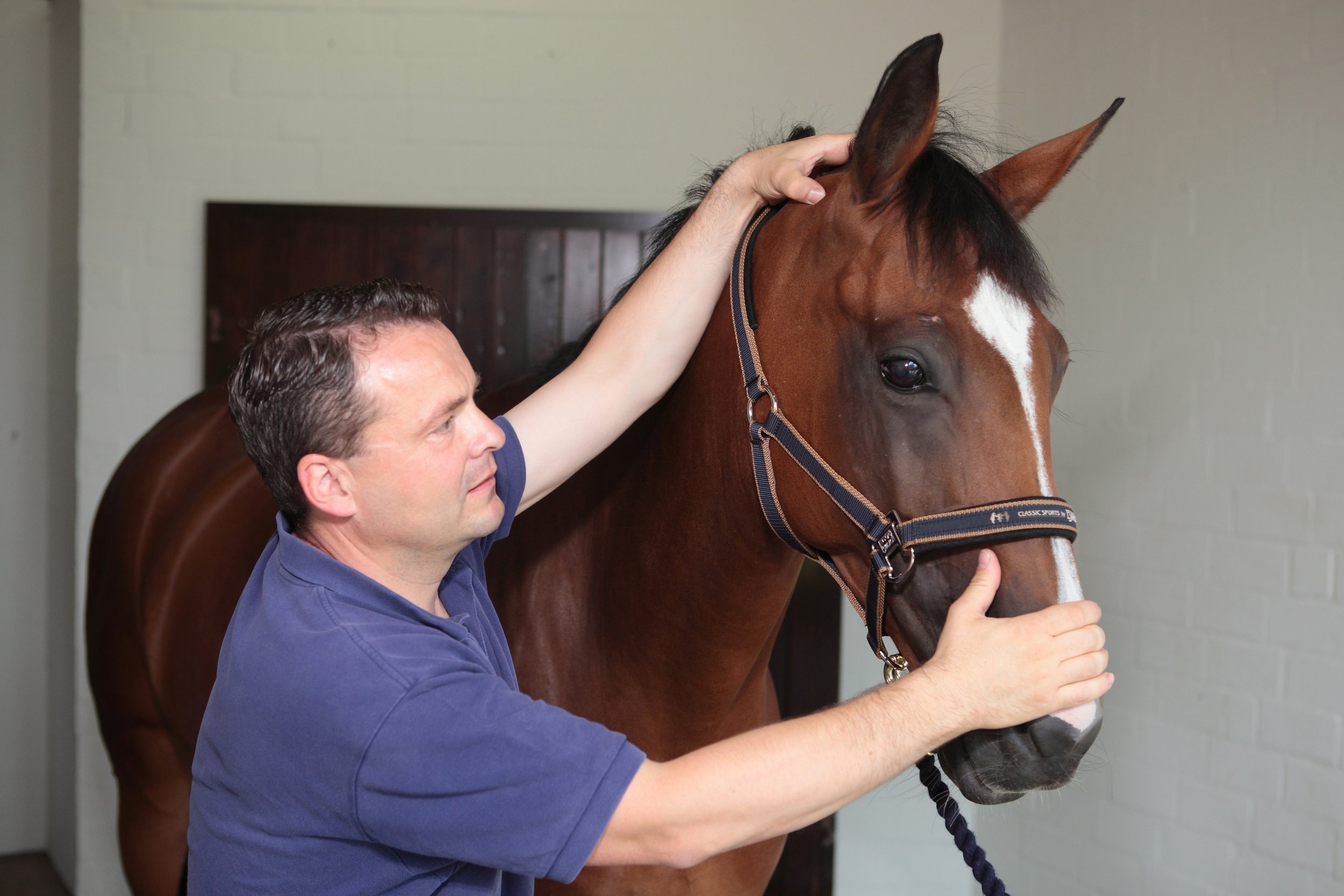
Horse positioned in ct scanner
At the milder end of the scale, signs may be subtle and easily missed, whilst still being responsible for discomfort and reduced performance. The recent ability to perform a computed tomography (CT) scan of a horse’s neck represents a major advancement in our ability to diagnose neck conditions. Timely and accurate diagnosis allows efficient and targeted treatment, the ability to plan schedules and improvement in welfare through the provision of appropriate treatment and earlier return to function.
In neurologic cases, an accurate diagnosis facilitates risk management for both the horse, their handlers and riders, while improving safety for all. As conditions and injuries of the neck are being better characterised using CT, new medical and surgical treatment options are being developed, giving the potential for improved outcomes and fewer losses from the racing industry.
This article summarises how CT is being increasingly used by vets to diagnose conditions of the neck and how it is revealing previously unknown information and providing exciting new treatment opportunities.
Presentation
Conditions of the neck can cause a range of signs in the horse, which are wide-ranging in their presentation and variable in their severity. The manner in which these conditions present depends on which anatomical structures are affected. Issues affecting the bones, joints and/or soft tissues of the neck can all cause neck pain, which can manifest in a number of ways. Cases of neck pain can be severe, resulting in a horse with a rigid, fixed neck carriage, an unwillingness to walk and struggling to eat, perhaps due to a traumatic event. Neck fractures are thankfully uncommon but can be catastrophic.
More moderate signs might be displayed as a stiff neck, with reduced range of movement and resentment of ridden work. There may be pain on palpation of the neck and changes in the neck musculature. Increasingly, we are seeing horses with far more subtle signs, which are ultimately revealed to be due to neck pain and neck pathology. Typically, these horses might have an acceptable range of motion of their neck under most circumstances, but they suffer pain or restriction in certain scenarios, resulting in poor performance. This may be seen as tension through the neck, resisting rein contact, a reluctance to extend the neck over fences, or they may struggle on landing.
Riders might report a feeling of restriction or asymmetry in the mobility of the neck. In addition, these horses may be prone to forelimb tripping or show subtle forelimb lameness.
Any condition, which causes injury or disease to the spinal cord or nerves within the neck, also causes a specific range of neurologic signs. Compression of the spinal cord is most commonly caused by malformations or fractures of the cervical vertebrae, or enlargement of the adjacent articular process (facet) joints. This results in classic ‘Wobbler’ symptoms, which can range from subtle weakness and gait abnormalities, through to horses that are profoundly weak, ataxic and uncoordinated. This makes them prone to tripping, falling, or they may even become recumbent.
Peripheral nerve deficits are uncommon but become most relevant if they affect the nerves supplying the forelimbs, which can result in tripping, forelimb lameness, or local sensory deficits. This lameness might be evident only in certain circumstances, such as when ridden in a rein contact. This lameness is difficult to pinpoint as there will be no abnormality to find in the lame limb, indeed a negative response to nerve and joint blocks (diagnostic analgesia) will usually be part of the diagnostic process.
Horses can present with varying combinations and severities of neck pain, neurologic signs and peripheral nerve deficits, creating a wide range of manifestations of neck related disease.
Diagnosis
A diagnosis of neck pain is based on careful static and dynamic clinical examination and may be supported by seeing a positive response to treatment. Neurologic deficits are noted during a specific neurologic assessment, which includes a series of provocation tests such as asking a horse to walk over obstacles, back up, turn circles and walk up and down a hill. Confirming neck pathology as the cause of signs can be difficult. Until recently, radiography has been the mainstay imaging modality. Radiographs are useful for assessment of the cervical vertebrae and continue to play an important role in diagnosis; however the complex 3-dimensional shape of these bones, the large size of the neck and an inability to take orthogonal (right-angled) x-ray views means that this 2-dimensional imaging modality has significant limitations. High quality, well-positioned images are essential to maximise the diagnostic potential of radiographs.
A turning point in our diagnostic ability and understanding of neck dysfunction has been the recent adaptation of human CT scanners to allow imaging of the horse’s neck. A number of equine hospitals across the United Kingdom and Northern Europe now offer this imaging modality. We have been providing this service at Liphook Equine Hospital since 2017, with over 150 neck scans performed to date.
The CT procedure
A computed tomography (CT) scan combines a series of x-ray images taken from different angles around the area of interest to create a 3-dimensional volume of imaging data. This data is presented as a grayscale image which can be viewed in any plane and orientation. It provides excellent bone detail, and post processing techniques can provide information on soft tissue structures too. Additional techniques can be employed such as positive contrast myelography to provide greater detail about soft tissue structures. Myelography delineates the spinal cord using contrast medium injected into the subarachnoid space and is indicated in any case showing neurologic signs suggestive of spinal cord compression.
Neck CT is performed under a short general anaesthetic. Scans without myelography typically take less than 20 minutes to complete. The entire neck is imaged, from the poll to the first thoracic vertebra. The procedure is non-invasive and low risk, with anaesthetic-related complication presenting the main risk factor to the procedure. We have not encountered any significant complications in our plain CT scan caseload to date. Horses showing ataxia, weakness and incoordination (Wobbler’s), undergo CT myelography which adds around 20 minutes to the procedure. These horses are exposed to a greater level of risk due to their neurologic condition, the injection of a contrast agent and the increased chance of destabilising a more severe lesion during the procedure.
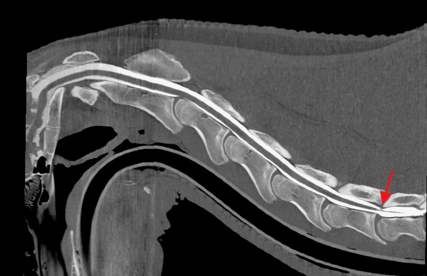
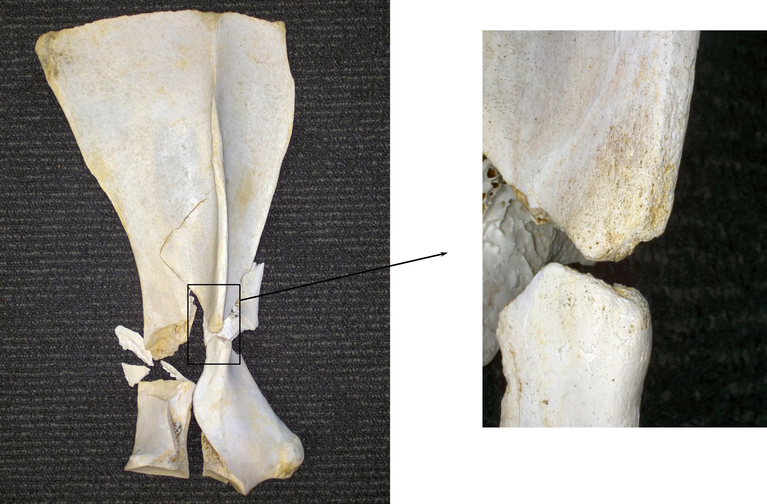
Sagittal and transverse CT myelogram images of a young racing thoroughbred showing neurologic (Wobbler) signs. shows narrowing of the spinal canal at the base of the neck (arrow)
CT is revealing more detailed information about ways that spinal cord compression can occur in Wobbler cases, about compression of spinal nerves resulting in forelimb gait deficits and precise detail about fracture configurations. It gives us detailed images of articular process joint disease, intervertebral disc disease, developmental conditions and anatomic variations. It is also revealing information about rare diseases such as vertebral abscesses or spinal neoplasia. As our caseload and confidence in the imaging modality grows, we are learning more about the value of CT in examining more subtle neck conditions. We are also bringing the benefit of a more accurate diagnosis, allowing precise targeted treatment and a better ability to provide a prognosis about outcomes—likely progression or safety factors. CT myelography allows circumferential imaging of the spinal canal and yields significantly more information than traditional x-ray myelography. As a result, we hope to enable better case selection of horses that may benefit from Wobbler surgery, with the goal of resulting in improved success rates of the surgery.
Innovations in treatment

image shows a fracture of the articular process of the 3rd cervical vertebrae in the mid neck, which was not visible on x-ray.
New treatment options are emerging as a result of our more accurate diagnoses of neck pathology. Of the first 55 horses which underwent neck CT at our hospital, we were surprised to discover that 13 (24%) had some form of osteochondral fragmentation within the articular process joints of their neck. Some of these horses were young Thoroughbreds, bred to race but showing Wobbler signs. These tended to have convincing CT evidence of type 1 CVM (Wobbler Syndrome) and osteochondrosis affecting their neck. Others had fragments which were larger and more discrete, with evidence of articular process joint enlargement/arthritis but no other bony lesions. These horses were typically older and of a range of breeds and uses.
In those horses presenting with signs of neck pain but no neurologic deficits, surgical removal of these fragments was proposed. Following further consideration and cadaver training, we have begun to offer this surgery for horses that fit the appropriate criteria and have surgically accessible fragments. We have performed arthroscopic or arthroscopic-assisted fragment removal from eight articular process joints in six horses to date. No intraoperative or postoperative complications have been encountered; and five of six horses showed complete resolution of neck pain. In the sixth horse, full recovery was not anticipated due to the presence of additional neck pathology, but partial improvement occurred for two years. Fragment removal has relieved signs of neck pain and stiffness and caused improved performance in these horses.
Two procedures that are emerging to treat spinal nerve root impingement are a targeted peripheral nerve root injection and a keyhole surgical procedure to widen the intervertebral foramen. Nerve root injection is performed in the standing sedated horse under ultrasound guidance. Surgery is performed under anaesthesia, using specialised minimally invasive equipment to widen the bony foramen using a burr. This surgery is in its infancy but offers an exciting treatment option.
Additionally, CT gives us the ability to better plan for fracture repair, undoubtedly improving our case selection for Wobbler surgery; it more accurately guides intra-articular injection of the articular process joints.
Summary
Computed tomography is transforming our ability to diagnose conditions of the horse’s neck. The procedure is low risk and now widely available in the UK and other parts of Europe. It is driving the innovation of novel treatment options with the goal of improving outcomes and reducing losses to conditions of the neck. Our CT findings are posing new questions about neck function, pain and neurologic disease and is an active area of ongoing research.
Anatomy of the cervical vertebrae

The neck vertebrae of the horse
The neck consists of seven cervical vertebrae which form a gentle S-shaped curve to link the skull at the poll to the thoracic vertebrae of the chest. Its primary functions are to protect the spinal cord, support the heavy weight of the head and to allow a large range of movement so that a horse can monitor his environment and run at speed, both being vital to this prey species.
The first (atlas) and second (axis) cervical vertebrae are highly adapted to allow head mobility. The third to sixth vertebrae are very similar in shape, whereas the seventh is shortened as it starts to show similar features to the vertebrae of the thorax. The typical cervical vertebra consists of a cylindrical column of bone (vertebral body) articulating with adjacent vertebrae via an intervertebral disc.
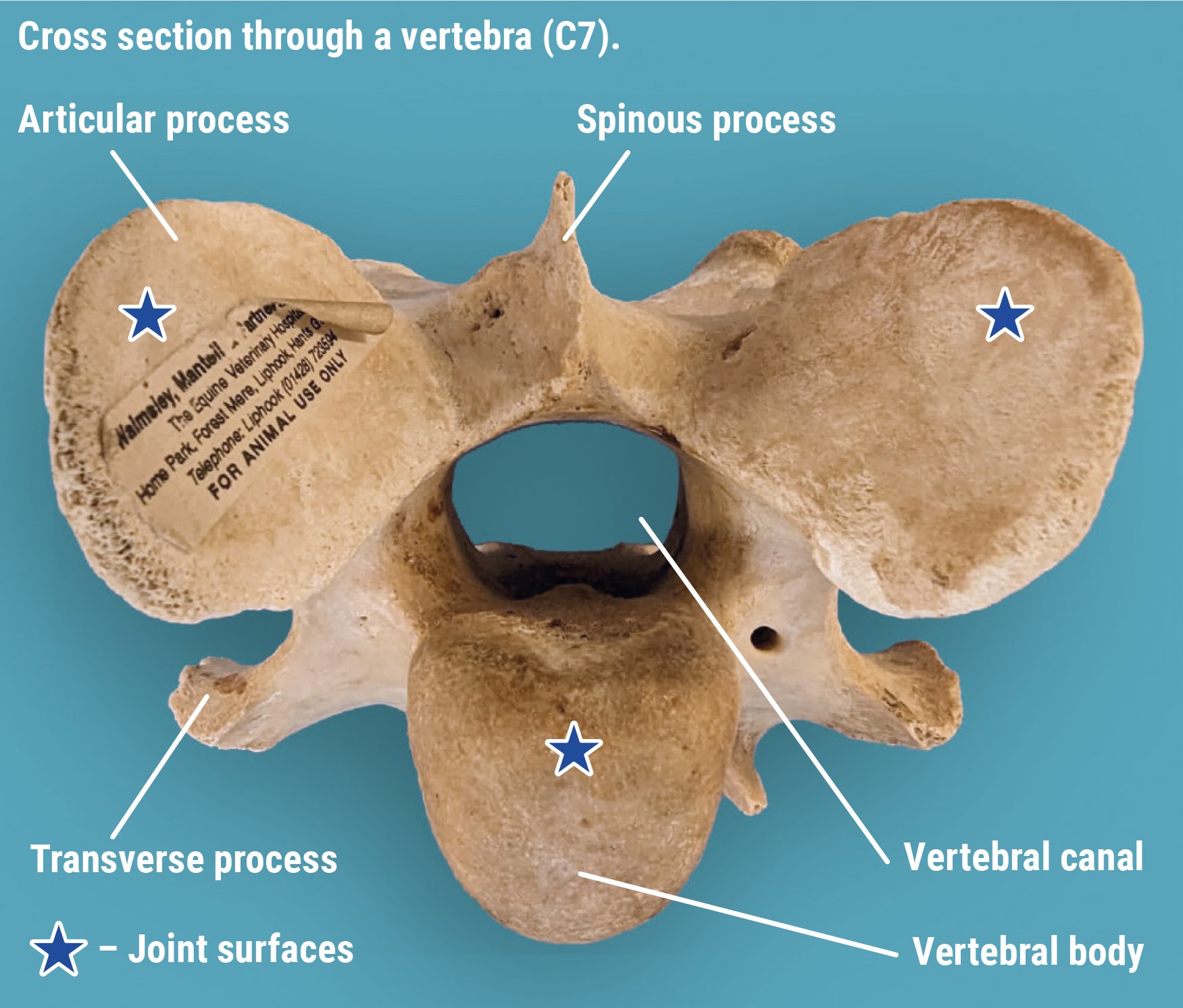
Running along the upper surface of this bony column is a bony canal created by the vertebral arch. This canal protects the spinal cord and associated structures that run through the centre. Above and to the side of the spinal canal sit the paired articular processes and below; and to the side sit the transverse processes. These are bony prominences to which muscles attach. The soft tissues of the neck are complex and intrinsically linked to the forelimbs and entire axial skeleton.
The articular processes also form the articular process joints (facet joints) which create an additional two joints between each vertebra. Although highly adapted, these joints are similar to others in the body consisting of cartilage-lined surfaces, a joint capsule and joint fluid. The joint surfaces are oval in shape, approximately 3-4cm in diameter and sit at an oblique angle to the neck. They provide important additional support and mobility to the neck.
References:
Schulze N., Ehrle, A., Beckmann, I and Lischer, C. (2021) Arthroscopic removal of osteochondral fragments of the cervical articular process joints in three horses. Vet Surg. ;1-9.
Swagemakers J-H, Van Daele P, Mageed M. Percutaneous full endoscopic foraminotomy for treatment of cervical spinal nerve compression in horses using a uniportal approach: Feasibility study. Equine Vet J. 2023.
Tucker R, Parker RA, Meredith LE, Hughes TK, Foote AK. Surgical removal of intra-articular loose bodies from the cervical articular process joints in 5 horses. Veterinary Surgery. 2021;1-9.
Wood AD, Sinovich M, Prutton JSW, Parker RA. Ultrasonographic guidance for perineural injections of the cervical spinal nerves in horses. Veterinary Surgery. 2021; 50:816–822.