How Equine Influenza viruses mutate

How equine influenza viruses mutateDebra Elton and Adam Rash Overview Equine influenza virus (EIV) causes equine influenza in horses, characterised by a raised temperature and harsh dry cough and rapid transmission amongst unprotected horses. It is …

By Debra Elton and Adam Rash

Overview

Equine influenza virus (EIV) causes equine influenza in horses, characterised by a raised temperature and harsh dry cough and rapid transmission amongst unprotected horses. It is a major threat to the thoroughbred racing industry as it has the potential to spread so quickly and can cause the cancellation of events and restriction of horse movement. The last major outbreak in Europe occurred in 2003, when over 1000 vaccinated horses in Newmarket became infected. The virus spread throughout the UK and outbreaks were also reported in Ireland and Italy. More recently, more than 50,000 horses were infected during the 2007 outbreak in Australia, large-scale outbreaks occurred in India during 2008 and 2009 and multiple countries were affected by widespread outbreaks in South America in 2012. At the time of writing, another widespread outbreak has been affecting South America, with reports from Chile, Argentina, Uruguay and Colombia to date. International transport of horses for events and breeding purposes means that equine influenza can spread readily from one country to another. Infected horses can shed the virus before they show any clinical signs of infection and vaccinated animals can be infectious without showing any obvious signs, adding to the risk.

Regular vaccination against equine influenza offers the best protection against infection. Three major vaccine manufacturers make products for the European market, each differing in the virus strains that are included in the vaccine. Sophisticated adjuvants are included in these vaccines, which help boost the horse’s immune response. However, EIV, like other influenza viruses, can mutate to change its surface proteins and can thereby escape from immunity generated by vaccination. It is important that vaccines contain relevant vaccine strains, to give them the best chance of working against current EIVs.

EIV belongs to the influenza A group of viruses, which infect a variety of other animals including humans, birds, pigs and dogs. The natural reservoir for most influenza A viruses is wild aquatic birds, from this pool some viruses go on to infect new hosts and adapt to spread in them. Influenza A viruses are subtyped according to two proteins found on the surface of the virus, haemagglutinin (HA) and neuraminidase (NA). Sixteen HA subtypes and 9 NA subtypes are found in aquatic birds, however only two subtypes are known to have become adapted to horses, H3N8 and H7N7. Equine H7N7 viruses were first isolated in 1956 but have not been isolated since the late 1970s and are now thought to be extinct. Equine H3N8 viruses were first isolated in 1963 when they caused an influenza pandemic in horses and continue to circulate today.

Antigenic drift and shift

International travel of horses means the virus can spread readily from one country to another.

The HA and NA proteins on the surface of the influenza virus particle induce antibodies in the host when the virus infects it. For EIV, these antibodies protect the horse against further infection provided the horse encounters similar viruses. A similar process occurs when horses are immunised with a vaccine, most vaccines contain virus proteins that induce the horse’s immune system to make protective antibodies. However, the response to the vaccine is not as good as to virus infection, so horses need to be vaccinated regularly to maintain a protective immune response.

To overcome the horse’s immune response and enable the virus to survive in the equine population, EIV gradually makes changes to its surface proteins. This process is called antigenic drift. The result is that eventually the horse’s antibodies no longer recognise the virus, which is then able to infect the animal. The two proteins that are important for antigenic drift are HA and NA. HA is involved in virus entry into target cells of the respiratory tract. Antibodies against HA block virus infection, either by preventing the virus from binding to the cell surface, or by preventing a later stage of the infectious cycle that occurs within the infected cell. Antibodies against HA are described as ‘neutralising’ because they prevent virus infection.

By changing the HA protein, equine influenza can avoid recognition by these neutralising antibodies. NA is also involved in virus entry, it is thought to help break through the mucus layer that protects the respiratory tract. It also plays a part in virus release, enabling newly formed virus particles to escape from the surface of the cell that made them. Antibodies against NA are thought to block this process, preventing the virus from spreading to new cells. By changing the NA protein, the virus can avoid inhibition by these antibodies and go on to infect new cells.

Equine influenza virus belongs to a family of viruses that have RNA as their genetic material rather than DNA. RNA viruses tend to mutate more rapidly than DNA viruses. The virus has an enzyme called RNA-dependent RNA polymerase that is responsible for making new RNA copies of the virus genetic material for packaging into new virus particles. This is an essential step during the virus life cycle. Compared to the polymerase enzymes found in DNA viruses, the influenza polymerase makes more mistakes when it is copying the virus RNA and this is how changes are made in the genes that code for HA and NA.

Figure 1.jpg

As well as undergoing antigenic drift, influenza viruses including equine influenza virus can change their genes by a process called antigenic shift. This is a much bigger rapid change, brought about by the virus-swapping sections of its genome with another influenza virus. This process is called reassortment and is possible because the virus genome is made up from eight separate segments of RNA, each individually packaged in a set of proteins. If a horse is infected with two different equine influenza viruses at the same time, the eight segments from each virus can be mixed up, generating progeny viruses with new combinations of segments compared to the two parent viruses. This can lead to new combinations of HA and NA that haven’t been seen before, meaning there is no immunity to the new virus. This has happened during the evolution of human influenza viruses and resulted in the influenza pandemics of 1957, 1968 and 2009. In two of these examples, human influenza viruses swapped genes with avian viruses, leading to viruses that replicated well in humans but had a new HA gene from an avian virus.

In the 2009 pandemic, a new reassortant virus was generated in pigs then transmitted to humans. Reassortment has also happened with equine influenza viruses. The two different subtypes of equine influenza viruses, H7N7 and H3N8, underwent reassortment resulting in viruses that had most of the internal components of the H3N8 virus but with the HA and NA surface proteins from the H7N7 virus. Eventually these viruses died out and the only equine influenza viruses now in circulation are H3N8. There has been reassortment amongst the different sublineages of equine H3N8 viruses too, for example several of the viruses isolated in the UK during 2009 had a mixture of Florida clade 1 and Florida clade 2 HA and NA. Fortunately these reassortant viruses do not contain a novel HA or NA that has not been seen in horses before, so have not resulted in a major epidemic threat to horses.

In addition to antigenic drift and antigenic shift, the other source of potential new influenza viruses is an animal reservoir, such as birds. We know that horses can be infected by viruses belonging to the H3N8 and H7N7 subtypes and both of these are found in wild aquatic birds. It is thought that the 1963 H3N8 equine pandemic probably arose as a result of cross-species transmission from birds to horses in South America. Such an event happened in China in 1989, when an avian H3N8 infected horses with a much higher mortality rate than is usual for equine influenza. This virus spread amongst horses within China but died out after a relatively short time. It is possible that further avian-equine cross species transmission events could take place, however the virus must then adapt to its new host in order to become established in horses and be able to transmit efficiently from horse to horse. This will require mutations in various virus genes that help the virus attach to and replicate in cells lining the horse’s respiratory tract and spread via droplet infection to other horses.

TO READ MORE --

BUY THIS ISSUE IN PRINT OR DOWNLOAD -

October - December 2018, issue 63 (PRINT)
£8.95

WHY NOT SUBSCRIBE?

DON'T MISS OUT AND SUBSCRIBE TO RECEIVE THE NEXT FOUR ISSUES!

Combating contamination in the racing yard

There is so much more to the Clean Sport initiative than the FEI Prohibited Substances List. Building relationships with local feed suppliers and developing a code of practice with staff will ensure your racehorse has a clean and clear system. Horse…

By Melissa Volpi

There is so much more to the Clean Sport initiative than the FEI Prohibited Substances List. Building relationships with local feed suppliers and developing a code of practice with staff will ensure your racehorse has a clean and clear system.

Horseracing is an inclusive sport. It was this ‘spirit of the sport’ that the FEI (Federation Equestre Internationale) wanted to preserve when it formed the Clean Sport commission on the 9th October 2009 in Copenhagen, Denmark.

But what is ‘Clean Sport’ and why is it necessary? According to the FEI, Clean Sport is about being honest and being true. It’s about having integrity and not letting our lust for winning cloud our judgement and respect for rules and laws. It’s about aiming for success, but accepting failure if it cannot be won fairly. The FEI believe that doping is contrary to the spirit of the sport and it has put together an initiative to ensure the end of positive drug testing for horses and their human athletes. Clean Sport is about being aware of prohibited substances, to include banned substances and controlled medication. It’s about being vigilant in feed management, medication management and stable management.

“In terms of applying the clean sport principles to racing yards, trainers need to be vigilant as it is so easy for prohibited substances to sneak in,” says Kate Hore, senior nutritionist at NAF (Natural Animal Feeds). “An obvious example may be an owner or staff member having a cup of coffee (source of caffeine) and a chocolate bar (source of theobromine) in the feed room, which then creates a potential risk of contamination and then for a positive drug test. The best advice is to avoid eating or drinking in the feed room or stable.”

The British Equestrian Trade Association (BETA) set up a Naturally Occurring Prohibited Substances (NOPS) scheme in 2009 that identifies the main NOPS and their sources, as well as providing a list of approved feed suppliers (companies that have signed up to be audited under the schemes new codes). “The BETA NOPS scheme is the only one of its kind and has cooperation from all corners of the world,” says Hore. “Contamination by NOPS is easier than people think, morphine being a good example. White poppies are grown commercially to produce morphine and the poppy seeds can be blown from one field to another, into a field of oats for example, which could then create a contamination issue further down the processing chain.”

TO READ MORE --

BUY THIS ISSUE IN PRINT OR DOWNLOAD -

October - December 2018, issue 63 (PRINT)
£8.95

WHY NOT SUBSCRIBE?

DON'T MISS OUT AND SUBSCRIBE TO RECEIVE THE NEXT FOUR ISSUES!

HBLB Research on Injuries in Flat Racing: Nature versus Nurture

HBLB Research on Injuries in Flat Racing: Nature versus NurtureNote: This research for this article, reprinted from European Trainer, was performed over a 14-year period in Great Britain and therefore only takes into account racing over turf and all…

By Kristien Verheyen & Sarah Rosanowski

Musculoskeletal injuries are an inherent risk of horseracing, and they are the primary cause of thoroughbreds failing to train and race, or even retiring altogether. In addition to the evident equine welfare concerns, racehorse injuries also have economic consequences and impact on jockey safety. The industry remains committed to investigating causes of injury and associated risk factors, which can inform strategies aimed at minimising their occurrence. Advancements in methods of identification, management, and prevention of musculoskeletal disease and injury in Thoroughbreds and improved training and racing environments to enhance the safety, health, and wellbeing of racehorses have long been strategic priorities of the Horserace Betting Levy Board (HBLB)’s veterinary research funding program in Great Britain.

In 2014, the HBLB funded a research team at the Royal Veterinary College in London to undertake a detailed study of injuries and other veterinary events occurring in flat racehorses on race day. The purpose of the project was to establish causes of fatal and non-fatal injuries occurring in British flat racing and to examine associated risk factors. The research also set out to measure heritability of common injury types and conditions, and to investigate genetic and environmental correlations between injury and race performance.

The study team had access to detailed race and performance data from all Thoroughbreds racing on the flat in Great Britain over a 14-year study period from 2000 – 2013. These were then linked to veterinary reports of injury or conditions attended to by a veterinary surgeon on race day over the same time period, provided by the British Horseracing Authority (BHA). Finally, extensive pedigree data were added to enable investigation of heritability of race day injury and genetic correlations between injury types, and between injury and performance.

Descriptive findings

The final 14-year dataset included nearly 68,000 horses making over 800,000 starts in around 77,000 flat races. The majority of races -- 67% of them -- were run on the turf, with 33% of races taking place on all-weather tracks.

Leading image (2).jpg

Just under 8,000 veterinary events were recorded over the study period, from which an incidence of nine events per 1000 starts was calculated. The most common incidents requiring veterinary attention on the racecourse were soft tissue injuries other than tendon and ligament injuries, e.g. wounds, lacerations, or muscle strains. Unspecified lameness and respiratory conditions were also common, accounting for around a fifth of veterinary reports each. Less than 10% of veterinary events had a fatal outcome, and the overall incidence of fatality was 0.8 per 1000 starts. Although bone injury was cited in only 14% of the veterinary reports overall, they accounted for the vast majority (77%) of the fatalities.

All-weather racing

Racing on all-weather tracks traditionally carries a higher risk of injury than racing on turf, which was reaffirmed in the current analyses. Therefore, the researchers also specifically investigated risk factors for fatality, distal limb fracture, and epistaxis (nose bleeds) in all-weather racing. These analyses were restricted to the ca. 258,000 all-weather starts in the dataset and included additionally collected information from the racecourse clerks on surface types and maintenance. The fatality incidence in all-weather racing was 0.9 per 1000 starts. Distal limb fracture occurred in around 1 in 1000 starts and epistaxis in 1.6 per 1000 starts. Risk factors varied for each outcome, although some factors were similar across outcomes including the going, racing intensity, horse age, age at first start, and horse and trainer performance variables. Generally, older horses and those that had started racing at an older age were at higher risk of an adverse outcome although for fatality, older horses that had started racing as two-year-olds were at highest risk. This association may be due to accumulation of microdamage in bone, which increases with increasing age as an effect of exercise accumulation over time and can ultimately lead to failure.  

TO READ MORE --

Order this issue in print or download 

July - September 2018, issue 62 (PRINT)
£8.95

Equine Herpesvirus-1 : An Elusive Target

Equine herpesvirus -1; an elusive targetInfectious diseases are not uncommon in racehorses in training, breeding stock, and pleasure horses. Some of the more serious diseases can be financially devastating to the animal’s owners and to the equine in…

By Neil Bryant 

Infectious diseases are not uncommon in racehorses in training, breeding stock, and pleasure horses. Some of the more serious diseases can be financially devastating to the animal’s owners and to the equine industry on the whole. Viruses belonging to the herpesvirus family cause some of the most well characterized equine infectious diseases, and the most problematic of these is equine herpesvirus 1 (EHV-1; species Equid alphaherpesvirus 1). EHV-1 is ubiquitous in most horse populations in the world. It is responsible for major economic and welfare problems causing respiratory disease, neurological disease (mainly seen in adult horses), and abortion and neonatal foal death in pregnant mares.
This was most notably highlighted by the multiple abortion outbreak recorded in Hertfordshire, England, between February and April 2016 in fully vaccinated animals (http://www.aht.org.uk/cms-display/interim-report16-april2.html). Studies have determined that EHV-1 is a common cause of abortion. Occasional cases have also been linked to EHV-4 infection, but this is much rarer and doesn’t account for episodes of multiple abortion, as is seen occasionally with EHV-1.

The virus

EHV-1 was first isolated from an equine abortion in the U.S. in the 1930s. At the time of first isolation the vets weren’t sure what it was, but they knew it was infectious. Subsequent genetic analysis much later led to the classification of the virus in the genus Varicellovirus (family Herpesviridae), together with its close relatives equine herpesvirus 4 (EHV-4; species Equid alphaherpesvirus 4) and equine herpesvirus 8 (EHV-8; species Equid alphaherpesvirus 8). Interestingly it is grouped with, and is therefore genetically similar to, the human herpesvirus responsible for chickenpox, the Varicella Zoster virus. Initial infection of horses was thought to occur around weaning, when virus-neutralizing antibodies transferred to the foal from the mare’s colostrum had declined enough to make them susceptible to infection. However, virus has been isolated from foals as young as seven days old with high antibody levels but without any significant clinical signs. Immunity to re-infection after primary infection is relatively short-lived, lasting between three-six months, but it is rare for naturally infected mares to abort in consecutive pregnancies.

TO READ MORE --

BUY THIS ISSUE IN PRINT OR DOWNLOAD -

April - June 2018, issue 61 (PRINT)
£8.95

Why not subscribe? 

Don't miss out and subscribe to receive the next four issues!

Castrating racehorses: A routine procedure not without its pitfalls

A recent study published in the Equine Veterinary Journal assessed the routine procedure of gelding and the complications associated with this procedure. The research was a retrospective study of horses castrated at the Sha Tin training complex in H…

By Tom O’Keeffe

A recent study published in the Equine Veterinary Journal assessed the routine procedure of gelding and the complications associated with this procedure.  The research was a retrospective study of horses castrated at the Sha Tin training complex in Hong Kong, between July 2007 and July 2012.

Hong Kong is a unique training and racing environment, and all horses training and racing there are imported, as there is no breeding in the region. Fillies are rarely imported. The majority of colts are castrated at some stage in their career, and open standing castration (OSC) is the method of choice by the vets of the Hong Kong Jockey Club (HKJC). Until now, nobody has looked at the prevalence of complications following castration of horses at the HKJC. This recently published study aimed to describe the prevalence and severity of complications in the 30 days following castration.

Reasons for gelding a racehorse in training

Most trainers perceive geldings as easier to train than colts, and if the horse has not shown enough ability for a stud career to beckon, there is little to lose by gelding.  In Hong Kong, due to the unique environment the horses live in, there is an added incentive to geld these horses sooner rather than later. Once gelded, their management becomes significantly more straightforward.

Castration Method Options

Three surgical techniques are commonly used for equine castration: 1) open, in which the parietal tunic surrounding the testicle is incised and, usually, retained; 2) closed, where the portion of the parietal tunic surrounding the testis and distal spermatic cord is removed, and 3) half closed, where an incision is made through the exposed parietal tunic at the cranial end of the testis or distal end of the spermatic cord allowing the testis and part of the spermatic vasculature to be prolapsed through the incision prior to removal.


In most cases, racehorse castration is done standing via the open technique under local anesthetic, with sedation and pain relief as necessary. The testicles and spermatic cords are first injected with local anesthetic to numb the region. Once the tissues are totally desensitized, a slash incision is made into the scrotum. The testicle is exteriorized, and it is removed with a surgical instrument called an emasculator. The emasculator has a set of interlocking crushing blades with a cutting blade placed at the bottom of the array. Once the testicular cord is clamped in the emasculator the testicle will usually fall off, but the cord is retained within the interlocking crushing blades for approximately one to two minutes. This creates trauma to the tissues, which causes them to swell once the crush is released, reducing blood flow. The second effect of the emasculators is for the blood to be held in position long enough to begin the clotting process, which carries on once the clamp is removed.

An alternative method of castration is to anesthetize the horse and carry out the procedure with the horse on its back, as a completely sterile operation in an operating room. This has the advantage of minimal post-castration swelling as there is no infection in the area, which can be a common problem with standing open castrations.  In horses who are cryptorchids (ridglings), which is when there is only one descended testicle in the scrotum, standard open standing castration is contraindicated. These horses require either castration under general anesthetic or testicle removal under standing surgery via laparoscopy (inserting a camera and instruments into the abdomen to remove testicle via a surgical incision).

Complications of Castration

As with all intrusive surgical procedures, there is the potential for things to go wrong. While the castration procedure is relatively straightforward, post-operative complications including excessive edema of the scrotum and surrounding tissues, infection and fever, hemorrhage, lameness, hydrocele formation, peritonitis, eventration, penile paralysis, scirrhous cord formation, and death have been recognized.

With castrations done under general anesthetic, there are all the attendant risks of putting a 1000lb animal on its back and up again. All anesthesia carries a risk of death in the horse. This has been calculated as approximately 1% in equine practice, and can be as low as 0.5% in the major well-equipped equine hospitals. In addition to this, occasional cases show prolonged bleeding after the surgery, which results in significant swelling that sometimes has to be resolved by opening the scrotal sac.

Intestine is prolapsing through the castration site - this severe castration complication requires immediate veterinary attention.

Intestine is prolapsing through the castration site - this severe castration complication requires immediate veterinary attention.

For standing castrations, some of the problems encountered include prolonged bleeding, which can occur irrespective of the length of time the cord has been clamped for. This can become serious enough to require a further surgery to identify the bleeding vessels and tie them off, but thankfully this is rare. Another rare complication is herniation of intestines through the potential space left in the inguinal canal with removal of the testicle. The intestines can either get trapped under the skin producing severe colic, or worse still, dangle out of the abdomen and become contaminated. This presents a very serious risk to the horse’s survival and requires immediate surgery to attempt to clean the exposed bowel and return it to the abdomen. Fortunately this is extremely rare in the Thoroughbred.

However, the most common complication is infection at the site of the castration. This procedure leaves an open wound and obviously the horse can lie down in bedding full of urine and feces on the same day it has been castrated, therefore potentially contaminating the open surgical site. Unfortunately many racehorses’ ability to be turned out in a paddock is often controlled by the training environment they reside in. Infection post-castration, and the added expense and lost training days associated with it, is a bugbear for trainers and vets, and this study reviews a common problem encountered worldwide.

Hong Kong Study

The Hong Kong training complex provides full-time stabling and training facilities to approximately 1250 horses spread out among 24 licensed trainers. The Department of Veterinary Clinical Services (DVCS) at the HKJC is the sole provider of veterinary care for this population. All clinical records of horses in training at the HKJC are collated within the Veterinary Medical Information System (VMIS). For a horse to be eligible for inclusion in the study, two testicles had to have been removed via an open standing castration. Veterinary records of all the horses that had been castrated were examined and any cases that did not meet the criteria were excluded.

Data on complications that occurred in the 30 days following castration was extracted from the clinical notes in the VMIS. The data was reviewed and the severity of complication was categorized into one of the five groups below:


Between July 2007 and July 2012, 280 racehorses in training were castrated. A total of 30 horses were omitted from the study, as they did not meet the inclusion criteria: 24 horses were castrated using general anesthetic, of which six were cryptorchid surgeries.  Horses included in the study were in the care of 24 different trainers, with thirteen different veterinarians performing the castrations.


Twenty-four hours after castration, this horse has mild scrotal swelling, which would be classed as Group C1 in the Hong Kong study.

TO READ MORE --

Buy this issue in print -

April - June 2018, issue 61 (PRINT)
£8.95

Why not subscribe?

Don't miss out and subscribe to receive the next four issues!

The On-going Effort to Minimise the Rate and Impact of Fractures

Published in European Trainer, January - March 2018, issue 60.

In thoroughbred racing, musculoskeletal injury is a major safety concern and is the leading reason for days lost to training.  Musculoskeletal injury is the greatest reason for horse turnover in racing stables, with financial implications for the owner and the racing industry. Injuries, particularly on race day, have an impact on public perception of racing.  

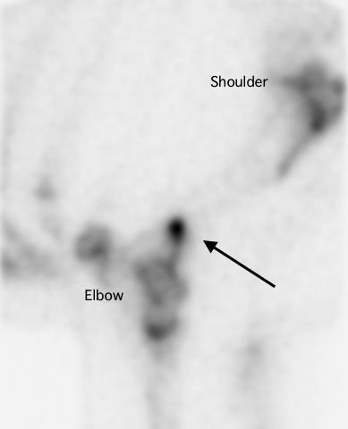
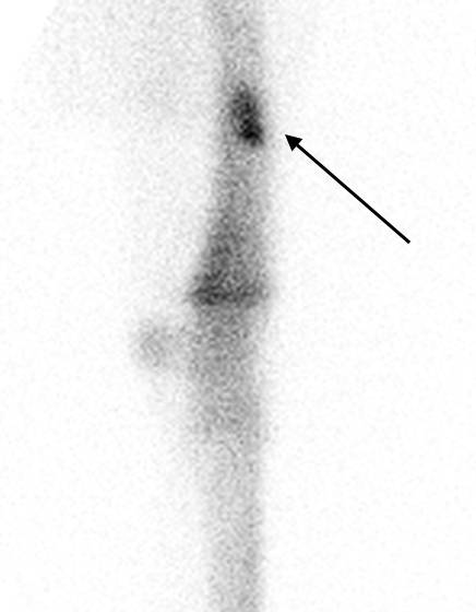
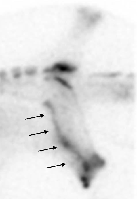
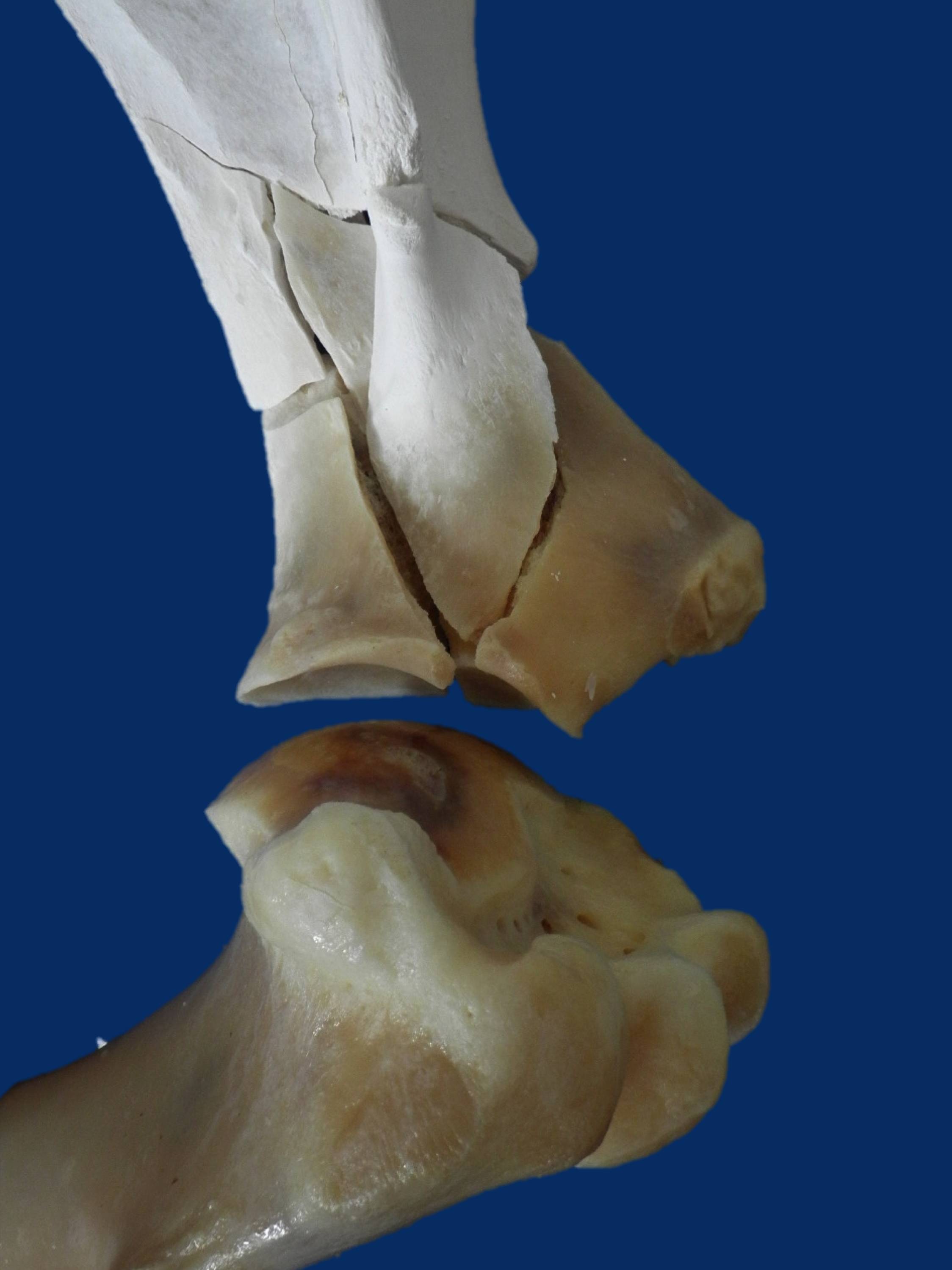
Upper limb and pelvis fractures are less common than lower limb fractures, but they can lead to fatalities. Reducing the overall prevalence of fractures is critical and, at the very least, improving the rate of detection of fractures in their early stages so the horse can be withdrawn from racing with a recoverable injury will be a big step forwards in racehorse welfare. Currently, we lack information on the outcomes following fracture, and an article recently published in the Equine Veterinary Journal (EVJ) from the veterinary team at the Hong Kong Jockey Club (HKJC) addressed this important knowledge gap.

Hong Kong Fracture Outcome Study

The HKJC veterinary team is in a unique position to carry out this work because their centralised and computerised database of clinical records, together with racing and retirement records, allows them to document follow-up, which is all but impossible elsewhere in the world. Dr Leah McGlinchey, working with vets in Hong Kong and researchers from the Royal Veterinary College, London, reviewed clinical records from 2003 to 2014 to identify racehorses that suffered a fracture or fractures to the bones of the upper limb or the pelvis during training or racing, confirmed by nuclear scintigraphy, radiography, ultrasonography, or autopsy....

To read more - subscribe now! 

Gallery

Getting to Grips with Strangles: Working Together to Break the Strangles-hold

Andrew S. WallerAnimal Health Trust, Kentford, Newmarket, Suffolk, CB8 7UU, UKStrangles, caused by a bacteria called Streptococcus equi, is one of the most frequently identified infectious diseases of horses worldwide. More than 600 outbreaks of Str…

Published in European Trainer, January - March 2018, issue 60.

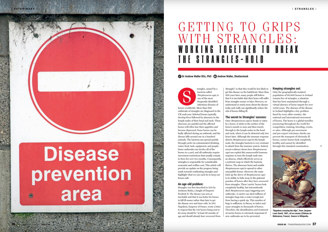
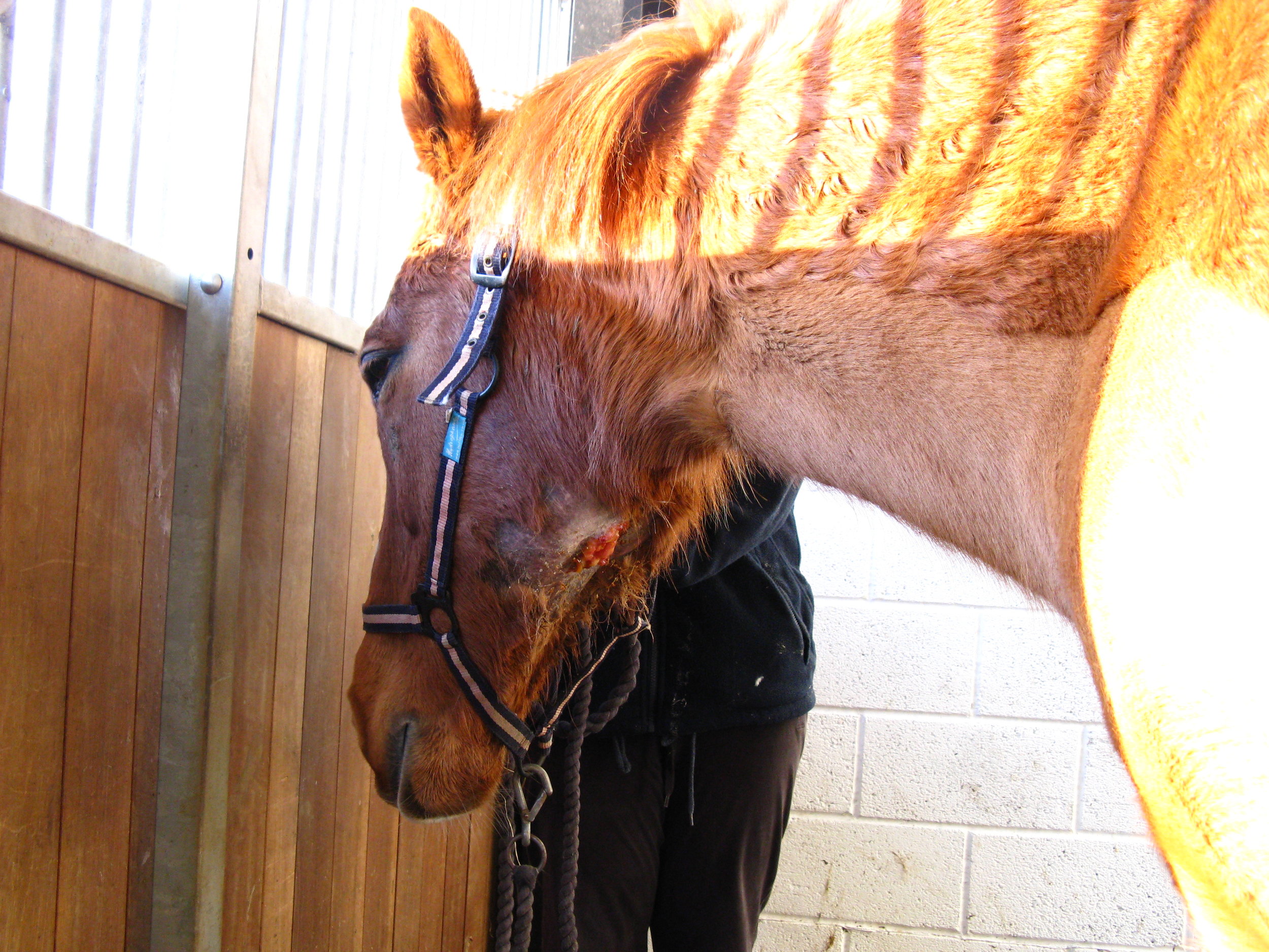
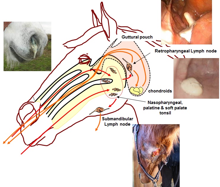
Strangles, caused by a bacteria called Streptococcus equi, is one of the most frequently identified infectious diseases of horses worldwide. More than 600 outbreaks of Strangles are diagnosed in the UK each year. Infected horses typically develop fever followed by abscesses in the lymph nodes of their head and neck.

These abscesses are painful and the affected horses will often lose their appetites and become depressed. Some horses can be badly affected during an outbreak and the disease kills around one in a hundred animals. The bacteria can spread quickly through yards via contaminated drinking water, food, tack, equipment and people. Some outbreaks can involve all of the horses on a yard and all outbreaks require movement restrictions that usually remain in force for over two months. Consequently, Strangles is responsible for considerable economic and welfare cost. This article will provide an update on the progress being made towards eradicating Strangles and highlight what we can each do to keep our horses safe.

An age-old problem:

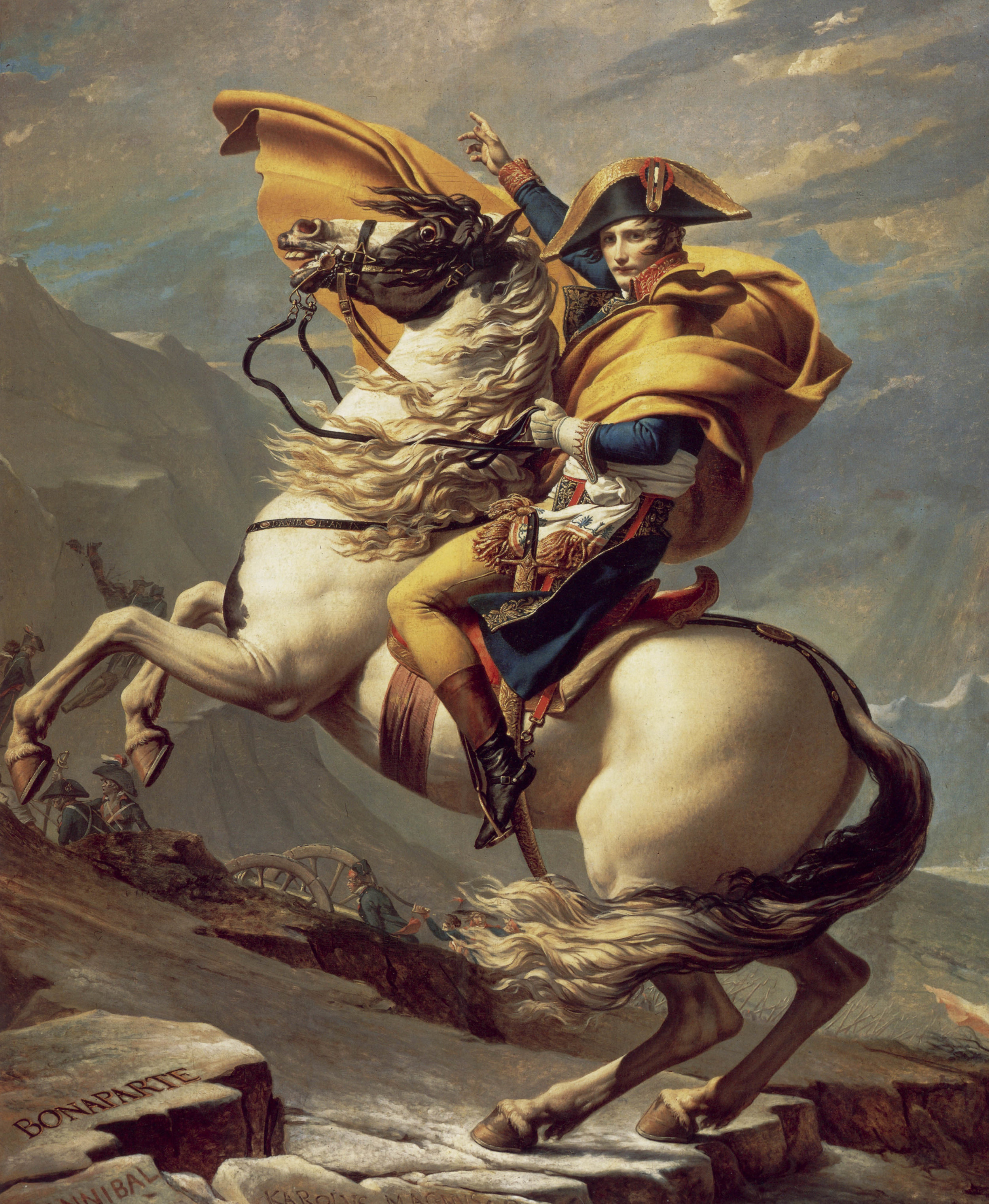
Strangles was first described in 1251 by Jordanus Rufus, a knight of Emperor Fredrick II. The disease was seen as inevitable and better for horses to fall ill sooner rather than later to get the disease over and done with. In 1811 Napoléon, Emperor of France, wrote a letter to request that the 543 horses being sent to his army should be “at least 60 months of age and should already have recovered from Strangles” so that they would be less likely to fall ill from this disease on the battlefront. More than 200 years later, many people still believe that it is inevitable that their horse will suffer from Strangles sooner or later. However, we understand so much more about the disease today and really can significantly reduce the risk of horses falling ill...

To read more - subscribe now! 

Gallery

A report from the Merial - Performance Horse CPD and Raceday at Gowran Park

Performance Horse CPD, Gowran Park RacecourseVets from all over Ireland congregated at Gowran Park racecourse in July for a continuing professional development event on the Performance Horse. The event, organised by European Trainer Magazine and Mer…

Becky James BSc, MSc - Haygain

Published in European Trainer - October - December 2017, issue 59

Click here to order this back issue!

Vets from all over Ireland congregated at Gowran Park racecourse in July for a continuing professional development event on the Performance Horse. The event, organised by European Trainer Magazine and Merial Animal Health, was the second in a series of veterinary CPD events for 2017 and featured a panel of expert speakers. The event was co-sponsored by Haygain and Connolly’s RED MILLS. 

Managing Inflammatory Airway Disease – Dr Emmanuelle Van Erck-Westergren

The first speaker Dr Van Erck-Westergren was due to fly in from Brussels on the morning of the event, so when her flight was cancelled at the last minute there was a moment of concern for the organisers but they arranged to bring her into the room via a video link so all was not lost!

Using her experience in practice at the Equine Sports Medicine Practice in Belgium, Dr Van Erck explained the importance of vets helping clients to manage the environment of the horses to prevent and manage Inflammatory Airway Disease (IAD). She described managing the horse’s environment to reduce exposure to noxious inhalable particles and improve hygiene and ventilation in the stable as the cornerstone to the success of treating IAD.

Important considerations for the environment include building design, bedding, stable activities and most critically, the forage, as this is in the horse’s breathing zone. Dr Van Erck explained that hay remains an important source of forage for horses but it is also a major source of dust and contaminants. Soaking hay is a cheap way of reducing airborne dust but it promotes bacterial proliferation and leaches out the nutritional value so well-made haylage or preferably steamed hay should only be fed to horses with IAD.

To read more of this report - subscribe here!

Epiduroscopy: an exciting window into back pain in horses

Back pain is a well known cause of lameness, gait alterations and poor performance in sport horses. Up to 25% of dressage horse owners report back problems in their animals, but not only sport horses are affected. Although racehorses compete at a yo…

Dr Timo Prange

Read full study online here: 2016 Jan;48(1):125-9. doi: 10.1111/evj.12470

Published in European Trainer - October - December 2017, issue 59

Click here to order this back issue!

Back pain is a well known cause of lameness, gait alterations and poor performance in sport horses. Up to 25% of dressage horse owners report back problems in their animals, but not only sport horses are affected.

Although racehorses compete at a younger age than other equine athletes, they might suffer from back pain more often than we think, autopsy studies have identified pathological changes in the back of the majority of examined young thoroughbreds. Until recently, it has been very difficult to investigate back pain and it is easy to overlook this as a cause of disappointing performance. A novel surgical technique which has recently been reported in Equine Veterinary Journal, may change all this.  

How can epiduroscopy help veterinarians to find the source of pain in a horse with back problems?

While the spinal cord sends and receives signals directly from the brain, the spinal nerves are the communication pathway between the spinal cord and the body. Spinal nerves are, among other things, responsible for the movements of the muscles in the back and legs and for sending information about pain in the back and legs back to the brain. Injury to a spinal nerve can result in dysfunction of muscles or in pain. Fortunately, the spinal nerve roots are well protected from injury, first by the surrounding bony vertebrae and, after leaving the spinal canal, by a thick muscle layer. However, the nerve can be injured at the point where it exits the spinal canal. The spinal nerves exit the canal through the openings between two adjacent vertebrae. The opening is called the intervertebral foramina. Each opening is in close proximity to the facet joints that connect the vertebrae. Inflammation of these joints (facet joint arthritis) causes back pain and results in irregular growth of bone around the arthritic joint.

While severe bony changes can be picked up on radiographs, subtle changes can easily be missed. Unfortunately, even relatively small bony growths can impinge on the passing nerve and cause irritation and inflammation. Nerve root impingement is a recognized cause of lower back and leg pain in people and now that we have epiduroscopy available for horses, it will be possible to identify the prevalence and importance of this condition in equine athletes.  This will be especially interesting for young racehorses, where facet joint arthritis can be found in the thoracolumbar spine of up to 97% of cases that undergo autopsy but currently there is no information about the effects of this arthritis on passing spinal nerves.

To read more of this article - subscribe now!

IF YOU LIKE THIS ARTICLE

WHY NOT SUBSCRIBE - OR ORDER THE CONTENT FROM THIS ISSUE IN PRINT?

Tendon function and failure: Recent advances

Tendon injuries continue to be one of the most problematic injuries that affect racehorses. One of major issues facing veterinarians and trainers is that we have little understanding of why tendons become injured in the first place, how such the SDF…
Published in European Trainer - October - December 2017, issue 59

Click here to order this back issue!

Tendon injuries continue to be one of the most problematic injuries that affect racehorses. One of major issues facing veterinarians and trainers is that we have little understanding of why tendons become injured in the first place, how such the SDFT in the horse acts like the spring of a pogo stick, stretching and storing energy as a horse lands, and releasing energy to aid a horse’s locomotion as the limb pushes off.
There is a lot of clinical and research focus on these “energy-storing” tendons (such as the equine SDFT), as it is these tendons which are most prone to injury, and it appears to be a property of the function of such high strain, elastic tendons which result in these significant injuries will lead to so much economic loss and welfare issues for the affected horses. Under such extreme mechanical demands, it is not surprising the SDFT is prone to overuse injury, particularly amongst racehorses. SDFT injuries are highly debilitating, requiring considerable rehabilitation periods and are often career-limiting.
There is little convincing evidence of efficacy for any current treatment, and even after extensive periods of rest and rehabilitation, re-injury rates are extremely high, with little knowledge of how best to safely reintroduce training.  In the horse, tendons are also extremely long, due to the length of a horse’s leg. In the horse’s forelimb, there is no muscle lower in the leg than the level of the knee (carpal) joints, and tendons mainly extend from the level of the knee down to the hoof.

To understand why tendons, such as the equine SDFT, become injured and how we may develop methods to allow better treatments, we and other researchers, have been developing an understanding of how elastic “energy-storing” tendons function and how do they fail.

To read more of this article - subscribe now!

IF YOU LIKE THIS ARTICLE

WHY NOT SUBSCRIBE - OR ORDER THE CONTENT FROM THIS ISSUE IN PRINT?

The Biome of the lung

Dr Emmanuelle van Erck – Westergren, DVM, PhD, ECEIM

Equine Sports Medicine Practice (www.esmp.be)

Published in European Trainer October - December 2017, issue 59.

Click here to order this back issue!

Of bugs and horses

A couple of weeks ago, I was on an emergency call to a training yard. Half of the horses had started coughing overnight, some had fever and, as you’d expect when bad karma decides to make a point, the two stars of the premises due to face their greatest challenge to date the following  week, were dull and depressed. A thick and yellow discharge was oozing from their noses. It was not long before the yard became the typical scene of a bad strangles nightmare. The bacteria involved in strangles outbreaks are Streptococcus equi equi, highly aggressive and contagious germs, that spread fast and cause disruption in days of training and mayhem in tight racing schedules.

So what inevitably comes to mind when you hear the words germs or bacteria? No nice and friendly terms. As veterinarians, we have been taught that microorganisms are responsible for an endless list of gruesome diseases and conditions: abscesses, pneumonia, septicaemia ... you name it. They need to be identified and eradicated. Thank heavens; we still have an arsenal of antibiotics to get rid of the damn bugs. But recent research in human “microbiome” is making us think twice, especially as we aim to hit hard and large with antibiotics.

Never alone

Your healthy and thriving self, likewise your horse, host millions and trillions of bacteria. The “microbiota” is that incredibly large collection of microorganisms that have elected you and your horse as their permanent home. The microbiota is constituted not only by an extremely diverse variety of resident bacteria, but also by viruses, fungi and yeasts that multiply in every part of your external and internal anatomy. The discovery of this prosperous microbial community has triggered fascinating new research. It has unveiled the unsuspected links that exist between health, disease and the microbiota. In simple words, these microorganisms are vital to your strength and healthiness.

To read more of the this article - subscribe now!

IF YOU LIKE THIS ARTICLE

WHY NOT SUBSCRIBE - OR ORDER THE CONTENT FROM THIS ISSUE IN PRINT?

The importance of identifying lower and upper limb lameness

In thoroughbred racing, injuries to the limbs are a major welfare and safety concern and are the leading reason for horses to be out of training. Lameness is the number one reason for a high turnover in racing stables and, as many trainers know, it …

First published in European Trainer issue 58 - July - September 2017

Click here to order this back issue!

In thoroughbred racing injuries to the limbs are a major welfare and safety concern and are the leading reason for horses to be out of training.

Lameness is the number one reason for a high turnover in racing stables and, as many trainers know, it has huge financial implications for the owner, trainer, and the racing industry in general. Previous investigators have found that just over 50% of horses in training in England and Germany experience lameness during training and approximately 20% of horses in the UK suffer lameness that prevents them from returning to training. With this amount of horses on lay-up, it can be difficult to run a profitable racing stable.

In addition to having an impact on the horse’s welfare and future career, severe musculoskeletal injury also poses a serious safety concern for jockeys. The main reason for a jockey to suffer injury in a race is a horse sustaining a catastrophic injury or sudden death.  Researchers in the US found that a jockey was 171 times more likely to be injured when a horse they were riding in a race died. In thoroughbred racing, the most common life-threatening injury to horses involves fractures of bones in the fetlock. Therefore, the best way we can improve safety and welfare of both horses and jockeys is to highlight risk factors for fractures in an attempt to prevent these catastrophic injuries from occurring.

IF YOU LIKE THIS ARTICLE

WHY NOT SUBSCRIBE - OR ORDER THE CONTENT FROM THIS ISSUE IN PRINT?

Coverage of the Merial Raceday - York 2017

Over 40 vets from around the UK attended the continuing professional development event titled ‘How to optimisethe respiratory effects on performance’ at York Racecourse this May. The event, organised by European TrainerMagazine and Merial Animal Hea…

First published in European Trainer issue 58 - July - September 2017

Click here to order this back issue!

Over 40 vets from around the UK attended the continuing professional development event titled ‘How to optimise the respiratory effects on performance’ at York Racecourse this May. The event, organised by European Trainer Magazine and Merial Animal Health, featured a panel of expert speakers and was co-sponsored by Connolly’s RED MILLS and Haygain. Louise Jones BSc, MSc attended the seminar and reports on the key messages as follows.

Functional Significance of Upper Airway Obstructions - Dr Kate Allen

Dr Kate Allen, from Langford Vets, commenced proceedings, explaining that whilst upper airway obstruction (UAO) is second to lameness as the most common cause of poor performance, it is difficult to quantify its significance on athletic performance.  

UAO is caused by a narrowing of the airways, often as a result of the collapse of the varying upper airway structures. However, Dr Allen emphasised that it is a complex condition and in almost half of the cases involves the concurrent collapse of multiple structures.

Horses suffering from UAO initially attempt to maintain airflow by increasing inspiration time and decreasing respiratory frequency. However, if this is unsuccessful then the amount of oxygen available for the muscles to work effectively will be reduced, resulting in impaired performance. The degree to which athletic performance is affected, especially in the elite horse, will obviously depend on several factors, including....

 CLICK HERE TO SUBSCRIBE!

IF YOU LIKE THIS ARTICLE

WHY NOT SUBSCRIBE - OR ORDER THE CONTENT FROM THIS ISSUE IN PRINT?

Developing the young foot

I remember my first yearling and two-year-old-in-training sales at Keeneland, Woodbine, and Tattersalls. To my untrained eye, and despite tracing backwards through the bloodlines, each and every horse appeared sound and fit, looking like a million b…

First published in European Trainer issue 58 - July - September 2017

Click here to order this back issue!

I remember my first yearling and two-year-old-in-training sales at Keeneland, Woodbine, and Tattersalls.To my untrained eye, and despite tracing backwards through the bloodlines, each and every horse appeared sound and fit, looking like a million bucks.

Although few horses ever actually sold for that amount, every inch of those young racehorse wannabes was gleaming from nose to toes. Even their feet were buffed and polished as perfectly as a pair of Usher’s coveted shoes.

Possibly because young horses for sale are primped and preened to the gills, few potential buyers actually ever pick their feet up to inspect them. Instead, buyers tend to focus on joints and throats, using the extensive repository to review joint radiographs (X-rays) and scoping prospects’ throats. “The horses in the September (yearling) sales are simply glamorous, including their feet. I would estimate that only 10% of buyers ever actually pick up at foot at those sales,” remarks Sam Christian, a Kentucky-based farrier servicing several top-level operations such as Shadwell Farm.

In general, the expectation appears to be that if the throat and joints are clear and the young horse appears straight, their feet must also be in good condition. While some horses may have hidden surprises once their party shoes are removed (indicating that some of those fancy feet are in fact simply mutton dressed as lamb), Mark Dewey, a highly sought-after racing farrier, attests this is not generally the case.

 CLICK HERE TO SUBSCRIBE!

IF YOU LIKE THIS ARTICLE

WHY NOT SUBSCRIBE - OR ORDER THE CONTENT FROM THIS ISSUE IN PRINT?

The benefits of salt

CLICK ON THE IMAGE ABOVE TO BUY THIS BACK ISSUE NOW!

First published in European Trainer issue 57 - April '17 - June '17

Diagnosis of laryngeal problems: hocus pocus or cutting edge science?

CLICK ON THE IMAGE ABOVE TO BUY THIS BACK ISSUE NOW!

First published in European Trainer issue 57 - April '17 - June '17

Acting up - the psychology of why horses can act up before a race

CLICK ON THE IMAGE ABOVE TO READ ONLINE

First published in European Trainer issue 56 - January '17 - March '17

IF YOU LIKE THIS ARTICLE

WHY NOT SUBSCRIBE - OR ORDER THE CONTENT FROM THIS ISSUE IN PRINT?

Gait and lameness analysis - the Singapore way

CLICK ON THE IMAGE ABOVE TO READ ONLINE

First published in European Trainer issue 56 - January '17 - March '17

Condylar Fractures - how early detection can reduce raceday breakdowns

CLICK ON THE IMAGE ABOVE TO READ ONLINE

First published in European Trainer issue 56 - January '17 - March '17

Testosterone and the thoroughbred - more than just about muscle

CLICK ON THE IMAGE ABOVE TO READ ONLINE

First published in European Trainer issue 56 - January '17 - March '17

IF YOU LIKE THIS ARTICLE

WHY NOT SUBSCRIBE - OR ORDER THE CONTENT FROM THIS ISSUE IN PRINT?