Osteochondrosis - genetic causes and early diagnosis

By Celia M. Marr

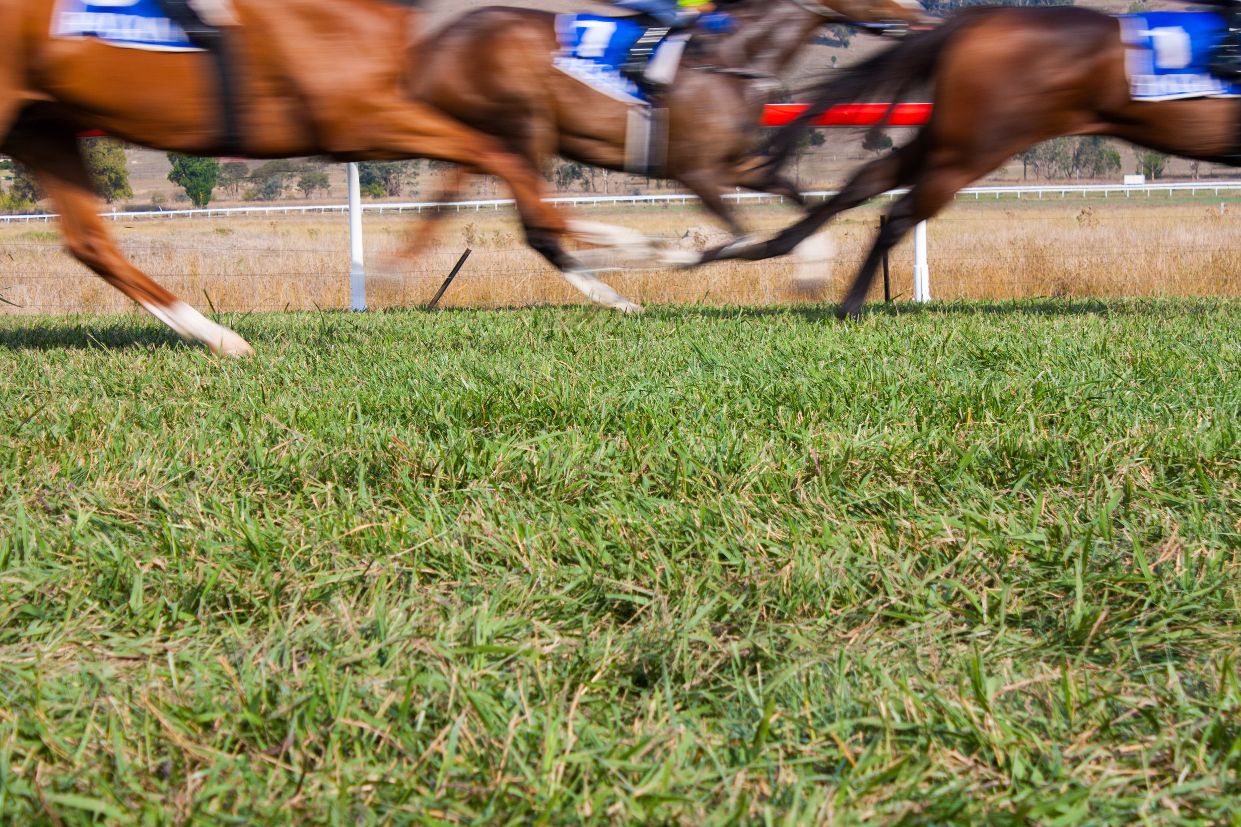
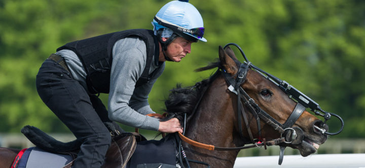
Osteochondrosis (OC) is a common lesion in young horses affecting the growing cartilage of the articular/epiphyseal complex of predisposed joints at specific predilection sites. In the young Thoroughbred, it commonly affects the stifles, hocks and fetlocks. As this condition has such important impact on soundness across many horse breeds, it is commonly discussed in Equine Veterinary Journal. Four recent articles covered causes of the disease, its genetic aspects, and a new and very practical approach to early diagnosis through ultrasound screening programs on stud farms.

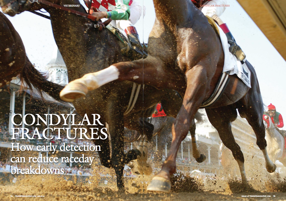
OC is a disease of joint cartilage. Cartilage covers the ends of bones in joints, and healthy cartilage is central to unrestricted joint movement. With OC, abnormal cartilage can be thickened, collapsed, or progress to cartilage flaps or osteochondral fragments separated from the subchondral bone leading to osteochondrosis dissecans (OCD). OC and OCD can be regarded as a spectrum rather than two discrete conditions.

Certain joints are prone to OC and OCD, and there is some variation between breeds on which joints have the highest prevalence. In Australian Thoroughbreds, 10% of yearlings had stifle OC, 8% had fetlock OC, and 6% had hock OC. The prevalence data may seem very high, but Thoroughbred breeders may take some comfort in learning that similar, and indeed slightly higher prevalences, are reported in the warmblood breeds, Standardbreds, and Scandinavian and French trotters. Heavy horse breeds have the highest prevalences.

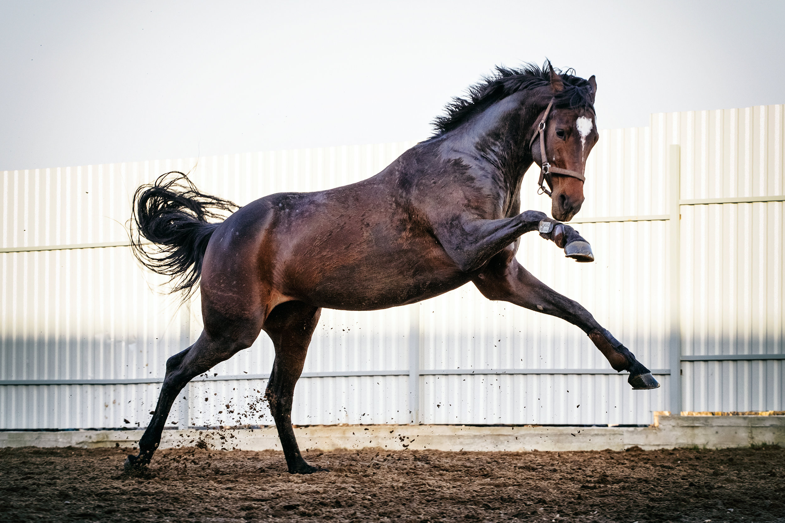
In an article discussing progress in OC/OCD research, Professor Rene Van Weeren concludes that the clinical relevance of OC is man made.  In feral horses, where there is no human influence on mating pairings, OC does occur but at much lower prevalence than in horse breeds selected for sports or racing. Similarly, in pony breeds where factors other than speed and size are desirable characteristics, OC is also rare. These facts suggest that sports and racehorse breeders have inadvertently introduced a trait for OC along with other desired traits. There is a strong link between height and OC, suggesting that one of the desired traits with unintended consequences is height. This is of particular relevance in sports horses: the Dutch warmblood has become taller at a rate of approximately 1 mm per year over the past decades, which might not seem much but it is still an inch in 25 years. Van Weeren points out that if the two-hands tall Eohippus or Hyracotherium and the browsing forest-dweller with which equine evolution started some 65 millions of years ago had evolved at this speed, the average horse would now have stood a staggering 40 miles at the withers.

Drs. Naccache, Metzger and Distal, based at the Institute for Animal Breeding and Genetics in Hannover, Germany, have worked extensively on heritability and the genetic aspects of OC in horses. Their work has shown that there is not one single gene involved. In fact, genes located on not less than 20 of the 33 chromosomes of the horse are relevant to OC.

These researchers use whole genome scanning—otherwise known as genome-wide association studies, or GWAS. This approach has only been possible since the equine genome was mapped. GWAS look at the entire genetic map to detect differences between subjects with and without a particular trait or disease. Millions of genetic variants can be read at the same time to identify genetic variants that are associated with the disease of interest. Based on the number of genetic markers already found in warmblood OC, it is unlikely that a simple single-gene test will prove to be useful for screening young Thoroughbreds for OC.

TO READ MORE —

BUY THIS ISSUE IN PRINT OR DOWNLOAD -

Breeders’ Cup 2018, issue 50 (PRINT)

$6.95

Pre Breeders’ Cup 2018, issue 50 (DOWNLOAD)

$3.99

WHY NOT SUBSCRIBE?

DON'T MISS OUT AND SUBSCRIBE TO RECEIVE THE NEXT FOUR ISSUES!

Print & Online Subscription

$24.95

European Research on Injuries in Flat Racing: Nature versus Nurture

By Kristien Verheyen and Sarah Rosanowski

Note: This research for this article, reprinted from European Trainer, was performed over a 14-year period in Great Britain and therefore only takes into account racing over turf and all-weather surfaces, but we feel that despite not including dirt statistics, the information is thought-provoking and of interest to our North American readers, especially with the increase in turf racing particularly in the U.S.

Musculoskeletal injuries are an inherent risk of horseracing, and they are the primary cause of Thoroughbreds failing to train and race, or even retiring altogether. In addition to the evident equine welfare concerns, racehorse injuries also have economic consequences and impact on jockey safety. The industry remains committed to investigating causes of injury and associated risk factors, which can inform strategies aimed at minimizing their occurrence. Advancements in methods of identification, management, and prevention of musculoskeletal disease and injury in Thoroughbreds and improved training and racing environments to enhance the safety, health, and wellbeing of racehorses have long been strategic priorities of the Horserace Betting Levy Board (HBLB)’s veterinary research funding program in Great Britain.

In 2014, the HBLB funded a research team at the Royal Veterinary College in London to undertake a detailed study of injuries and other veterinary events occurring in flat racehorses on race day. The purpose of the project was to establish causes of fatal and non-fatal injuries occurring in British flat racing and to examine associated risk factors. The research also set out to measure heritability of common injury types and conditions, and to investigate genetic and environmental correlations between injury and race performance.

The study team had access to detailed race and performance data from all Thoroughbreds racing on the flat in Great Britain over a 14-year study period from 2000 – 2013. These were then linked to veterinary reports of injury or conditions attended to by a veterinary surgeon on race day over the same time period, provided by the British Horseracing Authority (BHA). Finally, extensive pedigree data were added to enable investigation of heritability of race day injury and genetic correlations between injury types, and between injury and performance.

Descriptive findings

The final 14-year dataset included nearly 68,000 horses making over 800,000 starts in around 77,000 flat races. The majority of races -- 67% of them -- were run on the turf, with 33% of races taking place on all-weather tracks.

Just under 8,000 veterinary events were recorded over the study period, from which an incidence of nine events per 1000 starts was calculated. The most common incidents requiring veterinary attention on the racecourse were soft tissue injuries other than tendon and ligament injuries, e.g. wounds, lacerations, or muscle strains. Unspecified lameness and respiratory conditions were also common, accounting for around a fifth of veterinary reports each. Less than 10% of veterinary events had a fatal outcome, and the overall incidence of fatality was 0.8 per 1000 starts. Although bone injury was cited in only 14% of the veterinary reports overall, they accounted for the vast majority (77%) of the fatalities.

All-weather racing...

TO READ MORE --

BUY THIS ISSUE IN PRINT OR DOWNLOAD -

August - October 2018, issue 49 (PRINT)

$5.95

August - October 2018, issue 49 (DOWNLOAD)

$3.99

WHY NOT SUBSCRIBE?

DON'T MISS OUT AND SUBSCRIBE TO RECEIVE THE NEXT FOUR ISSUES!

Print & Online Subscription

$24.95

Castrating Racehorses: A routine procedure not without its pitfalls

By Tom O'Keeffe

A recent study published in the Equine Veterinary Journal assessed the routine procedure of gelding and the complications associated with this procedure.  The research was a retrospective study of horses castrated at the Sha Tin training complex in Hong Kong, between July 2007 and July 2012.

Hong Kong is a unique training and racing environment, and all horses training and racing there are imported, as there is no breeding in the region. Fillies are rarely imported. The majority of colts are castrated at some stage in their career, and open standing castration (OSC) is the method of choice by the vets of the Hong Kong Jockey Club (HKJC). Until now, nobody has looked at the prevalence of complications following castration of horses at the HKJC. This recently published study aimed to describe the prevalence and severity of complications in the 30 days following castration.

Reasons for gelding a racehorse in training

Most trainers perceive geldings as easier to train than colts, and if the horse has not shown enough ability for a stud career to beckon, there is little to lose by gelding.  In Hong Kong, due to the unique environment the horses live in, there is an added incentive to geld these horses sooner rather than later. Once gelded, their management becomes significantly more straightforward.

Castration Method Options

Three surgical techniques are commonly used for equine castration: 1) open, in which the parietal tunic surrounding the testicle is incised and, usually, retained; 2) closed, where the portion of the parietal tunic surrounding the testis and distal spermatic cord is removed, and 3) half closed, where an incision is made through the exposed parietal tunic at the cranial end of the testis or distal end of the spermatic cord allowing the testis and part of the spermatic vasculature to be prolapsed through the incision prior to removal.

In most cases, racehorse castration is done standing via the open technique under local anesthetic, with sedation and pain relief as necessary. The testicles and spermatic cords are first injected with local anesthetic to numb the region. Once the tissues are totally desensitized, a slash incision is made into the scrotum. The testicle is exteriorized, and it is removed with a surgical instrument called an emasculator. The emasculator has a set of interlocking crushing blades with a cutting blade placed at the bottom of the array. Once the testicular cord is clamped in the emasculator the testicle will usually fall off, but the cord is retained within the interlocking crushing blades for approximately one to two minutes. This creates trauma to the tissues, which causes them to swell once the crush is released, reducing blood flow. The second effect of the emasculators is for the blood to be held in position long enough to begin the clotting process, which carries on once the clamp is removed.

An alternative method of castration is to anesthetize the horse and carry out the procedure with the horse on its back, as a completely sterile operation in an operating room. This has the advantage of minimal post-castration swelling as there is no infection in the area, which can be a common problem with standing open castrations.  In horses who are cryptorchids (ridglings), which is when there is only one descended testicle in the scrotum, standard open standing castration is contraindicated. These horses require either castration under general anesthetic or testicle removal under standing surgery via laparoscopy (inserting a camera and instruments into the abdomen to remove testicle via a surgical incision).

Complications of Castration

As with all intrusive surgical procedures, there is the potential for things to go wrong. While the castration procedure is relatively straightforward, post-operative complications including excessive edema of the scrotum and surrounding tissues, infection and fever, hemorrhage, lameness, hydrocele formation, peritonitis, eventration, penile paralysis, scirrhous cord formation, and death have been recognized.

With castrations done under general anesthetic, there are all the attendant risks of putting a 1000lb animal on its back and up again. All anesthesia carries a risk of death in the horse. This has been calculated as approximately 1% in equine practice, and can be as low as 0.5% in the major well-equipped equine hospitals. In addition to this, occasional cases show prolonged bleeding after the surgery, which results in significant swelling that sometimes has to be resolved by opening the scrotal sac.

For standing castrations, some of the problems encountered include prolonged bleeding, which can occur irrespective of the length of time the cord has been clamped for. This can become serious enough to require a further surgery to identify the bleeding vessels and tie them off, but thankfully this is rare. Another rare complication is herniation of intestines through the potential space left in the inguinal canal with removal of the testicle. The intestines can either get trapped under the skin producing severe colic, or worse still, dangle out of the abdomen and become contaminated. This presents a very serious risk to the horse’s survival and requires immediate surgery to attempt to clean the exposed bowel and return it to the abdomen. Fortunately this is extremely rare in the Thoroughbred.

However, the most common complication is infection at the site of the castration. This procedure leaves an open wound and obviously the horse can lie down in bedding full of urine and feces on the same day it has been castrated, therefore potentially contaminating the open surgical site. Unfortunately many racehorses’ ability to be turned out in a paddock is often controlled by the training environment they reside in. Infection post-castration, and the added expense and lost training days associated with it, is a bugbear for trainers and vets, and this study reviews a common problem encountered worldwide.

Hong Kong Study....

TO READ MORE --

BUY THIS ISSUE IN PRINT OR DOWNLOAD -

August - October 2018, issue 49 (PRINT)

$5.95

August - October 2018, issue 49 (DOWNLOAD)

$3.99

Why not subscribe?

Don't miss out and subscribe to receive the next four issues!

Print & Online Subscription

$24.95

The Biome of the Lung

By Dr. Emmanuelle van Erck-Westergren, DVM, PhD, ECEIM

Of bugs and horses

A couple of weeks ago, I was on an emergency call to a training stable. Half of the horses had started coughing overnight, some had fever, and, as you’d expect when bad karma decides to make a point, the two stars of the premises, due to face their greatest challenge to date the following week, were dull and depressed. A thick and yellow discharge was oozing from their noses. It was not long before the barn became the typical scene of a bad strangles nightmare. The bacteria involved in strangles outbreaks are Streptococcus equi equi, highly aggressive and contagious germs that spread fast and cause disruption in days of training and mayhem in tight racing schedules.

So what inevitably comes to mind when you hear the words “germs” or “bacteria”? Certainly no nice and friendly terms. As veterinarians, we have been taught that microorganisms are responsible for an endless list of gruesome diseases and conditions: abscesses, pneumonia, septicemia ... you name it. All of these need to be identified and eradicated. Thank heavens we still have an arsenal of antibiotics to get rid of the damn bugs. But recent research in human “microbiome” is making us think twice, especially as we aim to hit hard and large with antibiotics.

Never alone

Your healthy and thriving self, and likewise your horse, hosts millions and trillions of bacteria. The “microbiota” is that incredibly large collection of microorganisms that have elected you and your horse as their permanent home. The microbiota is constituted not only by an extremely diverse variety of resident bacteria, but also by viruses, fungi, and yeasts that multiply in every part of your external and internal anatomy. The discovery of this prosperous microbial community has triggered fascinating new research. It has unveiled the unsuspected links that exist between health, disease, and the microbiota. In simple words, these microorganisms are vital to your strength and healthiness.


The microbes that compose the microbiota outnumber our own cells by 10 to one to the extent that the genetic information (or “genome”) you carry is over 99% microbial! And that is what researchers call the “microbiome” or “biome”: the collection of genetic information carried by your microbiota. Fortunately, the very large majority of bacteria is either beneficial or harmless, with only a very tiny fringe represented by potentially pathogenic strains. These microorganisms have evolved with us over thousands of years and the stability of this symbiotic ecosystem has important implications on our health status.

A gut feeling for biome

Research on the biome started with the study of the digestive ecosystem of mice. Researchers from Washington University showed that when they transplanted feces of obese mice in the gut of lean mice, these became obese, and vice versa. In other words, the composition of the gut biome could be said to influence morbid weight gain. Similar studies recently conducted in humans in the Netherlands came to the same conclusions.

We do not yet have all the keys to understanding the underlying processes, but we definitely know that gut microbes influence, amongst many other things, our metabolism, which is to say our capacity to process energy. This opened up tremendous possibilities to improving fitness and treating diseases. The research on the biome has since grown at an exponential rate, covering much larger areas. It was further discovered that problems in the gut biome leading to the proliferation of the wrong microorganisms were responsible for a very wide range of disorders or even chronic conditions that were far from the gut, such as arthritis, depression, and asthma.  The biome also seems to be critical in regulating our immune system to raise the alarm when enemies are identified and to modulate its response. The dramatic rise in autoimmune diseases could be a consequence of dietary changes that have disrupted our healthy microbiota.

TO READ MORE --

BUY THIS ISSUE IN PRINT OR DOWNLOAD -

August - October 2018, issue 49 (PRINT)

$5.95

August - October 2018, issue 49 (DOWNLOAD)

$3.99

Why not subscribe?

Don't miss out and subscribe to receive the next four issues!

Print & Online Subscription

$24.95


The ongoing effort to minimize the rate and impact of fractures

By Professor Celia Marr

In Thoroughbred racing, musculoskeletal injury is a major safety concern and is the leading reason for days lost to training.  Musculoskeletal injury is the greatest reason for horse turnover in racing stables, with financial implications for the owner and the racing industry. Injuries, particularly on race day, have an impact on public perception of racing.  

Upper limb and pelvis fractures are less common than lower limb fractures, but they can lead to fatalities. Reducing the overall prevalence of fractures is critical and, at the very least, improving the rate of detection of fractures in their early stages so the horse can be withdrawn from racing with a recoverable injury will be a big step forwards in racehorse welfare. Currently, we lack information on the outcomes following fracture, and an article recently published in the Equine Veterinary Journal (EVJ) from the veterinary team at the Hong Kong Jockey Club (HKJC) addressed this important knowledge gap.

Hong Kong Fracture Outcome Study

The HKJC veterinary team is in a unique position to carry out this work because their centralized and computerized database of clinical records, together with racing and retirement records, allows them to document follow-up, which is all but impossible elsewhere in the world. Dr. Leah McGlinchey, working with vets in Hong Kong and researchers from the Royal Veterinary College in London, reviewed clinical records from 2003 to 2014 to identify racehorses that suffered a fracture or fractures to the bones of the upper limb or the pelvis during training or racing, confirmed by nuclear scintigraphy, radiography, ultrasonography, or autopsy.

During these 11 racing seasons there were an average of 1468 horses in training each year, amounting to 102,785 starts over 8147 races, with 11% on dirt tracks and the rest on turf. McGlinchey found records of 108 racehorses that sustained 129 upper limb or pelvic fractures during 119 injury events. The most commonly fractured bone was the humerus at 50%, followed by the tibia at 30%. Nine horses sustained fractures that led to their immediate demise, five involving the scapula and four involving the humerus.

The majority (65%) of fractures occurred in training.  The overall incidence of upper limb and pelvic fractures in Hong Kong was three per 10,000 starts, and there were very similar incidences comparing both turf and dirt surfaces. The fatality rate due to upper limb and pelvic fracture was 0.8 per 10,000 starts. Over comparable time periods, race day upper limb and pelvic fracture rates were four per 10,000 starts in the UK, while race day fatalities were 1.8 per 10,000 starts in the UK and 1.9 per 10,000 in California; thus, rates of upper limb and pelvic fracture and fatality were lower in Hong Kong than in other racing jurisdictions.  Differences in training and racing regimens, racehorse surveillance, and veterinary care will vary across these racing centers, leading to different risk profiles for horses racing in these different locations.

This CT image taken during an autopsy, shows a comminuted fracture with multiple bone fragments.

All horses presented with lameness but importantly, the lameness grade was not necessarily very high. Indeed, 6.7% of the horses were Grade 1 of 5 lame, and 30.3% were Grade 2 of 5 lame, highlighting how important it is to rest and investigate mild new lameness. Typically, stress fractures cause acute lameness following fast work that soon eases in severity, and incipient fracture of the upper limb and pelvis can present as mild lameness with a subtle onset, which is all too easy to overlook. The degree of lameness associated with stress fracture is typically greatest when the scapula is involved and progressively less severe with the tibia, humerus, or radius. The diagnosis is all too obvious once severe, complete fracture has occurred.  In many cases, however, a diagnosis cannot be immediately made. Nuclear scintigraphy (also known as bone scanning) is the most sensitive method to detect stress fractures of the long bones and pelvis, although radiography and ultrasonography may also be useful.

Following fracture, all of the Hong Kong horses had a period of box rest followed by handwalking only. Three-quarters of these horses returned to racing a median of 169 days after sustaining the fracture; these made numerous starts, and 45 won.  In total, 59 horses had retired from training, 23 of which retired without returning to racing and, in 13 of these cases, with retirement directly attributable to the upper limb fracture.

TO READ MORE --

BUY THIS ISSUE IN PRINT OR DOWNLOAD -

August - October 2018, issue 49 (PRINT)

$5.95

August - October 2018, issue 49 (DOWNLOAD)

$3.99

Why not subscribe?

Don't miss out and subscribe to receive the next four issues!

Print & Online Subscription

$24.95

IF YOU LIKE THIS ARTICLE

WHY NOT SUBSCRIBE - OR ORDER THE CONTENT FROM THIS ISSUE IN PRINT?

Equine Herpes Virus-1: An elusive target

By Neil Bryant

Infectious diseases are not uncommon in racehorses in training, breeding stock, and pleasure horses. Some of the more serious diseases can be financially devastating to the animal’s owners and to the equine industry on the whole. Viruses belonging to the herpesvirus family cause some of the most well characterized equine infectious diseases, and the most problematic of these is equine herpesvirus 1 (EHV-1; species Equid alphaherpesvirus 1).

EHV-1 is ubiquitous in most horse populations in the world. It is responsible for major economic and welfare problems causing respiratory disease, neurological disease (mainly seen in adult horses), and abortion and neonatal foal death in pregnant mares. This was most notably highlighted by the multiple abortion outbreak recorded in Hertfordshire, England, between February and April 2016 in fully vaccinated animals (http://www.aht.org.uk/cms-display/interim-report16-april2.html). Studies have determined that EHV-1 is a common cause of abortion. Occasional cases have also been linked to EHV-4 infection, but this is much rarer and doesn’t account for episodes of multiple abortion, as is seen occasionally with EHV-1.

The virus

EHV-1 was first isolated from an equine abortion in the U.S. in the 1930s. At the time of first isolation the vets weren’t sure what it was, but they knew it was infectious. Subsequent genetic analysis much later led to the classification of the virus in the genus Varicellovirus (family Herpesviridae), together with its close relatives equine herpesvirus 4 (EHV-4; species Equid alphaherpesvirus 4) and equine herpesvirus 8 (EHV-8; species Equid alphaherpesvirus 8). Interestingly it is grouped with, and is therefore genetically similar to, the human herpesvirus responsible for chickenpox, the Varicella Zoster virus. Initial infection of horses was thought to occur around weaning, when virus-neutralizing antibodies transferred to the foal from the mare’s colostrum had declined enough to make them susceptible to infection. However, virus has been isolated from foals as young as seven days old with high antibody levels but without any significant clinical signs. Immunity to re-infection after primary infection is relatively short-lived, lasting between three-six months, but it is rare for naturally infected mares to abort in consecutive pregnancies.

Disease processes

The virus first enters the horses’ body via the respiratory tract, usually by direct contact with infected animals, contaminated surfaces, or equipment such as tack or veterinary instruments. Direct contact with infected aborted fetuses or placental tissues is also a major source of virus, which experience indicates can cause serious problems if they occur in open barns or large groups of horses.

Once the cells in the respiratory tract are infected, the virus spreads cell-to-cell until it finds its way to the regional lymph nodes, where it can infect white blood cells called lymphocytes. These lymphocytes circulate through the body carrying the virus with them, which is known as a “cell associated viraemia.” The infected lymphocytes can come into contact with and infect numerous cell types, including cells known as “endothelial cells,” which line the inside of blood vessels of the central nervous system and the pregnant uterus. With EHV-1 infection, these endothelial cells undergo an inflammatory response which can lead to bleeding, cell death, and blood clot formation, which in narrow veins disrupts blood supply. This process results in subsequent tissue damage and serious complications such as placental separation (occasionally with delivery of a virus-negative fetus) and/or leakage of virus across the separating placenta (most frequently with delivery of a virus-positive fetus).

Similar mechanisms play a role in neurological disease, a condition called equine herpesvirus myeloencephalopathy or EHM. This condition is also sometimes referred to as an equine stroke, as it is caused by the cellular inflammatory response rather than direct virus infection of nerve cells, which occurs with some other herpesviruses. Less serious clinical signs of infection can include fever, lethargy, inappetence, enlarged lymph nodes, and profuse clear nasal discharge, although not all infected animals will display clinical signs. Recently published work from the Irish Equine Centre has identified EHV-8 as also being occasionally responsible for abortions in mares.  Cases of EHV-8 abortion have also been detected retrospectively by the Animal Health Trust (AHT) among its pathology caseload, as this virus, which is genetically almost identical to EHV-1, triggers positive results in the EHV-1 tests. The frequency and clinical relevance of EHV-8 at this stage is unclear. Of 100 viruses presumed to be EHV-1 and whose genetic material were recently analyzed by the AHT, three were actually confirmed as EHV-8.

TO READ MORE --

BUY THIS ISSUE IN PRINT OR DOWNLOAD -

August - October 2018, issue 49 (PRINT)

$5.95

August - October 2018, issue 49 (DOWNLOAD)

$3.99

Why not subscribe?

Don't miss out and subscribe to receive the next four issues!

Print & Online Subscription

$24.95

IF YOU LIKE THIS ARTICLE

WHY NOT SUBSCRIBE - OR ORDER THE CONTENT FROM THIS ISSUE IN PRINT?

Tendon Function and Failure - Recent Advances

GET YOUR HANDS ON THIS ISSUE - VISIT OUR ONLINE SHOP HERE!

Are you a subscriber?

Don't miss out and subscribe to receive the print magazine now! CLICK HERE

IF YOU LIKE THIS ARTICLE

WHY NOT SUBSCRIBE - OR ORDER THE CONTENT FROM THIS ISSUE IN PRINT?

Epiduroscopy: An exciting window into back pain in horses

Back pain is a well-known cause of lameness, gait alterations, and poor performance in sport horses. Up to 25% of dressage horse owners report back problems in their animals, but not only sport horses are affected.

Although racehorses compete at a younger age than other equine athletes, they might suffer from back pain more often than we think, as autopsy studies have identified pathological changes in the back of the majority of examined young Thoroughbreds.

Until recently, it has been very difficult to investigate back pain and it is easy to overlook this as a cause of disappointing performance. A novel surgical technique that has recently been reported in Equine Veterinary Journal may change all this....

To read more of this article - subscribe now!

IF YOU LIKE THIS ARTICLE

WHY NOT SUBSCRIBE - OR ORDER THE CONTENT FROM THIS ISSUE IN PRINT?

The importance of identifying lower and upper limb lameness

In Thoroughbred racing, injuries to the limbs are a major welfare and safety concern, the leading reason for horses to be taken out of training. Lameness causes a high turnover in racing stables and, as many trainers know, it has huge financial implications for the owner, trainer, and the racing industry in general. Previous investigators have found that just over 50% of horses in training in England and Germany experience lameness during training, and approximately 20% of horses in the U.K. suffer lameness that prevents them from returning to training. With this amount of horses on lay-up, it can be difficult to run a profitable racing stable.

In addition to having an impact on the horse’s welfare, severe musculoskeletal injury poses a serious danger for riders, who are at risk when a horse sustains a catastrophic injury or suffers sudden death.  Researchers in the U.S. found that a jockey was 171 times more likely to be injured when a horse they were riding in a race died. In Thoroughbred racing, the most common life-threatening injury to horses involves fractures of bones in the fetlock. Therefore, the best way to improve safety and welfare of both horses and jockeys is to highlight risk factors for fractures in an attempt to prevent these catastrophic traumas.

To read more of this article - subscribe now!

IF YOU LIKE THIS ARTICLE

WHY NOT SUBSCRIBE - OR ORDER THE CONTENT FROM THIS ISSUE IN PRINT?

Laryngeal Problems - Hocus pocus or cutting edge science?

FIRST PUBLISHED IN NORTH AMERICAN TRAINER AUGUST - OCTOBER 2017 ISSUE 45

Click here to order this back issue!

PHOTO GALLERY

Recurrent laryngeal neuropathy (RLN) is the correct term for the condition better known as roaring or laryngeal hemiplegia. It is extremely common in Thoroughbreds and represents one of the major causes of poor performance or jockey-reported noise.

Because it is so important, many young horses are scoped at sales looking for laryngeal asymmetry to try to identify those that may be at risk of having this condition. But scoping at rest is fraught with difficulty – the larynx may be normal at rest only to show signs of weakness during exercise, so positive cases can be missed.

Conversely, tired young horses can have apparently poor laryngeal function at rest that in fact is of little significance. Many question whether subjecting foals and young horses to endoscopy at sales is reasonable, although in part this latter concern is reduced now that videoendoscopy is available.

Furthermore, the dynamic endoscopy, overground or on a treadmill, is widely accepted as the best way to evaluate horses suspected of having upper airway disorders leading to dynamic obstruction of the airway during exercise, a population that may or may not have obviously abnormal throats when examined at rest.

Nevertheless, there is a need for better tools to evaluate the larynx for clinical application and also to allow researchers to study the condition in more detail. Two recent studies published in Equine Veterinary Journal have addressed this issue..

To read more - subscribe here!

IF YOU LIKE THIS ARTICLE

WHY NOT SUBSCRIBE - OR ORDER THE CONTENT FROM THIS ISSUE IN PRINT?

Developing the two-year-old's foot

Lameness the Singapore way: Monitoring gait

IF YOU LIKE THIS ARTICLE

WHY NOT SUBSCRIBE - OR ORDER THE CONTENT FROM THIS ISSUE IN PRINT?

Compounding Pharmacies

IF YOU LIKE THIS ARTICLE

WHY NOT SUBSCRIBE - OR ORDER THE CONTENT FROM THIS ISSUE IN PRINT?

The benefits of Salt

IF YOU LIKE THIS ARTICLE

WHY NOT SUBSCRIBE - OR ORDER THE CONTENT FROM THIS ISSUE IN PRINT?

How rife is contamination on the backstretch?

2017 Media Eclipse Award winner

Click on the image to read this article online now!

Both first published in North American Trainer issue 43 - February to April '17

IF YOU LIKE THIS ARTICLE

WHY NOT SUBSCRIBE - OR ORDER THE CONTENT FROM THIS ISSUE IN PRINT?

Treating Fetlock Condylar Fractures

IF YOU LIKE THIS ARTICLE

WHY NOT SUBSCRIBE - OR ORDER THE CONTENT FROM THIS ISSUE IN PRINT?

Why do horses act up - the psychology of starting gate jitters / tail swishing / kicking out

CLICK ON THE IMAGE ABOVE TO READ ONLINE

First published in North American Trainer issue 42 - November '16 to January '17

IF YOU LIKE THIS ARTICLE

WHY NOT SUBSCRIBE - OR ORDER THE CONTENT FROM THIS ISSUE IN PRINT?

Testosterone and the thoroughbred

CLICK ON THE IMAGE ABOVE TO READ ONLINE

First published in North American Trainer issue 42 - November '16 to January '17

IF YOU LIKE THIS ARTICLE

WHY NOT SUBSCRIBE - OR ORDER THE CONTENT FROM THIS ISSUE IN PRINT?

Equine flu - research and development into the treatment of the disease

CLICK ON THE IMAGE ABOVE TO READ ONLINE

First published in North American Trainer issue 42 - November '16 to January '17

IF YOU LIKE THIS ARTICLE

WHY NOT SUBSCRIBE - OR ORDER THE CONTENT FROM THIS ISSUE IN PRINT?

International horse movements - disease risk

CLICK ON THE IMAGE ABOVE TO READ ONLINE

First published in North American Trainer issue 42 - November '16 to January '17

IF YOU LIKE THIS ARTICLE

WHY NOT SUBSCRIBE - OR ORDER THE CONTENT FROM THIS ISSUE IN PRINT?