Minimizing serious fractures of the racehorse fetlock
By VA Colgate, PHL Ramzan & CM Marr
Minimizing serious fractures of the racehorse fetlock
In March 2020, a symposium was held in Newmarket, UK, aiming to devise measures which could be used internationally to reduce the risk of catastrophic fracture associated with the fetlock joint. The meeting was supported by the Gerald Leigh Charitable Trust, the Beaufort Cottage Charitable Trust and the Jockey Club with additional contributions from a number of industry stakeholders. On the first day a panel of international experts made up of academic professors, Chris Whitton (Melbourne, Australia), Sue Stover (Davis, California), Chris Kawcak (Colorado), Tim Parkin (Glasgow) and Peter Muir (Wisconsin); experienced racehorse clinicians, Ryan Carpenter (Santa Anita) and Peter Ramzan (Newmarket); imaging experts, Sarah Powell (Newmarket) and Mathieu Spriet (Davis, California); and vets with experience in racing regulatory bodies, Scott Palmer (New York) and Chris Riggs (Hong Kong) joined forces to discuss risk assessment protocols, particularly those based on imaging features which might indicate increased risk of imminent fracture. This was followed by a wider discussion with a diverse invited audience of veterinary and industry stakeholders on how our current knowledge of fracture pathophysiology and risk factors for injury could be used to target risk assessment protocols. A report of the workshop outcomes was recently published in Equine Veterinary Journal.
The importance of risk reduction
With the ethics of the racing industry now in the public spotlight, there is recognition that together veterinary and horseracing professionals must strive to realise an improvement in equine injury rates. Intervention through risk profiling programmes, primarily based on training and racing metrics, has a proven track record; and the success of a racing risk management program in New York gives evidence that intervention can and will be successful.
The fetlock of the thoroughbred racehorse is subjected to very great loads during fast work and racing, and over the course of a training career this can result in cumulative changes in the bone underlying the articular cartilage (‘subchondral’ bone) that causes lameness and may in some circumstances lead to fracture. Fracture propagation involving the bones of the fetlock (cannon, pastern or proximal sesamoid bones) during fast work or racing can have catastrophic consequences, and while serious musculoskeletal injuries are a rare event when measured against race starts, there are obviously welfare and public interest imperatives to reduce the risk to racehorses even further. The dilemma that faces researchers and clinicians is that ‘fatigue’ injuries of the subchondral bone at some sites within the fetlock can be tolerated by many racehorses in training while others develop pathology that tips over into serious fracture. Differentiating horses at imminent risk of raceday fracture from those that are ‘safe’ to run has not proven particularly easy based on clinical grounds to date, and advances in diagnostic imaging offer great promise.
Profiling to inform risk assessment
Risk profiling examines the nature and levels of threat faced by an individual and seeks to define the likelihood of adverse events occurring. Catastrophic fracture is usually the end result of repetitive loading, but currently there are no techniques that can accurately determine that a bone is becoming fatigued until some degree of structural failure has actually occurred. However, diagnostic imaging has clear potential to provide information about pathological changes which indicate the early stages of structural damage.
Previous research has identified a plethora of epidemiological factors associated with increased risk of serious catastrophic musculoskeletal injury on the racetrack. These can be distilled into race, horse and management-related risk factors that could be combined in statistical models to enable identification of individual horses that may be at increased risk of injury.
In North America, the Equine Injury Database compiles fatal and non-fatal injury information for thoroughbred racing in North America. Since 2009, equine fatalities are down 23%; and important risk factors for injury have been identified, and this work has driven ongoing improvement.
The problem with all statistics-based models created so far for prediction of racehorse injury is that they have limited predictive ability due to the low prevalence of racetrack catastrophic events. If an event is very rare, and a predictive tool is not entirely accurate, many horses will be incorrectly flagged up as at increased risk. At the Newmarket Fetlock workshop, Prof Tim Parkin shared his work on a model which was based on data from over 2 million race starts and almost 4 million workout starts. Despite the large amount of data used to formulate the model, Tim Parkin suggested that if we had to choose between two horses starting in a race, this model would only correctly identify the horse about to sustain a fracture 65% of the time. Furthermore, the low prevalence of catastrophic injury means it will always be difficult to predict, regardless of which diagnostic procedure is employed.
Where do the solutions lie?
A radiograph showing a racing thoroughbred’s fetlock joint. The arrow points to a linear radiolucency in the parasagittal groove of the lower cannon bone—a finding that is frequently detectable before progression to serious injury.
One possible strategy to overcome the inherent challenge of predicting a rare event involves serial testing. Essentially with this approach, a sequence of tests is carried out to refine sub-populations of interest and thus improve the predictive ability of the specific tests applied. An additional consideration in the design of any such practical profiling system would have to be the ability to speedily come to a decision. For example, starting with a model based on racing and training metrics such as number of starts and length of lay-off periods, as well as information about the risk associated with any particular track or racing jurisdiction, entries could be screened to separate those that are not considered to be at increased risk of injury from a smaller sub-group of horses that warrant further evaluation and will progress to Phase 2. The second phase of screening would be something relatively simple. Although not yet available, there is hope that blood tests for bone biomarkers or genetic profiles could be used to further distil horses into a second sub-group. This second sub-group might then be subjected to more detailed veterinary examination, and from that a third sub-group, involving a very small and manageable number of horses flagged as potentially at increased risk, would undergo advanced imaging. The results of such diagnostic imaging would then allow vets to make evidence-based decisions on whether or not there is sufficient concern to prompt withdrawal of an individual from a specific race from a health and welfare perspective. Of course there are other considerations which limit the feasibility of such a system, including availability of diagnostic equipment and whether or not imaging can be quickly and safely performed without use of sedation or other drugs, which are prohibited near to a race start.
Diagnostic techniques for fetlock injury risk profiling
Currently there is no clear consensus on the interpretation of images from all diagnostic imaging modalities, and important areas of uncertainty exist. Although a range of imaging modalities are available, each has its own strengths and weaknesses, and advances in technology currently outstrip our accumulation of published evidence on which to base interpretation of the images obtained.
Interpretation is easy when the imaging modality shows an unequivocal fracture such as a short fissure in a cannon bone. Here the decision is simple: the horse has a fracture and must stop exercising. Many cases, however, demonstrate less clearly defined changes that may be associated with bone fatigue injury.
Currently radiography remains the most important imaging modality in fetlock bone risk assessment. With wide availability and the knowledge gained by more advanced imaging techniques refining the most appropriate projections to use; radiography represents a relatively untapped resource that through education of primary care vets could immediately have a profound impact on injury mitigation. The most suitable projection with which to detect prodromal condylar fracture pathology in the equine distal limb is the flexed dorsopalmar (forelimb) or plantarodorsal (hindlimb) projection. On this projection, focal radiolucency in the parasagittal groove, whether well or poorly defined, with or without increased radio-opacity in the surrounding bone, should be considered representative of fracture pathology unless evidence from other diagnostic imaging modalities demonstrates otherwise.
Computed Tomography (CT) excels at identification of structural changes and is better than radiography at showing very small fissures in the bone. However, additional research is needed to determine specific criteria for interpretation of the significance of small lesions in the parasagittal groove with respect to imminent risk of serious injury. There are good indications that fissure lesion size and proximal sesamoid bone volumetric measurements have the potential to be useful criteria for prediction of condylar and proximal sesamoid bone fractures respectively. With technological advancement, it is likely that CT will be more widely used in quantitative risk analysis in the future.
Magnetic Resonance Imaging (MRI) has the ability to detect alterations in the fluid content of bones, which allows assessment of acute, active changes. Indeed standing, low-field MRI has been shown to be capable of detecting bone abnormalities not readily identifiable on radiography and has been successfully used for injury mitigation in racehorse practice for some time. However, when used for evaluation of cartilage and subchondral bone lesions, there is a relatively high likelihood of false positive results.
PET is the most recent advance in diagnostic imaging. It is being developed in California and, when combined with CT, provides information on bone activity and structure. In these three images of the same fetlock, from different aspects, the orange spots indicate increased activity in the proximal sesamoid bone, which is a potential precursor to more serious injury.
Image courtesy of Dr M. Spriet, University of California, Davis.
CLICK HERE to return to issue contents
ISSUE 57 (PRINT)
$6.95
ISSUE 57 (DIGITAL)
$3.99
WHY NOT SUBSCRIBE?
DON'T MISS OUT AND SUBSCRIBE TO RECEIVE THE NEXT FOUR ISSUES!
Four issue subscription - ONLY $24.95
Thoroughbred Sales Assessment
By Tom O’Keeffe
The Beaufort Cottage Educational Trust Gerald Leigh Memorial Lectures took place this year at the National Horseracing Museum in Newmarket and a host of international and local veterinary specialists and industry leaders were present to discuss the veterinary aspects of the sales selection of the thoroughbred.
Gerald Leigh was a prominent breeder and racehorse owner until his death in 2002; and his friend and vet Nick Wingfield Digby opened the seminar and introduced the speakers. The Gerald Leigh Charitable Trust has established this annual lecture series to provide a platform for veterinary topics relating to the thoroughbred to be discussed amongst vets and prominent members of the industry.
Sir Mark Prescott described his take on the sales process and some of the changes he has noted since his early involvement in the industry. He recalled how the first Horses in Training sale he attended had only 186 horses. In those early days, his role was to sneak around the sales ground stables late at night on the lookout for crib biters. Back then, there was no option to return horses after sale, and as a result, trainers preferred to buy horses from studs they were familiar with—a policy Sir Mark still follows to this day.
Sir Mark went on to explain that he believes strongly that the manner in which an animal is reared has a strong bearing on their ability to perform at a later date. Sir Mark also mentioned that horses can cope with many conformational faults nowadays that would have been deemed unacceptable in his early years. He attributed this to improvements in ground conditions, such as watering and all-weather surfaces.
Mike Shepherd, MRCVS, of Rossdales Equine Practice in Newmarket had been tasked with describing and discussing the sales examination from a veterinary viewpoint and in particular attempting to define what vets are trying to achieve in this process.
Shepherd’s key message was that the physical exam is the cornerstone of any veterinary evaluation. A vet examining a horse on the sales ground is not a guarantee that the horse will never have an issue—there is no crystal ball. Owners and trainers should be aware there are several limitations of the vetting process, and it is helpful to think in terms of a “pre-bid inspection” rather than a “pre-purchase examination”. The horse is away from its home environment, and this puts a lot of stress on the animal. In most cases, pre-purchase exercise is not possible, so conditions that are only apparent when the horse is exercising and in training may go undetected.
Time is a major challenge, with both vendors and prospective purchasers pushing for everything to be done as quickly as possible. A busy sales vet may have a long list of horses to examine, and information on each must be transferred to their client coherently and clearly—all before the horse is presented for sale. It can be challenging to acquire a detailed veterinary history. Previous surgeries, medication and vices displayed by the animal ought to be reported, but in many cases the person with the horse is not in a position to accurately answer questions on longer-term history.
At Sales, ultrasonography of the heart (echocardiography) can be used to estimate heart size.
Everyone involved—the vendor, the prospective purchaser, and the auction house—wants the process to go ahead. The horse to be bought/sold and the vet can be seen as a stumbling block. Prospective purchasers may want the horse to be examined clinically, its laryngeal function examined by endoscope, radiographs of the horse’s limbs either reviewed or taken, ultrasound examinations of their soft tissue structures and heart performed. The role of the vet is to help the purchaser evaluate all this information and make an evidence-based decision on whether to purchase the horse.
Examining vets can face conflicts of interest when examining horses that are under the ownership or care of one of their clients. Shepherd explained how Rossdales, and some other practices involved in sales work, have a protocol that an examining vet will not perform a vetting on a horse in the care of one of their own clients, and will disclose to the prospective purchaser if the vendor is a client of the practice. It is crucial to avoid working for both buyer and seller as a conflict of interest becomes unavoidable.
It is also essential that the vet understands exactly what the horse is expected to do following the sale. Thoroughbred horses in flat racing have short timescale targets and, as a result, certain parts of the examination carry more weight than others. For example, the knees and fetlock joints are commonly implicated in lameness in flat racehorses; thus particular attention must be paid to these joints when examining yearlings. Soft tissue injuries are impactful in all young thoroughbreds, but there is a particular emphasis on tendon integrity in the National Hunt racehorse because career-threatening tendon injuries are particularly prevalent in these horses. When evaluating potential broodmares, good feet are very relevant, and overall conformation is particularly important if the aim is to breed to sell.
Vetting horses for clients aiming to pin hook their purchases places different requirements on the examining vet. These horses need to be able to cope with the preparation required for another sale, and they must also stand up to the scrutiny of vets at a later sale. The horse’s walk and conformation rank high in the foal/ yearling stage but may be judged to be less significant if the horse breezes in a fast time at a breeze up sale.
It is also critical that purchasers recognise that many of the common veterinary issues encountered in training are not detectable at the Sales stage. For example, subchondral fetlock pain (bone bruising), which is common in a large subset of thoroughbreds in training, is not accurately practicable in young thoroughbred prior entering training.
For assessing laryngeal function, there is often concern that this may be too subjective. Dr Justin Perkins, of the Royal Veterinary College, has shown good agreement on endoscopic grading between experienced vets. However, horses’ laryngeal scores vary significantly day to day, and more worrying in the sales setting, one horse examined several times on the same day can have different scope grades. The gold standard in assessing the horse laryngeal function is an overground exercising endoscopy. This is not feasible on sales grounds, and purchasers should be aware that the limited purpose of a resting laryngeal scope is to exclude specific serious hereditary conditions and not to rule out problems which are only evident during exercise. Dorsal displacement of the soft palate cannot be accurately predicted at rest. Similarly, exercise-induced pulmonary haemorrhage or bleeding can be career limiting, yet once again in the juvenile thoroughbred, there is no way to predict this condition before the horse enters training.
Some conditions can affect some individuals yet be of little consequence in others. An example is kissing spines (impinging spinous processes), and whilst it can be a clinical issue in some individuals, it is commonly encountered in normal horses and therefore is very easy to over-interpret its significance.
The Hong Kong vetting process is considered the gold standard in terms of assessing and examining a racehorse in training. Shepherd highlighted that some new rules have recently been introduced. There are now guidelines on measuring tendons, and horses with a superficial digital flexor tendon cross-sectional area of greater than 1.6cm2 are not allowed to be imported into Hong Kong because of a potential increased risk of tendon injury.
The use of medication in horses at the sales ground and early in their life is currently an area of controversy. The concern is that there may be longer-term impact. Penalties associated with the use of anabolic steroids are well documented: the horse will be placed under a lifetime ban if these drugs have been used. The British Horseracing Authority will also place a lifetime ban on horses which have received bisphosphonates. Potentially, these drugs may have been given without the knowledge of the vendor, therefore client education and sales conditions will have to be adapted unless a sensible compromise can be reached to prevent a high-profile embarrassment for the authorities.
TO READ MORE - BUY THIS ISSUE IN PRINT OR DOWNLOAD -
ISSUE 54 (PRINT)
$6.95
ISSUE 54 (DIGITAL)
$3.99
WHY NOT SUBSCRIBE?
DON'T MISS OUT AND SUBSCRIBE TO RECEIVE THE NEXT FOUR ISSUES!
Four issue subscription - PRINT & ONLINE - ONLY $24.95
Wobbler Syndrome and the thoroughbred
By Celia M. Marr
Wobbler syndrome, or spinal ataxia, affects around 2% of young Thoroughbreds. In Europe, the most common cause relates to narrowing of the cervical vertebral canal in combination with malformation of the cervical vertebrae. Narrowing in medical terminology is “stenosis,” and “myelopathy” implies pathology of the nervous tissue, hence the other name often used for this condition is cervical vertebral stenotic myelopathy (CVSM).
Wobbler syndrome was the topic of this summer’s Gerald Leigh Memorial Lectures, an event held at Palace House, Newmarket. Gerald Leigh was a very successful owner/breeder, and these annual lectures, now in their second year, honor Mr. Leigh's passion for the Thoroughbred and its health and welfare. The lectures are attended by vets, breeders and trainers, and this year because of the importance and impact of wobbler syndrome on Thoroughbred health, several individuals involved in Thoroughbred insurance were also able to participate.
Dr. Steve Reed, of Rood and Riddle Equine Hospital, Kentucky and international leader in the field of equine neurology gave an overview of wobbler syndrome. Affected horses are ataxic, which means that they have lost the unconscious mechanisms that control their limb position and movement. Young horses with CVSM will generally present for acute onset of ataxia or gait abnormalities, however, mild ataxia and clumsiness may often go unnoticed. Trainers often report affected horses are growing rapidly, well-fed, and large for their age. It is common for riders to describe an ataxic horse as weak or clumsy. Sometimes, a horse that has been training normally will suddenly become profoundly affected, losing coordination and walking as though they were drunk, or in the most severe cases stumbling and falling. Neurological deficits are present in all four limbs, and are usually, but not always more noticeable in the hindlimbs than the forelimbs. In horses with significant degenerative joint disease, lateral compression of the spinal cord may lead to asymmetry of the clinical signs.
When the horse is standing still, it may adopt an abnormal wide-based stance or have abnormal limb placement, and delayed positioning reflexes. At the walk, the CVSM horse’s forelimbs and hindlimbs may not be moving on the same track, and there can be exaggerated movement of the hindlimbs when the horse is circled. Detailed physical examination may reveal abrasions around the heels and inner aspect of the forelimbs due to interference and short, squared hooves due to toe-dragging. Many young horses affected with CVSM have concurrent signs of developmental orthopedic disease such as physitis or physeal enlargement of the long bones, joint effusion secondary to osteochondrosis, and flexural limb deformities.
Radiography is generally the first tool which is used to diagnose CVSM. Lateral radiographs of the cervical vertebrae, obtained in the standing horse, reveal some or all of five characteristic bony malformations of the cervical vertebrae: (1) “flare” of the caudal vertebral epiphysis of the vertebral body, (2) abnormal ossification of the articular processes, (3) malalignment between adjacent vertebrae, (4) extension of the dorsal laminae, and (5) degenerative joint disease of the articular processes. Radiographs are also measured to document the ratio between the spinal canal and the adjacent bones and identify sites where the spinal canal is narrowed.
Dr. Reed also highlighted myelography as the currently most definitive tool to confirm diagnosis of focal spinal cord compression and to identify the location and number of lesions. The experts presenting at the Gerald Leigh Memorial Lectures agreed that myelography is essential if surgical treatment is pursued. However, an important difference between the U.S. and Europe was highlighted by Prof. Richard Piercy of the Royal Veterinary College, University of London. In Europe, protozoal infection is very rare, whereas in U.S., equine protozoal myeloencephalitis can cause similar clinical signs to CVSM. Protozoal myeloencephalitis is diagnosed by laboratory testing of the cerebral spinal fluid, but there is also a need to rule out CVSM. Therefore, spinal fluid analysis and myelography tends to be performed more often in the U.S. Prof. Piercy pointed out that in the absence of this condition, vets in Europe are often more confident to reach a definitive diagnosis of CVSM based on clinical signs and standing lateral radiographs.
Figure 4
Dr. Reed went on to discuss medical therapy in horses with CVSM, which is aimed at reducing cell swelling and edema formation with subsequent reduction of the compression on the spinal cord. In the immediate period following an acute onset of neurologic disease, anti-inflammatory therapy is important. Thereafter, depending on the type of CVSM and the age of the horse, different therapeutic options exist. A diet that is aimed at reducing protein and carbohydrate intake and, thus, reducing growth will allow the vertebral canal to “catch up”. The three most important nutritional factors appear to be excessive dietary digestible energy, excessive dietary phosphorus, and dietary copper deficiency. However, it is important that restricted diets are carefully managed with professional supervision. Dietary supplementation with vitamin E/selenium is also recommended. In adult horses, the options for medical therapy are restricted to stabilizing a horse with acute neurological deterioration and injecting the articular joints in an attempt to reduce soft tissue swelling and stabilize or prevent further bony proliferation.
The aim of surgical treatment is to stop the repetitive trauma to the spinal cord, which is caused by narrowing of the vertebral canal, and thereby, to allow the inflammation in and around the spinal cord to resolve. Surgical treatment of CVSM is controversial, mainly due to concerns regarding safety of the horse after surgery and potential heritability of the disease. Ventral interbody fusion through the use of a stainless steel “basket” is currently the most commonly used surgery for CVSM. The prognosis of horses following surgical treatment depends on the age of the horse, the grade of neurological deficits that were present prior to surgery, the time the horse has demonstrated neurologic disease for, the number of compressed sites, the severity of the lesions, and the post-operative complications encountered. Following surgery, an improvement of 1-2 out of 5 grades is expected, although some affected horses improve more than 3 grades.
Whether horses are treated medically, surgically or not treated (i.e., just turned out), the response and the prognosis depend on the age of the horse, the severity of the neurological deficits, the duration of neurological signs, and what level of performance is expected from the horse. Without treatment the prognosis in all types of CVSM is poor, as there is continued damage to the cervical spinal cord with an increasing chance of severe cord damage.
TO READ MORE —
BUY THIS ISSUE IN PRINT OR DOWNLOAD -
Spring 2019, issue 51 (PRINT)
$6.95
Spring 2019, issue 51 (DOWNLOAD)
$3.99
WHY NOT SUBSCRIBE?
DON'T MISS OUT AND SUBSCRIBE TO RECEIVE THE NEXT FOUR ISSUES!
Print & Online Subscription
$24.95
Ulcer medication: are the products different?
By Celia M. Marr
Stomach Ulcers Are Not All the Same
Racehorse trainers and their vets first began to be aware of stomach ulcers over 20 years ago. The reasons why we became aware of ulcers are related to technological advances, which produced endoscopes long enough to get into the equine stomach. At that time, scopes were typically about eight feet long and were most effective in examining the upper area of the stomach, which is called the squamous portion. Once this technology became available, it was quickly appreciated that it is very common for racehorses to have ulcers in the squamous portion of the stomach.
Figure 1
The equine stomach has two main areas: the squamous portion and the glandular portion. The stomach sits more or less in the middle of the horse, immediately behind the diaphragm and in front of and above the large colon. Imagine the stomach as a large balloon with the esophagus—the gullet—entering halfway up the front side and slightly to the left of the balloon-shaped stomach and the exit point also coming out the front side but slightly lower and to the right side. The tissue around the exit—the pylorus—and the lower one-third, the glandular portion, has a completely different lining to the top two-thirds, the squamous portion.
The stomach produces acid to start the digestive process. Ulceration of the squamous portion is caused by this acid. Like the human esophagus, the lining of the squamous portion has very limited defenses against acid. But, the acid is actually produced in the lower, glandular portion. The position of the stomach is between the diaphragm, which moves backwards as the horse breathes in and the heavy large intestine which tends to push forwards as the horse moves. During exercise, liquid acid produced at the bottom of the stomach is squeezed upwards onto the vulnerable squamous lining. It makes sense then that the medications used to treat squamous ulcers are aimed at blocking acid production.
Lesions in the glandular portion of the stomach are less common than squamous ulcers. The acid-producing glandular portion has natural defenses against acid damage including a layer of mucus and local production of buffering compounds. At this point, we actually know relatively little about the causes of glandular disease, but it is becoming increasingly obvious that disease in the glandular portion is very different from squamous disease. Often, it is more difficult to treat.
Figure 2
Stomach ulcers can cause a wide range of clinical signs. Some horses seem relatively unaffected by fairly severe ulcers, but other horses will often been off their feed, lose weight, and have poor coat quality. Some will show signs of abdominal discomfort, particularly shortly after eating. Other horses may be irritable—they can grind their teeth or they may resent being girthed. Additional signs of pain include an anxious facial expression, with ears back and clenching of the jaw and facial muscles and a tendency to stand with their head carried a little low.
Assessing Ulcers
Ulcers can only be diagnosed with endoscopy. A grading system has been established for squamous ulcers, which is useful in making an initial assessment and in documenting response to treatment.
Grade 0 = normal intact squamous lining
Grade 1 = mild patches of reddening
Grade 2 = small single or multiple ulcers
Grade 3 = large single or multiple ulcers
Grade 4 = extensive, often merging with areas of deep ulceration
Although it is used for research purposes, this grading system does not translate very well to glandular ulcers where typically, lesions are described in terms of their severity (mild, moderate or severe), distribution (focal, multifocal or diffuse), thickness (flat, depressed, raised or nodular) and appearance (reddening, hemorrhagic or fibrinosuppurative). Fibrinosuppurative suggests that inflammatory cells or pus has formed in the area. Focal reddening can be quite common in the absence of any clinical signs. Nodular and fibrinosuppurative lesions may be more difficult to treat than flat or reddened lesions. Where the significance of lesions is questionable, it can be helpful to treat the ulcers and repeat the endoscopic examination to determine whether the clinical signs resolve along with the ulcers.
Medications for Squamous Ulcers
Because of the prevalence and importance of gastric ulcers, Equine Veterinary Journal publishes numerous research articles seeking to optimize treatment. The most commonly used drug for treatment of squamous ulcers is omeprazole. A key feature of products for horses is that the drug must be buffered in order to reach the small intestine, from where it is absorbed into the bloodstream in order to be effective. Until recently only one brand was available, but there are now several preparations on the market and researchers have been seeking to show whether new medicines are as effective as the original brand. There is limited information comparing the new products, and this information is essential to determine whether the new, and often cheaper, products should be used. A team of researchers formed from Charles Sturt University in Australia and Louisiana State University has compared two omeprazole products given orally. A study reported by Dr. Raidal and her colleagues, showed that not only were plasma concentrations of omeprazole similar with both products, but importantly, the research also showed that gastric pH was similar with both products, and both products reduced summed squamous ulcer scores. Both the products tested in this trial are available in Australia and, although products on the market in other regions have been shown to achieve similar plasma concentrations and it is therefore reasonable to assume that they will be beneficial, as yet, not all of them have been tested to show whether products are equally effective in reducing ulcer scores in large-scale clinical trials. Trainers should discuss this issue with their vets when deciding which specific ulcer product they plan to use in their horses.
Avoiding drugs altogether and replacing this with a natural remedy is appealing. There is a plethora of nutraceuticals around and anecdotally, horse owners believe they may be effective. One such option is aloe vera that has antioxidant, anti-inflammatory and mucus stimulatory effects, which might be beneficial in a horse’s stomach. Another research group from Australia, this time based in Adelaide, has looked at the effectiveness of aloe vera in treating squamous ulcers and found that, although 56% of horses treated with aloe vera improved and 17% resolved after 28 days, this is compared to 85% improvement and 75% resolution in horses given omeprazole. Therefore, Dr. Bush and her colleagues from Adelaide concluded treatment with aloe vera was inferior to treatment with omeprazole.
Medications for Glandular Ulcers….
TO READ MORE —
BUY THIS ISSUE IN PRINT OR DOWNLOAD -
Spring 2019, issue 51 (PRINT)
$6.95
Spring 2019, issue 51 (DOWNLOAD)
$3.99
WHY NOT SUBSCRIBE?
DON'T MISS OUT AND SUBSCRIBE TO RECEIVE THE NEXT FOUR ISSUES!
Print & Online Subscription
$24.95
Osteochondrosis - genetic causes and early diagnosis
By Celia M. Marr
Osteochondrosis (OC) is a common lesion in young horses affecting the growing cartilage of the articular/epiphyseal complex of predisposed joints at specific predilection sites. In the young Thoroughbred, it commonly affects the stifles, hocks and fetlocks. As this condition has such important impact on soundness across many horse breeds, it is commonly discussed in Equine Veterinary Journal. Four recent articles covered causes of the disease, its genetic aspects, and a new and very practical approach to early diagnosis through ultrasound screening programs on stud farms.
OC is a disease of joint cartilage. Cartilage covers the ends of bones in joints, and healthy cartilage is central to unrestricted joint movement. With OC, abnormal cartilage can be thickened, collapsed, or progress to cartilage flaps or osteochondral fragments separated from the subchondral bone leading to osteochondrosis dissecans (OCD). OC and OCD can be regarded as a spectrum rather than two discrete conditions.
Certain joints are prone to OC and OCD, and there is some variation between breeds on which joints have the highest prevalence. In Australian Thoroughbreds, 10% of yearlings had stifle OC, 8% had fetlock OC, and 6% had hock OC. The prevalence data may seem very high, but Thoroughbred breeders may take some comfort in learning that similar, and indeed slightly higher prevalences, are reported in the warmblood breeds, Standardbreds, and Scandinavian and French trotters. Heavy horse breeds have the highest prevalences.
In an article discussing progress in OC/OCD research, Professor Rene Van Weeren concludes that the clinical relevance of OC is man made. In feral horses, where there is no human influence on mating pairings, OC does occur but at much lower prevalence than in horse breeds selected for sports or racing. Similarly, in pony breeds where factors other than speed and size are desirable characteristics, OC is also rare. These facts suggest that sports and racehorse breeders have inadvertently introduced a trait for OC along with other desired traits. There is a strong link between height and OC, suggesting that one of the desired traits with unintended consequences is height. This is of particular relevance in sports horses: the Dutch warmblood has become taller at a rate of approximately 1 mm per year over the past decades, which might not seem much but it is still an inch in 25 years. Van Weeren points out that if the two-hands tall Eohippus or Hyracotherium and the browsing forest-dweller with which equine evolution started some 65 millions of years ago had evolved at this speed, the average horse would now have stood a staggering 40 miles at the withers.
Drs. Naccache, Metzger and Distal, based at the Institute for Animal Breeding and Genetics in Hannover, Germany, have worked extensively on heritability and the genetic aspects of OC in horses. Their work has shown that there is not one single gene involved. In fact, genes located on not less than 20 of the 33 chromosomes of the horse are relevant to OC.
These researchers use whole genome scanning—otherwise known as genome-wide association studies, or GWAS. This approach has only been possible since the equine genome was mapped. GWAS look at the entire genetic map to detect differences between subjects with and without a particular trait or disease. Millions of genetic variants can be read at the same time to identify genetic variants that are associated with the disease of interest. Based on the number of genetic markers already found in warmblood OC, it is unlikely that a simple single-gene test will prove to be useful for screening young Thoroughbreds for OC.
TO READ MORE —
BUY THIS ISSUE IN PRINT OR DOWNLOAD -
Breeders’ Cup 2018, issue 50 (PRINT)
$6.95
Pre Breeders’ Cup 2018, issue 50 (DOWNLOAD)
$3.99
WHY NOT SUBSCRIBE?
DON'T MISS OUT AND SUBSCRIBE TO RECEIVE THE NEXT FOUR ISSUES!
Print & Online Subscription
$24.95
The ongoing effort to minimize the rate and impact of fractures
By Professor Celia Marr
In Thoroughbred racing, musculoskeletal injury is a major safety concern and is the leading reason for days lost to training. Musculoskeletal injury is the greatest reason for horse turnover in racing stables, with financial implications for the owner and the racing industry. Injuries, particularly on race day, have an impact on public perception of racing.
Upper limb and pelvis fractures are less common than lower limb fractures, but they can lead to fatalities. Reducing the overall prevalence of fractures is critical and, at the very least, improving the rate of detection of fractures in their early stages so the horse can be withdrawn from racing with a recoverable injury will be a big step forwards in racehorse welfare. Currently, we lack information on the outcomes following fracture, and an article recently published in the Equine Veterinary Journal (EVJ) from the veterinary team at the Hong Kong Jockey Club (HKJC) addressed this important knowledge gap.
Hong Kong Fracture Outcome Study
The HKJC veterinary team is in a unique position to carry out this work because their centralized and computerized database of clinical records, together with racing and retirement records, allows them to document follow-up, which is all but impossible elsewhere in the world. Dr. Leah McGlinchey, working with vets in Hong Kong and researchers from the Royal Veterinary College in London, reviewed clinical records from 2003 to 2014 to identify racehorses that suffered a fracture or fractures to the bones of the upper limb or the pelvis during training or racing, confirmed by nuclear scintigraphy, radiography, ultrasonography, or autopsy.
During these 11 racing seasons there were an average of 1468 horses in training each year, amounting to 102,785 starts over 8147 races, with 11% on dirt tracks and the rest on turf. McGlinchey found records of 108 racehorses that sustained 129 upper limb or pelvic fractures during 119 injury events. The most commonly fractured bone was the humerus at 50%, followed by the tibia at 30%. Nine horses sustained fractures that led to their immediate demise, five involving the scapula and four involving the humerus.
The majority (65%) of fractures occurred in training. The overall incidence of upper limb and pelvic fractures in Hong Kong was three per 10,000 starts, and there were very similar incidences comparing both turf and dirt surfaces. The fatality rate due to upper limb and pelvic fracture was 0.8 per 10,000 starts. Over comparable time periods, race day upper limb and pelvic fracture rates were four per 10,000 starts in the UK, while race day fatalities were 1.8 per 10,000 starts in the UK and 1.9 per 10,000 in California; thus, rates of upper limb and pelvic fracture and fatality were lower in Hong Kong than in other racing jurisdictions. Differences in training and racing regimens, racehorse surveillance, and veterinary care will vary across these racing centers, leading to different risk profiles for horses racing in these different locations.
This CT image taken during an autopsy, shows a comminuted fracture with multiple bone fragments.
All horses presented with lameness but importantly, the lameness grade was not necessarily very high. Indeed, 6.7% of the horses were Grade 1 of 5 lame, and 30.3% were Grade 2 of 5 lame, highlighting how important it is to rest and investigate mild new lameness. Typically, stress fractures cause acute lameness following fast work that soon eases in severity, and incipient fracture of the upper limb and pelvis can present as mild lameness with a subtle onset, which is all too easy to overlook. The degree of lameness associated with stress fracture is typically greatest when the scapula is involved and progressively less severe with the tibia, humerus, or radius. The diagnosis is all too obvious once severe, complete fracture has occurred. In many cases, however, a diagnosis cannot be immediately made. Nuclear scintigraphy (also known as bone scanning) is the most sensitive method to detect stress fractures of the long bones and pelvis, although radiography and ultrasonography may also be useful.
Following fracture, all of the Hong Kong horses had a period of box rest followed by handwalking only. Three-quarters of these horses returned to racing a median of 169 days after sustaining the fracture; these made numerous starts, and 45 won. In total, 59 horses had retired from training, 23 of which retired without returning to racing and, in 13 of these cases, with retirement directly attributable to the upper limb fracture.
TO READ MORE --
BUY THIS ISSUE IN PRINT OR DOWNLOAD -
August - October 2018, issue 49 (PRINT)
$5.95
August - October 2018, issue 49 (DOWNLOAD)
$3.99
Why not subscribe?
Don't miss out and subscribe to receive the next four issues!
Print & Online Subscription
$24.95
Laryngeal Problems - Hocus pocus or cutting edge science?
FIRST PUBLISHED IN NORTH AMERICAN TRAINER AUGUST - OCTOBER 2017 ISSUE 45
Click here to order this back issue!
PHOTO GALLERY
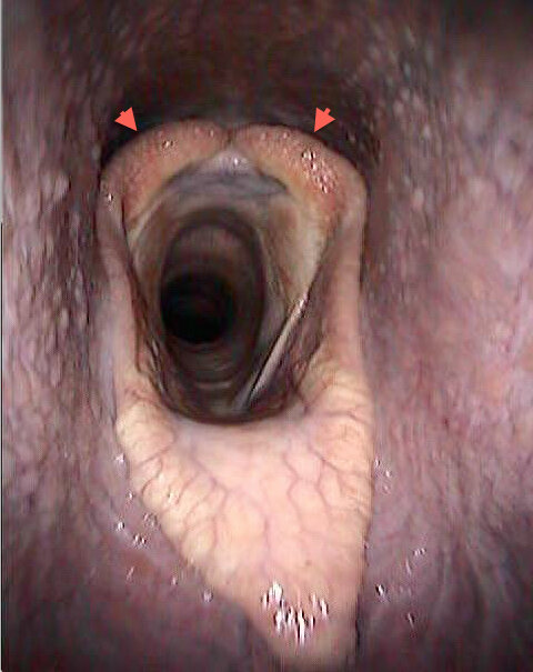
Recurrent laryngeal neuropathy (RLN) is the correct term for the condition better known as roaring or laryngeal hemiplegia. It is extremely common in Thoroughbreds and represents one of the major causes of poor performance or jockey-reported noise.
Because it is so important, many young horses are scoped at sales looking for laryngeal asymmetry to try to identify those that may be at risk of having this condition. But scoping at rest is fraught with difficulty – the larynx may be normal at rest only to show signs of weakness during exercise, so positive cases can be missed.
Conversely, tired young horses can have apparently poor laryngeal function at rest that in fact is of little significance. Many question whether subjecting foals and young horses to endoscopy at sales is reasonable, although in part this latter concern is reduced now that videoendoscopy is available.
Furthermore, the dynamic endoscopy, overground or on a treadmill, is widely accepted as the best way to evaluate horses suspected of having upper airway disorders leading to dynamic obstruction of the airway during exercise, a population that may or may not have obviously abnormal throats when examined at rest.
Nevertheless, there is a need for better tools to evaluate the larynx for clinical application and also to allow researchers to study the condition in more detail. Two recent studies published in Equine Veterinary Journal have addressed this issue..
To read more - subscribe here!
New research into the treatment of tendon and ligament damage in the Thoroughbred
Around 35% of the veterinary research and education budget is spent on projects to understand musculoskeletal disorders, improve their treatment, and prevent and minimize injury to racehorses.


