Equine Neck CT: Advancing diagnostic precision in racehorses
Words - Rachel Tucker MRCVS

Introduction
When considering neck disorders in the racehorse, we most commonly think of severe conditions such as acute neck trauma and cervical vertebral myelopathy (Wobbler Syndrome). These represent the most severe end of the scale of orthopedic and neurologic injury to the neck; and a diagnosis, or at least prognosis, is usually clear. However, neck conditions encompass a far wider range of clinical presentations.
At the milder end of the scale, signs may be subtle and easily missed, whilst still being responsible for discomfort and reduced performance. The recent ability to perform a computed tomography (CT) scan of a horse’s neck represents a major advancement in our ability to diagnose neck conditions. Timely and accurate diagnosis allows efficient and targeted treatment, the ability to plan schedules and improvement in welfare through the provision of appropriate treatment and earlier return to function.
In neurologic cases, an accurate diagnosis facilitates risk management for both the horse, their handlers and riders, while improving safety for all. As conditions and injuries of the neck are being better characterized using CT, new medical and surgical treatment options are being developed, giving the potential for improved outcomes and fewer losses from the racing industry.
This article summarizes how CT is being increasingly used by vets to diagnose conditions of the neck and how it is revealing previously unknown information and providing exciting new treatment opportunities.
Presentation

Conditions of the neck can cause a range of signs in the horse, which are wide-ranging in their presentation and variable in their severity. The manner in which these conditions present depends on which anatomical structures are affected. Issues affecting the bones, joints and/or soft tissues of the neck can all cause neck pain, which can manifest in a number of ways. Cases of neck pain can be severe, resulting in a horse with a rigid, fixed neck carriage, an unwillingness to walk and struggling to eat, perhaps due to a traumatic event. Neck fractures are thankfully uncommon but can be catastrophic.
More moderate signs might be displayed as a stiff neck, with reduced range of movement and resentment of ridden work. There may be pain on palpation of the neck and changes in the neck musculature. Increasingly, we are seeing horses with far more subtle signs, which are ultimately revealed to be due to neck pain and neck pathology. Typically, these horses might have an acceptable range of motion of their neck under most circumstances, but they suffer pain or restriction in certain scenarios, resulting in poor performance. This may be seen as tension through the neck, resisting rein contact, a reluctance to extend the neck over fences, or they may struggle on landing.
Riders might report a feeling of restriction or asymmetry in the mobility of the neck. In addition, these horses may be prone to forelimb tripping or show subtle forelimb lameness.
Any condition, which causes injury or disease to the spinal cord or nerves within the neck, also causes a specific range of neurologic signs. Compression of the spinal cord is most commonly caused by malformations or fractures of the cervical vertebrae, or enlargement of the adjacent articular process (facet) joints. This results in classic ‘Wobbler’ symptoms, which can range from subtle weakness and gait abnormalities, through to horses that are profoundly weak, ataxic and uncoordinated. This makes them prone to tripping, falling, or they may even become recumbent.
Peripheral nerve deficits are uncommon but become most relevant if they affect the nerves supplying the forelimbs, which can result in tripping, forelimb lameness, or local sensory deficits. This lameness might be evident only in certain circumstances, such as when ridden in a rein contact. This lameness is difficult to pinpoint as there will be no abnormality to find in the lame limb, indeed a negative response to nerve and joint blocks (diagnostic analgesia) will usually be part of the diagnostic process.
Horses can present with varying combinations and severities of neck pain, neurologic signs and peripheral nerve deficits, creating a wide range of manifestations of neck related disease.
Diagnosis

A diagnosis of neck pain is based on careful static and dynamic clinical examination and may be supported by seeing a positive response to treatment. Neurologic deficits are noted during a specific neurologic assessment, which includes a series of provocation tests such as asking a horse to walk over obstacles, back up, turn circles and walk up and down a hill. Confirming neck pathology as the cause of signs can be difficult. Until recently, radiography has been the mainstay imaging modality. Radiographs are useful for assessment of the cervical vertebrae and continue to play an important role in diagnosis; however the complex 3-dimensional shape of these bones, the large size of the neck and an inability to take orthogonal (right-angled) x-ray views means that this 2-dimensional imaging modality has significant limitations. High quality, well-positioned images are essential to maximize the diagnostic potential of radiographs.
A turning point in our diagnostic ability and understanding of neck dysfunction has been the recent adaptation of human CT scanners to allow imaging of the horse’s neck. A number of equine hospitals across the United Kingdom and Northern Europe now offer this imaging modality. We have been providing this service at Liphook Equine Hospital since 2017, with over 150 neck scans performed to date.
The CT procedure

A computed tomography (CT) scan combines a series of x-ray images taken from different angles around the area of interest to create a 3-dimensional volume of imaging data. This data is presented as a grayscale image which can be viewed in any plane and orientation. It provides excellent bone detail, and post processing techniques can provide information on soft tissue structures too. Additional techniques can be employed such as positive contrast myelography to provide greater detail about soft tissue structures. Myelography delineates the spinal cord using contrast medium injected into the subarachnoid space and is indicated in any case showing neurologic signs suggestive of spinal cord compression.
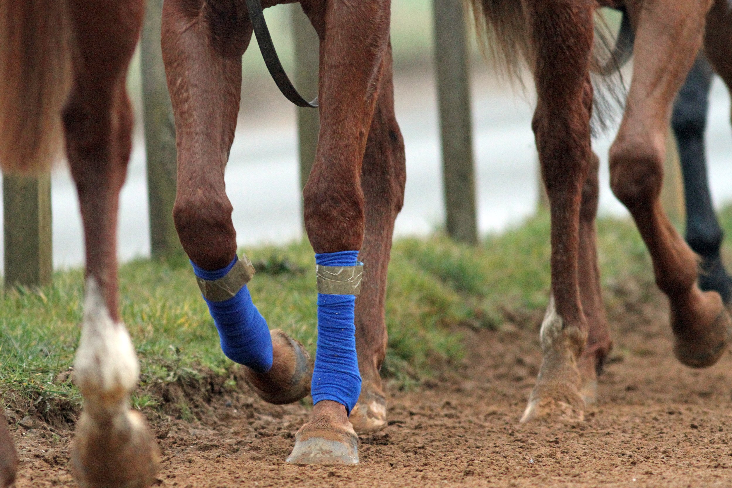
Neck CT is performed under a short general anesthetic. Scans without myelography typically take less than 20 minutes to complete. The entire neck is imaged, from the poll to the first thoracic vertebra. The procedure is non-invasive and low risk, with anesthetic-related complication presenting the main risk factor to the procedure. We have not encountered any significant complications in our plain CT scan caseload to date. Horses showing ataxia, weakness and incoordination (Wobbler’s), undergo CT myelography which adds around 20 minutes to the procedure. These horses are exposed to a greater level of risk due to their neurologic condition, the injection of a contrast agent and the increased chance of destabilizing a more severe lesion during the procedure.
CT is revealing more detailed information about ways that spinal cord compression can occur in Wobbler cases, about compression of spinal nerves resulting in forelimb gait deficits and precise detail about fracture configurations. It gives us detailed images of articular process joint disease, intervertebral disc disease, developmental conditions and anatomic variations. It is also revealing information about rare diseases such as vertebral abscesses or spinal neoplasia. As our caseload and confidence in the imaging modality grows, we are learning more about the value of CT in examining more subtle neck conditions. We are also bringing the benefit of a more accurate diagnosis, allowing precise targeted treatment and a better ability to provide a prognosis about outcomes—likely progression or safety factors. CT myelography allows circumferential imaging of the spinal canal and yields significantly more information than traditional x-ray myelography. As a result, we hope to enable better case selection of horses that may benefit from Wobbler surgery, with the goal of resulting in improved success rates of the surgery.
Innovations in treatment

New treatment options are emerging as a result of our more accurate diagnoses of neck pathology. Of the first 55 horses which underwent neck CT at our hospital, we were surprised to discover that 13 (24%) had some form of osteochondral fragmentation within the articular process joints of their neck. Some of these horses were young Thoroughbreds, bred to race but showing Wobbler signs. These tended to have convincing CT evidence of type 1 CVM (Wobbler Syndrome) and osteochondrosis affecting their neck. Others had fragments which were larger and more discrete, with evidence of articular process joint enlargement/arthritis but no other bony lesions. These horses were typically older and of a range of breeds and uses.
In those horses presenting with signs of neck pain but no neurologic deficits, surgical removal of these fragments was proposed. Following further consideration and cadaver training, we have begun to offer this surgery for horses that fit the appropriate criteria and have surgically accessible fragments. We have performed arthroscopic or arthroscopic-assisted fragment removal from eight articular process joints in six horses to date. No intraoperative or postoperative complications have been encountered; and five of six horses showed complete resolution of neck pain. In the sixth horse, full recovery was not anticipated due to the presence of additional neck pathology, but partial improvement occurred for two years. Fragment removal has relieved signs of neck pain and stiffness and caused improved performance in these horses.
Two procedures that are emerging to treat spinal nerve root impingement are a targeted peripheral nerve root injection and a keyhole surgical procedure to widen the intervertebral foramen. Nerve root injection is performed in the standing sedated horse under ultrasound guidance. Surgery is performed under anesthesia, using specialized minimally invasive equipment to widen the bony foramen using a burr. This surgery is in its infancy but offers an exciting treatment option.
Additionally, CT gives us the ability to better plan for fracture repair, undoubtedly improving our case selection for Wobbler surgery; it more accurately guides intra-articular injection of the articular process joints.
Summary
Computed tomography is transforming our ability to diagnose conditions of the horse’s neck. The procedure is low risk and now widely available in the UK and other parts of Europe. It is driving the innovation of novel treatment options with the goal of improving outcomes and reducing losses to conditions of the neck. Our CT findings are posing new questions about neck function, pain and neurologic disease and is an active area of ongoing research.
References:
Schulze N., Ehrle, A., Beckmann, I and Lischer, C. (2021) Arthroscopic removal of osteochondral fragments of the cervical articular process joints in three horses. Vet Surg. ;1-9.
Swagemakers J-H, Van Daele P, Mageed M. Percutaneous full endoscopic foraminotomy for treatment of cervical spinal nerve compression in horses using a uniportal approach: Feasibility study. Equine Vet J. 2023.
Tucker R, Parker RA, Meredith LE, Hughes TK, Foote AK. Surgical removal of intra-articular loose bodies from the cervical articular process joints in 5 horses. Veterinary Surgery. 2021;1-9.
Wood AD, Sinovich M, Prutton JSW, Parker RA. Ultrasonographic guidance for perineural injections of the cervical spinal nerves in horses. Veterinary Surgery. 2021; 50:816–822.
Enhancing horse safety in training and racing
The Gerald Leigh Memorial Lectures 2023

Article by Adam Jackson MRCVS
The Gerald Leigh Memorial Lectures, is an annual gathering devoted to the racing industry and the health and wellbeing of the horses involved.
This year, equine veterinarians, researchers, students and industry professionals from around the world attended the event, held June 8, 2023, at the historic Tattersalls Sales in Newmarket, England.
There were insightful and informative lectures that educated the attendants but also instigated a healthy, lively debate on the health and welfare of the training and competing of horses. The underlying theme that was present during the whole event was all members of the conference had a deep passion and commitment to continuously progress and improve on managing the welfare and wellbeing of the horses in the industry, both on and off of the track.
Two very special guest speakers, Sir Mark Prescott and Luca Cumani, wonderfully illustrated these sentiments as they described their reflections on the improvement and enhancement of horse safety.
Horse racing may be regarded as an elite sport, and all activities involving horses have an element of risk. All stakeholders in the racing industry must continuously work to ensure that the risks are minimized in order to reduce the number of injuries and fatalities that may occur in training and on the racecourse. There are now well-publicized concerns regarding the acceptability of exposing horses to risk in racing. These lectures and all of the attendees embraced the values of the public will so that there can be continued acceptance of horse sports.
Reducing the Incidence of Fractures in Racing
Christopher Riggs of The Hong Kong Jockey Club clearly outlined the various strategies to reduce the risk of fractures in racehorses. There are two principal strategies that may used to reduce the incidence of severe fractures in horses while racing and training:
1.Identifying extrinsic factors that increase risk and take action to minimize them.
An example would be investigating different racing surfaces in order to determine which may provide the safest racing surface. However, studies have provided limited evidence and support for subtle extrinsic factors.
2.Identifying individuals that are at increased risk and prevent them from racing or minimize that risk until the risk has subsided.

There are many research routes that are being undertaken to identify those horses that may be at a higher risk of fractures. There are investigations involving heritability and molecular studies that may provide evidence of genetic predisposition to fracture. However, Dr. Riggs explained that further understanding of the relationship between genetic, epigenetic and environmental factors is required before genetic screening is likely to be of practical use.
Pre-race screening of horses by diligent clinical examination is poor at reducing the incidence of fracture. Dr. Riggs described another strategy that may assist with a clinical examination that is the use of biomarkers in blood and urine.
Unfortunately, the precision to be of practical value has so far remained relatively unrewarding. Wearable technology that records biometric parameters, including stride characteristics, has shown some promise in identifying horses that are at increased risk of fracture; although Dr. Riggs explained that this work requires further development.
Finally, Dr. Riggs described both the use and current limitations of diagnostic imaging in identifying pre-fracture pathology in order to identify a horse at imminent risk of fracture. He conceded that further knowledge of the significance of the range of abnormalities that can be detected by imaging is incomplete.
Dr. Riggs concluded his lecture by expressing that the implementation of diagnostic imaging to screen “high-risk” horses identified through genetic, epidemiology, biomarkers and/or biometrics may be the best hope to reduce the incidence of racing fractures. This field can be advanced with further studies, especially of a longitudinal nature.
Professor Tim Barker of Bristol Veterinary School discussed the need for further investment in welfare research and education. One avenue of investment that should be seriously considered is the analysis of data related to (fatal) injuries in Thoroughbred racing over the last 25 years.
It was expressed, with the abundance of data that has been collected, that some risk factors would be relatively simple to identify. An encouraging example in the collection and use of data to develop models in predicting and potentially preventing injury has been conducted by the Hong Kong Jockey Club funded by the Hong Kong Jockey Club Equine Welfare Research Foundation. This may provide an opportunity to pilot the use of risk profiling to contribute to decision-making about race entries. In addition, the results of the pilot study combined with other sources of data may encourage race authorities to mandate the collection of veterinary and training data in order to help in risk mitigation.
Horse racing is an international sport, and there are different governing bodies that ensure racing integrity. However, the concept of social license equestrian sports and Thoroughbred horse racing continues to gain significant public attention. Therefore, racing governing bodies are increasingly aiming to provide societal assurances on equine welfare.
Dr. Ramzan of Rossdales Veterinary Surgeons provided an eloquent and clear message during his lecture that race yard veterinarians and trainers are instrumental in ensuring good horse health and welfare and reducing serious injury of the horse both while training or racing, which will provide sufficient trust and legitimacy from the public and society. This feasible goal can be reached with good awareness of members involved in the care and training of each individual horse and conveying this information and any concerns to their veterinarian. The veterinarian can also contribute by honing their knowledge and skills and working closely with yard staff in order to make appropriate and better targeted veterinary intervention.
In the last two decades, there has been an incredible evolution and exciting developments in diagnostic imaging in the veterinary profession. It is believed that these technologies can provide a significant contribution to helping in mitigating fracture risks to racehorses on the course and in training.
Professor Mathieu Spriet of University of California, Davis, described how these improvements in diagnostic imaging has led to the detection of early lesions as well as allowing the monitoring of the lesions’ evolution.

He continued by explaining the strengths and limitations of different imaging modalities such as computed tomography (CT), magnetic resonance imaging (MRI) and positron emission tomography (PET). Being one of the leaders in the use of PET in equine veterinary medicine, he presented further insight on how this particular modality provides high-resolution 3-D bone scans while being very sensitive to the identification of bone turn-over prior to the development of structural changes and allowing one to distinguish between active and inactive processes when structural changes are present.
He concluded his impressive lecture by providing evidence with amazing PET images that the role of imaging is not merely for diagnostic purposes to characterize clinical abnormalities, but can also be used as a screening tool in certain horse populations for fracture risk assessment or for the monitoring of lesions to provide clearance for racing.
Fractures, due to bone overloading rather than direct trauma occur commonly in Thoroughbred racehorses and are the leading cause of euthanasia on the racecourse. Despite many changes to race conditions, the number of catastrophic fractures has remained relatively static, with approximately 60 horses a year having a fatal fracture during a race in the UK.
Against this backdrop, there have been great developments in the diagnosis and treatment of fractures in the last 40 years. Prevention of racecourse and training fractures would be ideal so the development of efficacious techniques to screen horses at risk may reduce the incidence and preserve social licensing.

Dr. Ian Wright
One technique discussed by Dr. Ian Wright of Newmarket Equine Referrals was to help mitigate the impact of racecourse fractures, which would be acute immobilization of racecourse fractures, thus, reducing associated pain and anxiety while optimizing clinical outcome and reducing on course fatality rates. Because of our increased understanding of fracture pathogenesis and their associated biomechanics, effective fracture immobilization has been made possible. The majority of fractures that occur in flat racing and between obstacles in jump racing, are a result of stress or fatigue failure of the bone and not associated with trauma.
In addition, fractures seen on the racecourse are often found in the same specific sites (i.e., metacarpal/metatarsal condyles and the proximal sesamoid bones of the fetlock) and have repeatable configurations. With this understanding and knowledge, racecourse veterinarians can optimally immobilize a fracture in a logical and pre-planned manner.
As Dr. Wright expressed, this allows the fracture patient to have reduced pain and anxiety and enable the horse to be moved from the course comfortably so that it can be further examined. Ultimately, this allows the veterinarian and all stakeholders to make effective and judicious decisions for the sake of the horse’s welfare and wellbeing. As Dr. Wright concluded, this benefits both horses and racing.
Dr. Debbie Guest of the Royal Veterinary College discussed a different approach in mitigating the risk of fractures during training and racing by developing novel tools to reduce catastrophic fractures Thoroughbreds. Because it has been found that some horses are more inherently predisposed to fractures than other horses, Dr. Guest and her team have developed a genome-wide polygenic risk score so that one can potentially calculate an individual horse’s risk of fracturing during training or racing compared to the population as a whole.
This strategy may contribute in identifying genetically high-risk horses so that additional monitoring of the patients can be exercised during their careers and also leading to fracture risk, which are found to be the cause of approximately half of these incidents.
The system of using DNA testing to identify biological processes that may or may not be present ultimately leading to fracture risk may be a powerful tool in lowering the risk of catastrophic fracture and requires further research and application.
Cardiac events & sudden cardiac death in training and racing

In racehorses, sudden death that is associated with exercise on the racetrack or during training is a serious risk to jockeys and adversely affects horse welfare and the public perception of the sport. It is believed 75% of race day fatalities result from euthanasia following a catastrophic injury. The other 25% of fatalities is due to sudden deaths and cardiac arrhythmias are found to be the cause of approximately half of these incidents. The lectures focused on this area of concern by providing three interesting lectures on cardiac issues in the racehorse industry.
Dr. Laura Nath of the University of Adelaide, explained the difficulties in identifying horses that are at risk of sudden cardiac death. It is believed that part of the solution to this difficult issue is the further development and use of wearable devices including ECG and heart rate monitors.
With the use of these technologies, the goal would be to recognize those horses that are not progressing appropriately through their training and screen these horses for further evaluation. This course of action has been seen in human athletes that develop irregular rhythms that are known to cause sudden cardiac death with the use of computational ECG analysis, even when the ECGs appear normal on initial visual inspection.
Knowing that ECGs and particularly P-waves are used as a non-invasive electrocardiographic marker for atrial remodeling in humans, Dr. Nath recently completed a study on the analysis variations in the P-wave seen on ECGs in athletic horses and found that increases of P-waves in racehorses are associated with structural and electrical remodeling in the heart and may increase the risk of atrial fibrillation (cardiac event).
Dr. Celia Marr of Rossdales Veterinary Surgeons continued the discussion of cardiac disease in both the training and racing of horses. Unfortunately, cardiac disease knowledge does lag compared to musculoskeletal and respiratory diseases when considering the causes of poor performance in racehorses. Due to the fact that cardiac rhythm disturbances are fairly common, occurring in around 5–10% of training sessions in healthy horses in Newmarket and over 50% of horses investigated for poor performance, Dr. Marr expressed the need for further research and investigation in this area.
In addition, this research needs to determine if there is indeed a link between heart rhythm disturbances and repeated episodes of poor performance and sudden cardiac arrest. ECGs and associated technologies are helpful, but there are limitations such as the fact that rhythm disturbances do not always occur every time the horse is exercised. Therefore, it would be of great value that a robust criterion is established when evaluating ECGs in racehorses. The Horserace Betting Levy Board has provided funding for investigation by initially exploring the natural history of paroxysmal atrial fibrillation (self-correcting form) to understand risk factors and predict outcomes for affected horses.
Continuing the theme of the lectures on irregular heart rhythms and associated sudden cardiac death (SCD) in training and racing, Professor Kamalan Jeevaratnam described his exciting research in using artificial intelligence (AI) to identify horses at increased risk of developing irregular rhythms that may cause SCD.

AI is an exciting and rapidly expanding field of computer science that is beginning to be implemented in veterinary medicine. With funding by the Horserace Betting Levy Board and the Grayson Jockey Club Research Foundation, Professor Jeevaratnam of the University of Surrey, has piloted three novel algorithms that help predict horses with rhythm abnormalities through the analysis of horses’ ECGs.
It was acknowledged that further research is required to develop this technology by using data collected from multiple sources, but the initial results are promising in the development of an useful AI tool to identify horses at risk of SCD and prevent catastrophic events, thus, ensuring the welfare of the horse in racing.
Conclusion
The Gerald Leigh Memorial Lectures was a thoroughly successful and enjoyable event attended by a variety of different members of the horse racing industry. Not only did the lecturers provide interesting and valuable information but also excitement for the future of racing. It was very clear that all the lecturers and attendees were passionate and committed to the racehorse welfare and wellbeing as well as retaining the social license for an exciting sport.