Assessing the approaches to diagnosing and treating proximal suspensory desmitis
/Article by Connor Parsons DipWCF

Diagnosing proximal suspensory desmitis in the hind limb can be difficult. However, the modern diagnostic modalities available to the industry today makes it possible to isolate injuries, allowing both veterinarians and farriers to work together to achieve the best diagnosis and prognosis possible for the equine in question.
In this article, Connor Parsons reviews the anatomy and function of the suspensory ligament, causes and signs of proximal suspensory desmitis and whether there is an ideal procedure for diagnosing, treating and formulating a prognosis for the horse as part of his DipHE Farriery studies.
ANATOMY
The equine limb is complex yet effective. The suspensory ligament is made up of dense white fibrous connective tissue which suspends the fetlock and prevents hyperextension.

Originating at the proximal, plantar aspect of the third metatarsal/carpal attaching to two palmar depressions distal to the carpometacarpal and tarsometatarsal joints descending the channel formed by the 2nd, 3rd and 4th metatarsal/carpal, bifurcating two thirds of the way down the 3rd metatarsal/carpal, making a firm attachment to the palmar aspect of the proximal sesamoids, pulling the sesamoids proximally, then travelling dorsally and distally at an oblique angle to merge with the common digital extensor tendon. This forms a sling to support the fetlock joint. The ligament and its branches are strong but only slightly elastic (Devereux, 2006).
The suspensory ligament also forms a part of the hindlimb stay apparatus which is a system of ligaments, tendons and muscles that work together to allow the horse to stand and doze with minimal muscular effort. Also known as the fright and flight mechanism (Colles & Ware, 2020).
DAMAGE TO THE SUSPENSORY LIGAMENT
Suspensory ligament damage can affect horses of all breeds and ages. However, it is most common in competition horses. Proximal suspensory desmitis (PSD) is inflammation or damage of the main body at the origin of the ligament at the proximal end of the third metacarpal/metatarsal.
The suspensory ligament can be inflamed or there can be changes to the fibre pattern of the ligament. These cases will present with lack of performance, being worse on soft surfaces. In more severe cases a core lesion (hole) can be seen on an ultrasound scan, where a number of fibres have ruptured. This type of injury will have a more sudden onset of lameness (Dyson, 1994). Injury can be solely within the ligament, involve tearing of the fibres of the ligament or be connected to avulsion fractures at the origin, involving the proximal 3rd metacarpal/tarsal (Baxter, 2020). Complete rupture is possible, however, very rare. The prognosis for a complete rupture is not favourable (Dyson, 1994).
Although the suspensory ligament has a slight elasticity to its make-up, if it is stretched it tends to heal with a loss of elasticity making it susceptible to recurrent damage (Colles & Ware, 2020).
SIGNS OF PROXIMAL SUSPENSORY DESMITIS

Proximal suspensory desmitis is a difficult condition to diagnose as the hind limb is complex and many of the functioning structures work in unison. A horse suffering with inflammation or damage to the main body of its hind suspensory can present one of three ways. It may have a unilateral lameness, a bilateral lameness or just a general decrease in performance (Dyson,1994).
CAUSES OF PROXIMAL SUSPENSORY DESMITIS OF THE HINDLIMB
Although there has been extensive research into proximal suspensory desmitis, there is no primary cause in all cases.
Proximal suspensory desmitis is a common injury in both front and hind limbs of the equine athlete. Usually bilateral in the hind limb (Dyson, 2016). All types and breeds of horses are susceptible to this type of injury. Poor conformation is a contributing factor to proximal suspensory desmitis.
Conformational defects such as straight hocks, sloping pasterns and long-toe, low-heel conformations would be at higher risk to injury. These conformational defects will all apply unnecessary pressure to the suspensory ligament. Horses that have suffered with this condition will be predisposed to a repetitive strain injury of this ligament (Devereux, 2006). Overextension of the tarsus as a result of overextension of the fetlock has been linked to proximal lesions. The higher the severity of trauma, the higher the severity of ligamentous lesion. Working horses on deep, soft surfaces will increase the risk of this injury (Baxter, 2020).
The hindlimbs are more frequently affected with this condition than the forelimbs with a much lower success rate of the horse returning back to performance prior to rest (69% hind vs 80% forelimb) (Colles & Ware, 2020).
DISCUSSION

In a study of six horses, this is an extremely small cohort of horses to be able to state an average age a horse is likely to present with this condition. This study also shows that all of the horses studied were of varying fitness levels, therefore stating that this does not affect the likelihood of injuring the hind suspensory ligament. There was only one horse in this study that was unfit and overweight. The rest were all competition fit with good muscle mass, showing that fitness doesn’t necessarily decrease the risk of this injury happening. The case history of the six horses studied did not include which discipline or level the horse was working at. This would be an interesting factor to consider when looking at which horses would be more susceptible to proximal suspensory desmitis.
Each individual case was being looked after by different veterinarians, giving a clear picture of different approaches on how to diagnose and treat this condition. Although for the purpose of a study the varying opinions will make the comparison more difficult. All horses presented with a reduction in performance prior to veterinary contact. Only one horse was reported with a bilateral lameness behind. Flexion testing appeared to aggravate the lameness making it more prominent to see. Local analgesia has been shown to be effective in isolating the area to be investigated. Also, showing lameness on the other hind once the worse limb has been blocked out.
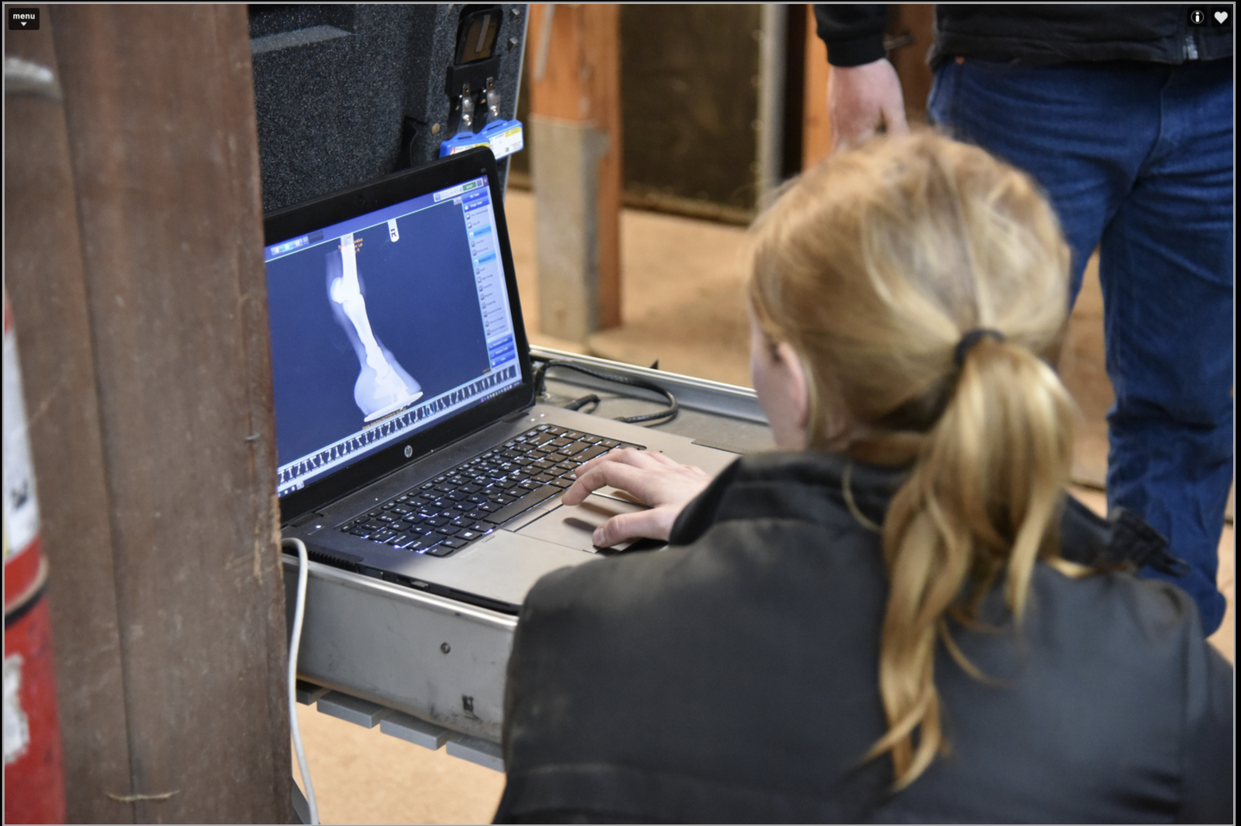
Using digital diagnostic modalities such as ultrasonography to diagnose this condition allows the veterinarian to study the changes in the fibre pattern of the suspensory ligament. This will allow the veterinarian to see the severity of damage caused and allow them to provide the best treatment plan possible. In this study only one horse had a lesion while the other five horses had thickening and slight changes to the fibre pattern. Horse 2 had lesions on both hind limbs however the veterinarian didn’t medicate, box rest was recommended. His prognosis was guarded.
Although radiographs of the feet don’t directly help with the diagnosis of proximal suspensory desmitis, they do allow the farrier to trim accordingly to restore the hoof back to correct hoof pastern axis and mediolateral foot balance. This will reduce lever arm forces thus reducing any unnecessary pressures on the plantar aspect of the limb.

Horses were radiographed for foot balance to aid with remedial trimming and shoeing. This will increase the equines prognosis allowing the farrier to have a clear picture of what is being dealt with. All of the horses that were radiographed presented with a negative sole plane and weak heels.
The question is whether this foot conformation is because the horses are wanting to apply more pressure to the caudal aspect of the hoof in the landing phase, reducing the movement of the metacarpophalangeal articulation. This is an attempt to reduce the loading forces applied to the suspensory ligament. However, it will also cause the heels to become weak. Or, if this conformational defect has caused the suspensory ligament to become inflamed or damaged, thus causing proximal suspensory desmitis.
Proximal suspensory desmitis can be secondary to other conditions such as hock conditions or sacroiliac problems which cause the horse to adopt a different gate. Therefore causing unnecessary loading on the suspensory ligament. It is important that the primary cause is diagnosed and treated when treating proximal suspensory desmitis. This is where scintigraphy can be a useful tool to get a clear picture of the cause involved in individual cases. Scintigraphy is an expensive diagnostic modality which carries significant health and safety risks, this must be taken into consideration when dealing with cases.
All horses studied were worse on a soft surface where it is harder for the horse to guard itself from soft tissue injuries. Horses that are worse on soft surfaces generally are suffering from soft tissue pain. However, nerve blocks will help the veterinarian pinpoint the structures involved when diagnosing lameness.

Although it is possible to have a unilateral lameness with proximal suspensory desmitis in the hind limb it is most common for the lameness to be bilateral. All of the horses in this study had a bilateral lameness, generally worse on one limb than the other. Although presenting prior to veterinary contact as lack of power or struggling to strike off on the correct canter lead.
When a veterinarian is deciding on a treatment plan, the horse is looked at carefully including its previous history as some treatments come with higher risks, although can be extremely effective for reducing inflammation. Shockwave treatment comes with minimal risks involved and is effective; however, many racing authorities require a mandatory 5 day Stand-Down period from racing following the administration of extra-corporeal shockwave therapy. Findings from this study show that the horses with the best prognosis of getting back to competitive work have undergone surgery. Understandably this is the last resort treatment as it is invasive and expensive for the client.
Only one horse from this study did not have any medical intervention and this horse had the least favourable prognosis. This would suggest that box rest alone is not generally enough if the horse is expected to get back to full athletic fitness. The most common veterinary treatment is steroidal injections into the area of interest and shockwave therapy with rest. However, the use of corticosteroids in horses in training often adopt a clear 14-day exclusion on the use of intra-articular (joint) injections before racing in line with different racing authority regulations.

Water based therapy can also be considered as part of the recovery process when bringing the horse back into work. It’s known to reduce limb oedema, stimulate nerves, and improve circulation, which speeds the healing process and provides pain relief. It also aids in joint stability, providing all-around support to the limbs.
Cold water therapy is typically prescribed when the goal is to reduce heat and inflammation. Applying cold water or ice reduces the amount of accumulating fluid to an injured area and can somewhat numb the area, causing a topical analgesic effect.
Underwater treadmills are often used for horses with tendon and ligament injuries to provide a gradual transition back into exercise and regain the range of motion. Swimming is also used to condition the horse without putting a load on the skeletal system. It is often used in the early stages of tendon and suspensory injuries due to no pressure being placed on the lower limb. Trainers who use swimming as part of their routine often find that, in addition to the cardiovascular workout, it also helps the horse relax and settle its mind.
This is not always successful and horses are then admitted for surgery. While the surgery for this condition is successful, there must be consideration taken into the fact that it is not legal to compete at certain levels once this surgery has taken place.
The study shows that the farriery treatment involved when dealing with this condition is varied, depending on which veterinarian the horse is being looked after by. However, the author has had positive results from many different shoeing styles. The main importance of trimming and shoeing for this condition has been shown to restore the best possible hoof pastern axis through trimming, supporting the entire limb and fitting a shoe with an early breakover. This will reduce the lever arm on the metacarpophalangeal articulation, thus minimising unnecessary pressure on the suspensory ligament.
CONCLUSION
Having such a small cohort of horses in a study makes it difficult to finish with a conclusive result. This small study however, has given a positive result in the diagnosis stages of dealing with this condition. At this stage nerve blocks are invaluable along with ultrasonography. In less obvious cases MRI is useful to gain a diagnosis and occasionally scintigraphy will be used to locate the problem. Radiography is a useful tool when dealing with PSD and checking the origin area for avulsion fractures.
This study has also shown that there is a link between a negative solar angle and proximal suspensory desmitis. However, this would need to be studied further and on a greater scale to determine why there is a link between this conformational defect and this condition.
It is paramount that correct foot balance is achieved by the farrier. To achieve this foot balance radiographs are required. This study has shown that there is no definitive way to shoe for this condition, however it has shown a positive result from an early breakover shoe, allowing the horse relieve pressures on the caudal aspect of its hoof. Horses that had the best prognosis underwent surgery, allowing them to get back to competitive fitness.
REFERENCES
Baxter, G. M., 2020. Adams and Stashak's Lameness in Horses. 7th Edition ed. Hoboken, NJ: John Wiley & Sons.
Colles, C. & Ware, R., 2020. The Principles of Farriery. 2nd edition ed. Marlborough: J.A.Allen.
Devereux, S., 2006. The Veterinary Care Of The Horse. 2nd Edition ed. London: J.A.Allen. Dyson, S., 1994. Proximal suspensory desmitis in the hindlimb: 42 cases. British Veterinary Journal, 150(3), pp. 279-291.
Dyson, S., 2016. American Association of Equine Practitioners. [Online] Available at: https://aaep.org/horsehealth/lowdown-high-suspensory-disease-proximal-Suspensory-desmitis [Accessed 19 11 2022].
Smith, M., 2022. Newmarket Equine Hospital. [Online] Available at: https://www.newmarketequinehospital.com/media/pm1beabc/hah349-Vet_susp_desmitis-final.pdf [Accessed 9 April 2023].