There's more to it than meets the eye!
/Article by Adam Jackson MRCVS

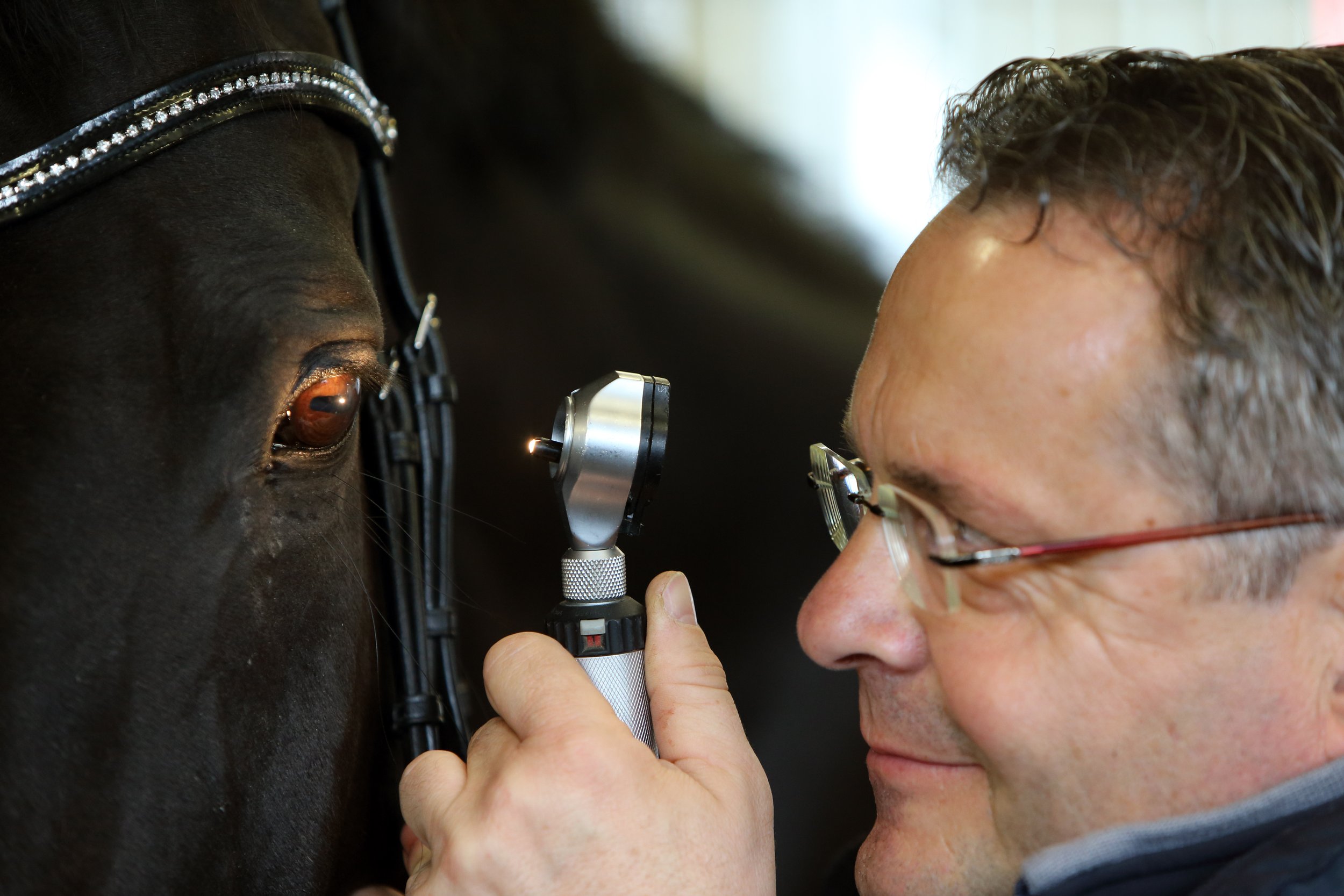
The horse’s eyesight has evolved to scan its environment rather than picking up sharp details, in order to survive from predators. As a prey animal, the horse’s eyes are eight times larger than a human’s eye; however, this makes them more vulnerable to injury and disease that may be catastrophic. Horses develop many of the same eye problems as humans such as glaucoma, corneal ulcers, cataracts and other issues.
The working of the eye
Vision is provided by light entering the eye, which is made into an image by the brain through various complex biomechanical and physical processes.
As light enters the eye, it is targeted to the retina by the cornea and the lens bending the light. This light reaches the sensory tissue at the back of the eye. In fact, the retina or nervous tunic is made of cells that are extensions of the brain coming off the optic nerve. The retina consists of 2 types of photoreceptors called rods and cones. The rod cells are more light-sensitive, thus providing night vision, whereas the cones are less light sensitive but provide visual acuity and the ability to see colour. The optic disc in the retina does not contain photoreceptors and is the location the optic nerve leaves the eye to transmit the visual information to the visual cortex of the brain.
Visual field of the horse
Because the horse’s eyes are positioned on the side of the head, the range of vision is roughly 350 degrees, thus, allowing the horse to spot potential predators. Due to the positioning of the eyes, the horse has two blind spots that include in front of the face and behind its head extending over its back and behind the tail.
The horse has both binocular and monocular vision. Monocular vision means vision in one eye only and binocular vision means seeing with two eyes. 65 degrees of the 350 degree vision consists of binocular vision while the remaining 285 degrees is monocular vision. As a result, the horse has a smaller field of depth perception compared to a human. The horse must raise or lower its head in order to increase its range of binocular vision. By introducing a bit and making the horse hold its head perpendicular to the ground, the binocular vision becomes less focused on distant objects and more focused on what is immediately in front of the horse. Show jumpers and jump jockeys allow the horse to raise its head a few strides before a jump so that the horse can properly assess the jumps to allow appropriate take-off.
Sensitivity to light
Horses’ eyes have evolved to allow them to have good vision in dim light and due to this evolution they have better vision on slightly cloudy days compared to sunny, bright days. There are two particular structures that allow them to have superior night vision, which include a high proportion of rods to cones (20:1) and the presence of the tapetum lucid.
The horse’s large pupils allow a large amount of light to enter and the size of the retina allows a high number of cells to be involved in the capturing of light. In addition to the rods and cones, the horse's tapetum lucidum is a reflective structure in the back of the eye that bounces light back to the photoreceptors for a second time, thus further increasing the ability to capture more light. Ultimately, this structure allows greater night vision.
Interestingly, horses have also evolved structures to protect their eyes from photic damage during bright sunny days. The pupil has the ability to significantly constrict in order to reduce the amount of light entering the eye. In addition, there is a structure referred to as the corpora nigra, which is a bulbous structure extending from the iris into the space of the pupil that acts as a shade.
Colour Vision
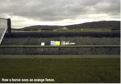
Horses have dichromatic vision; therefore, they are not colour blind but they have a smaller spectrum than humans typically do. Horse’s dichromatic vision means they see in the green-blue spectrum and the ocular variations based upon them. They cannot distinguish red and are often thought to have a red-green colour blindness. The horse’s colour vision must be taken into account when designing obstacles for horses to jump.
Eyelids
There are three layers to the eyelids that include a thin layer of skin covered in hair, a layer of muscles that allow the opening and closing of the eyelid and the palpebral conjunctiva, which lies against the eyeball. The horse also has a third eyelid, also known as the nictitating membrane which has the function of protecting the cornea.
Non-pigmented third eyelids are more susceptible both to solar-induced inflammation and to squamous cell carcinoma. Therefore, careful scrutiny of this structure is important. Prominence of the third eyelid may be a result of inflammation caused by solar-induced inflammation or conjunctivitis (inflammation of the conjunctiva). Inflammation and neoplasia should be differentiated on the basis of clinical appearance. For example, squamous cell carcinoma has a plaque-like appearance and erosion. Conjunctivitis is the inflammation with thickening and reddening of the transparent membrane that lines the eyelid and eyeball. Any suspected tumour should be excised and undergo histopathology to determine if it is indeed neoplasia or a type of inflammation. Other neoplasia that may occur in the eyelids are melanomas or periocular sarcoids.
Entropion is the inversion of the eyelid margin and lashes. Often seen in foals as a consequence of either anatomical imperfection or of dehydration and debility, it is the inward rotation of the eyelid that leads to the rubbing of hair in the cornea leading to keratitis. Later onset entropic is usually a consequence of a traumatic injury and can result if primary repair of an eyelid laceration has not been performed.
Trauma to the eyelids may result in bruising or a laceration. If bruising has occurred, a warm compress may be helpful if the horse will tolerate it. If a laceration has occurred it should always be repaired.
Lacrimal system
The horse has a pair of nasolacrimal ducts that carry lacrimal secretions, commonly known as tears, from the eye to the nasal cavity.

Keratoconjunctivitis sicca is a deficiency in the acqueous portion of the tear film and is relatively rare. If it occurs, it is a result of damage to the facial nerves or direct damage to the lacrimal gland or duct. With the lack of tears the cornea appears dull and lacklustre and may lead to corneal ulceration. It is often associated with a mucopurulent eye discharge as well as pain and inflammation. This condition can be managed with regular cleaning and the application of a tear replacement solution.

Acquired stenosis/occlusion of the lacrimal drainage system may be a consequence of infectious, trauma, neoplastic or inflammatory disease within the drainage system or external to it. It is often presented with epiphora (tear overflow) or a mucopurulent discharge if infection is involved. Following treatment of the underlying cause, the goal is to re-establish the drainage system with flushing of the duct with saline solution, or a combination of steroid, antibiotic (if required) and saline solution.
Conjunctiva/Sclera
The sclera is the white of the eye which is the relatively tough outer layer of the eye and is covered by a thin mucous membrane, referred to as the conjunctiva, and runs from the edge of the cornea and covers the inside of the eyelid.
Conjunctivitis is the inflammation and swelling of the conjunctiva and includes a primary conjunctivitis or a secondary conjunctivitis. Primary conjunctivitis is inflammation caused directly by irritants, chemicals, toxins and bacteria. However, conjunctivitis may be secondary to another ocular disease such as disorders of the lacrimal system, eyelid problems, and keratitis. In addition, conjunctivitis may be a non-specific symptom of other systemic diseases such as a respiratory viral infection. Conjunctivitis presents with a reddened inflamed conjunctiva with mould, purulent, serous or a combination of these discharges. The horse will have discomfort of the eye with this ailment.
Conjunctival foreign bodies are often acute and unilateral and caused by organic material resulting in excessive tearing, inflammation of the conjunctiva and ocular discomfort.
Conjunctival neoplasia is most often a squamous cell carcinoma (SCC) as this tumour usually affects areas of epithelial transition such as the mucocutaneous junction of the eyelids. The extent and appearance of the lesion is variable but SCC should always be considered especially in those horses lacking pigment in those areas. The symptoms range from mild ocular discomfort with discharge to plaque-like and cauliflower-like masses without ulceration.

Cornea
The cornea is the transparent front part of the eye that covers the iris, pupil and anterior chamber. It is a domed-shaped structure that acts as the eye’s windshield protecting the eye from insult such as an infection. Along with the tear film, it provides a proper anterior refractive surface for the eye, in fact, it contributes two-thirds of the refractive power of the eye. Congenital problems of clinical significance are rare in horses but acquired corneal problems as a result of trauma are common in horses.
Traumatic keratitis due to lacerations or penetrating injuries are common and in most cases involve full thickness penetration, acqueous loss and iris prolapse. This condition presents with sudden and severe pain accompanied with excess tearing and blepharospasm (involuntary tight closure of the eyelids). The extent of the damage to the cornea can be determined by the use of fluorescein dye. If the wound is not repaired quickly then the iris may become incarcerated and the restoration of the normal eye anatomy is difficult.
Abrasions to the surface of the cornea is a common condition seen by equine practitioners. Some simple scratches heal quickly while others may become more complex, involving fungal or bacterial infections resulting in a protracted recovery.

Corneal ulcers are a defect in the surface of the epithelium of the cornea that involves the underlying stroma. They are often described as sores on the cornea. It is important that they are diagnosed and treated promptly as there is potential that the horse’s vision may be affected. The clinical symptoms are often ocular discomfort with excessive tearing, squinting or blepharospasms. Discolouration and swelling of the cornea and the eventual development of blood vessels around the ulcer and an irregularity of the cornea. The depth of the ulcer must be established and it may range from superficial to deep.
Liquefactive stromal necrosis (melting ulcers) are not an uncommon condition in the horse and may present acutely or as a progression from a corneal ulcer. It should be deemed as an emergency because corneal perforation may result. This disease may be accompanied by uveitis.
Corneal foreign bodies are usually organic material and present with blepharospasm, excess tearing and pain. Various illuminations, magnifications as ophthalmic stains may be used to identify it and aid in removal.
Bacterial keratitis is often seen after a corneal injury especially if an ulcer is present. The horse will demonstrate acute eye pain with serous discharge that quickly becomes mucopurulent or purulent. The clinical appearance is not usually diagnostic and cultures and scrapings should be taken from the edge of the ulcer. This procedure ensures the correct selection of treatment and pain relief.
Mycotic keratitis is uncommon in the UK but with the changing climate it may become more prominent. This type of keratitis is a result of fungal growth so tends to occur in climates supportive of this type of growth. Diagnosis is based on the history, clinical appearance and the demonstration of fungal hyphae and positive fungal culture. This disease may be a consequence of inappropriate drug therapy (such as corticosteroids) or from previous corneal trauma. Following the identification of the fungus, topical treatments can be used but may take weeks to months.
Uveal Tract
The uveal tract consists of three parts that include; The choroid which is the tissue layer filled with blood vessels; The ciliary body that is the ring of tissue containing muscles that change the shape of the lens as well as producing the clear fluid that fills the space between the cornea and the iris; The iris which is the coloured part of the eye.
Persistent pupillary membranes are vascular arcades and developing tissue of the eye that fail to atrophy as the eye matures. These are very common in horses and usually have no consequence and no treatment is needed.
Cysts may arise in various parts of the uveal tract and are not uncommon in the horse. Irrespective of their origin, they may vary between pigmented to unpigmented and are smooth, round and do not invade neighbouring tissue. No treatment is required except on rare occasions when they interfere with the horse’s vision.
Neoplasia of the uvea is not common but may arise and are often melanomas that are locally invasive but without cellular malignancy.
Trauma of the iris may result from direct trauma, or a secondary consequence of corneal perforation or a whiplash injury. Any uveitis that is caused by trauma can be treated medically. If there is an iris prolapse, then the iris is placed back into the anterior chamber provided they are not contaminated. Any foreign bodies must be removed and any hyphaema (bleeding in the anterior chamber between the cornea and iris) is usually left to be resorbed naturally.

Uveitis is inflammation of the uveal tract and can cause eye pain and alterations in vision. There are many causes of uveitis that include trauma, lens-associated uveitis, general viral infections (such as equine viral arteritis) and bacterial disease (such as Rhodococcus equi in foals). However, many situations of uveitis are an immune-mediated uveitis often referred to as equine recurrent uveitis (also known as moon blindness). Uveitis may present as an acute or a chronic condition.
This disease can be treated medically often with the use of a sub-palpebral or nasolacrimal lavage system. In addition, the patient should be placed in a quiet fly-free and dust-free environment.
Lens
The lens is a clear curved disk that sits behind the iris and in front of the vitreous of the eye, which bends light as it enters the eye to develop an image. The horse’s lens is large and minor opacities associated with embryonic remnants are common.
Cataracts are the most common lens abnormality to be encountered causing an opacity of the lens. Cataracts may be acquired from trauma or post-inflammation situations. However, cataracts may be congenital commonly seen in Arab and thoroughbred foals. These opacities can be classified in various ways:
Age of onset – juvenile, senile or congenital

Cause - post inflammation (uveitis) or trauma
Location – cortical, capsular, nuclear, polar, equatorial
Stage of development – immature, mature, hypermature
Most cataracts cause no obvious visual deficits unless they are dense and obstruct the visual axis. in which cataract surgery may be considered.
Acqueous drainage
The acqueous humor is a transparent water-like fluid similar to blood plasma but containing low protein levels. It is secreted from the ciliary body (a supporting structure of the lens) and fills both the anterior and posterior chamber of the eye.
Glaucoma is a pathological elevation of the intraocular pressure resulting in the optic nerve becoming damaged . Primary glaucoma in horses is exceptionally rare while secondary glaucoma is uncommon but may occur after anterior segmental inflammation. Often there is little to no pain but an enlarged globe and raised intraocular pressure with the lack of pupillary light reflex may be seen. Treatment may be attempted if the vision is present with various medications to reduce the intraocular pressure. If the horse is blind it may be left without treatment.
Conclusion
Good eye care is vital as the horse relies on its site to receive a great deal of information on its surrounding environment. Even with the horse holding its head forward it has remarkable peripheral vision but the horse’s vision is a little blurrier and less colourful compared to humans. In addition, both the strengths and weaknesses of the visual abilities of the horse must be seriously considered when looking at various techniques for training.
